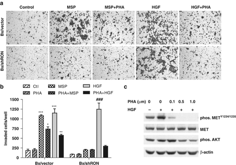
Figure 5.
MET inhibition blocks HGF-induced cell invasion. Bx/vector and Bx/shRon cells were seeded in 24-well Matrigel invasion chambers and treated with 200 ng/ml of MSP or 50 ng/ml of HGF for 24 h in the presence or absence of 0.5 μM MET inhibitor PHA-665752. (a) Representative images of Matrigel invasion assays (6 × 10⁴ cells/well plated). (b) The quantitative graph of invasion assays (3 × 10⁴ cells/well plated). The data represent mean±s.d. from repeated duplicate experiments. *P<0.05; **P<0.01; ***P<0.001 compared with Bx/vector control; ###P<0.001 compared with Bx/shRon control. (c) Subconfluent BxPC-3 cells were serum starved for 16 h and pretreated cells with different doses of PHA-665752 for 4 h, then stimulated with 50 ng/ml of HGF for 30 min. Cell lysates were analyzed by western blotting. Inhibition of phosphorylation of MET and AKT by PHA-665752 was detected with phos-METY1234/1235 or phos-AKT antibodies, respectively.

