Abstract
Since the discovery of the “starburst polymer”, later renamed as dendrimer, this class of polymers has gained considerable attention for numerous biomedical applications, due mainly to the unique characteristics of this macromolecule, including its monodispersity, uniformity, and the presence of numerous functionalizable terminal groups. In recent years, dendrimers have been studied extensively for their potential application as carriers for nucleic acid therapeutics, which utilize the cationic charge of the dendrimers for effective dendrimer-nucleic acid condensation. siRNA is considered a promising, versatile tool among various RNAi-based therapeutics, which can effectively regulate gene expression if delivered successfully inside the cells. This review reports on the advancements in the development of dendrimers as siRNA carriers.
Keywords: dendrimer, siRNA, delivery
1. Introduction
Interference of the cellular pathway of protein synthesis by a RNA interference mechanism has been considered the most powerful approach by which over-expression of aberrant proteins can be successfully minimized [1,2,3]. RNA interference (RNAi) is the mechanism adopted by most eukaryotic cells, where by small double-stranded RNA (dsRNA) molecules control gene expression by potent degradation of its complementary messenger RNA (mRNA) sequence, commonly referred to in plants as post-transcriptional gene silencing (PTGS) [4,5]. While transcriptional gene silencing involves mutations to specific regions of the genes that results in the production of proteins with non-functional domains, PTGS involves repression of gene expression through specific mRNA degradation [1,6].
Since their discovery less than a decade ago, RNAi-based therapeutics have found their way into clinical trials [7,8,9]. Gene silencing at the post-transcriptional level can be achieved by three basic mechanisms, including the use of antisense oligo-deoxyribonucleic acids (ODN), ribozymes and dsRNA [10]. RNAi by double-stranded small interfering RNA (siRNA) is considered the most powerful technique since-specific siRNA complementary to the target mRNA elicits potent target-specific knockdown of any mRNA [10,11]. Since the discovery of their ability to interfere with a disease-causing aberrant protein at the level of gene expression, siRNAs have garnered considerable attention as potential therapeutic agents for the treatment of cancers and other related diseases [2,12]. However, the successful use of this molecule faces challenges for in vivo delivery [13,14,15]. Low resistance against enzymatic degradation, limited translocation across the cell membrane and a substantial liver clearance limit the applications [16]. Therefore, development of an efficient delivery system requires protection of siRNA from degradation, limitation of rapid renal clearance, and promotion of targeted intracellular delivery for effective knockdown of protein synthesis with minimal side-effects.
For decades, cationic polymers, lipids and polyamino acids, have been used extensively as carriers for nucleic acid therapeutics [17,18,19,20,21,22,23,24]. The cationic charge of the carriers allows electrostatic interaction with the anionic nucleic acid molecules that leads to effective condensation. These nanosized siRNA-polymer complexes can protect nucleic acids from non-specific interactions and enzymatic degradation in the systemic circulation. The cationic polymers utilized for nucleic acid delivery applications include low-and high-molecular weight poly(ethyleneimines), cationic poly-saccharides, such as chitosan, dendrimers, polypeptides such as poly-L-lysines, polyarginines and various cationic lipids [25,26,27,28,29,30,31,32,33].
Dendrimers are a relatively new class of cationic polymers, which have been studied extensively as potential nucleic acid carriers [34,35,36,37,38]. Since the synthesis of a “cascade molecule” in 1978, and later, a starburst polymer in 1985, such hyperbranched macromolecules with a defined core and repetitively attached exterior units have been synthesized with a variety of chemical structures. Dendrimers are highly symmetric, spherical, hyperbranched macromolecules having a tunable structure, molecular size, and surface charge. Their unique structural features including chemical homogeneity, the possibility of increasing the generation by repeated attachment of chemical groups, and a high density of functional groups on the surface for numerous ligand attachments make dendrimers an excellent polymer candidate for numerous biomedical applications.
Polycationic forms such as poly(amidoamine) (PAMAM) and poly(propyleneimine) (PPI) dendrimers have been studied extensively as drug/gene carriers [36,37,38,39,40]. However, the potential of dendrimers as a siRNA delivery vector remains relatively unexplored. This paper reviews the major obstacles for translating RNAi from a genomic tool into clinical practice and the recent progress made in developing nano-scale siRNA delivery systems utilizing dendrimers as the cationic polymer.
2. siRNA-Mediated Gene Silencing
Small interfering RNA (siRNA) with 19-21 base pairs has been recognized as a therapeutic agent for effectively silencing a disease-related gene on a post-transcriptional level [3,4,9]. siRNA therapeutics interfere with the RNAi pathway by inhibiting the translation of a complementary mRNA. In cellular systems, RNAi can be triggered by two basic pathways in which the ultimate effector molecule is a small 21–23 nucleotide antisense RNA. One approach utilizes a relatively long dsRNA which is processed by the cellular Dicer enzyme into short 21–23 nucleotide dsRNA, referred to as siRNA. Synthetically prepared exogenous siRNA, complementary to the mRNA of a specific disease-causing protein can be transferred to the cells. The other cellular approach uses short hairpin RNAs (shRNAs) that are transported from the nucleus to the cytoplasm via the microRNA (miRNA) machinery. In the nucleus, transcription of a long primary miRNA by RNA polymerase II produces a stem-loop structured miRNA of ~70 nucleotides which is termed precursor miRNA (pre-miRNA) [41]. dsRNA binding protein, Exportin-5 chaperones this pre-miRNA to the cytoplasm. Once in the cytoplasm, Dicer, an endoribonuclease in the RNAse III family, cleaves pre-mRNA to a mature miRNA duplex of 22 nucleotide (nt) length with 5'-phosphorylated ends and 2-nt 3' overhangs. A ribonucleoprotein complex, RNA-induced silencing complex (RISC) unwinds the RNA duplex and discards the sense strand [42]. The antisense strand of miRNA guides RISC to its target mRNA and binds partially with the mRNA transcript in a complementarity-dependent manner [43,44].
siRNA used for therapeutic application is a chemically synthesized RNA duplex, 19-23 nt in length. siRNA has a 2-nt 3' overhang, similar to endogenous miRNA that allows Dicer to recognize and further process them in the same way as miRNA. Generally, siRNA consists of two separate, annealed single strands of 21 nucleotides, where the terminal two 3'-nucleotides are unpaired (3' overhang). siRNA can also be in the form of a single stem-look, termed a short hairpin RNA (shRNA). Unlike miRNA, the antisense strand of siRNA is completely complementary to the mRNA target site. Typically, but not always, the antisense strand is complementary to the sense strand of the siRNA. Binding of siRNA strand-incorporated RISC to the target mRNA complementary to a single siRNA strand initiates the cleavage of the mRNA strand within the target site, which leads to translational repression followed by mRNA degradation. The siRNA strand can be recycled to bind with RISC for a repetitive action of mRNA degradation. Even though direct use of siRNA is simple and efficient for gene silencing, the effect is transient and so requires repeated administration for effectiveness. The DNA-based RNAi approach for gene therapy is stable and thus requires only a single treatment with shRNA genes.
3. Advantages of siRNA for Therapy
Generally, RNAi can be considered an effective strategy to combat disease progression over conventional therapeutics because of its ability to repress translation of any disease-causing protein via gene silencing. The Watson-Crick base pairing interactions of the RNA molecules with the target specifically discriminate between target and off-targets [7]. Among all the RNA molecules causing gene-specific silencing via the RNAi pathway including siRNA, shRNA and miRNA, the antisense strand of siRNA is completely complementary to the mRNA target and has higher target recognition and binding compared to other RNA molecules which are partially complementary to the target mRNA [7,45]. With our currently improved understanding of the intricate molecular mechanisms of many diseases and the regulation of disease-related biomarkers, it is possible to determine the molecular signature of each patient's specific disease to enable development of a tailored therapy with increased efficacy and safety profiles [9]. Complete understanding of the methodology for siRNA synthesis enables preparation of a siRNA that selectively turns off a targeted disease-causing gene. In general, antisense drugs can be designed for synthesis based on the target mRNA sequence and, theoretically, any gene can be silenced using an antisense strategy. Owing to the high potency and minimum off-target interaction of siRNA among all the antisense molecules, use of siRNA has been considered the most promising tool being applied to personalized medicine [46].
Gene silencing using RNAi technology has advantages over conventional therapeutic. The inhibitory effect of conventional drugs is achieved mainly by blocking a protein’s function by binding to the protein's active site, where the receptor-ligand interaction takes place leading to downstream activation of signaling pathways. However, the small molecule-mediated drug action may not be achieved if the disease-related protein has a conformation that is not accessible to small molecules, which is then referred to as a “non-druggable” target [9,47]. Design and synthesis of small molecule ligands for inhibition of a target protein is challenging and there is always a possibility for discovery of novel ligands, lead molecules and optimization to achieve higher inhibition than an existing ligand. On the other hand, RNAi technology allows blocking of the gene expression of the target protein rather than blocking the activity of the target protein [48,49]. Synthesis of RNAi molecules complementary to any gene is relatively easy and can be applied using well demonstrated strategies compared to widely varied small molecule synthesis.
4. Challenges
Therapeutic application of all RNAi molecules, including siRNA, faces challenges related to their delivery. The naked siRNA is highly unstable and rapidly degraded by serum nucleases when administered systemically [7,50]. Chemical modification in the siRNA molecule may be used to overcome this limitation [50]. However, modification should be tolerated by the RNAi machinery. Since the 2'-OH group of the ribose in the RNA molecule is not essential for siRNA recognition by RNAi machinery, diverse modifications at the 2'-position in both the strands have been done extensively, and are referred to as 2'-modifications [51]. Examples of such modifications include an ether linkage as a 2'-methylnucleoside (2'-O-Me) and replacement of the -OH with its bioisostere, F, as in 2-deoxyfluridine (2'-F) [51]. Another modification of the ribose at 2'-position involves introducing a methylene group between the 2'- and 4'-positions by an ether linkage (-O-CH2-O-), which is termed a locked nucleic acid (LNA) modification [52]. The modifications done on the phosphate backbone at the 3'-end provides another effective approach to prevent siRNA degradation. Phosphothionate, boranophosphate, phosphoroamidate and methylphosphonate modifications are made between two riboses [50,53,54]. These modifications of siRNA enhance the stability in serum and thermal stability without compromising the efficiency of RNAi [53,54,55]. Chemical modifications of siRNA by attachment of bulky groups at the 2'-position adversely affect interaction with Dicer and loading into RISC [50,56].
Apart from the recognition by nucleases in the systemic circulation, naked, unmodified RNAi molecules undergo rapid renal clearance. Molecules of formula weight less than 50 KDa (~10 nm) appear in the glomerular filtrate of the kidney and are excreted [57]. Systemically administered siRNA accumulates preferentially in the kidney at a 40-fold higher concentration than in other organs and is excreted in the urine within an hour [13,58]. Naked siRNA also induces an immune response and activates circulating mononuclear phagocytosis as a defense mechanism against viral infection [59]. Stimulation of an immune response by siRNA leads to the production of pro-inflammatory cytokines including IL-6, TNF-α and triggers activation of the type I interferon pathway [60]. siRNA exhibits off-target gene silencing effects by interfering with other endogenous miRNA pathways. Different siRNA sequences against the same gene can generate similar gene silencing signatures. Off-target gene silencing occurs when other mRNA transcripts partially hybridize with the administered siRNA [61].
Application of siRNA therapeutics faces major challenge for delivery to the desired site of action [7]. Nanoparticle-mediated siRNA delivery offers advantages and has the potential for safe and effective delivery of siRNA with improvement of immune tolerance, pharmacokinetics and biodistribution profile [62]. Complexation of negatively charged siRNA with cationic polymers or nanoparticles is the basis for vector-mediated siRNA delivery. However, there is a distinct difference between the vector-mediated delivery of DNA and siRNA molecules. Due to the smaller size of siRNA compared to therapeutic plasmid-sized DNA, this molecule interacts less efficiently with cationic polymers [63]. siRNA is also less flexible compared to plasmid DNA, as indicated by its persistence length [64]. DNA behaves as a rigid rod if its length does not exceed 50 nm [65]. A double-stranded RNA molecule has a length of 70 nm, which makes it less flexible compared to plasmid DNA [64]. Generally, 260 base pairs is the minimum requirement for optimum flexibility for DNA [66,67]. Therefore, unlike plasmid DNA, an approximately 21–23 base-paired rigid rod-structured therapeutic siRNA does not compact efficiently when complexed with a vector and leads to incomplete encapsulation and the formation of undesirably large complexes [68,69]. Consequently, the usual cationic polymers proven effective for gene delivery are not necessarily optimal as siRNA delivery vectors [70].
5. Polymeric siRNA Delivery Systems
Carriers for siRNA are divided broadly into two categories: viral and non-viral. Non-viral carriers typically involve complexation of siRNA with positively charged vectors such as cationic polymers, cationic cell penetrating peptides, dendrimers and cationic lipids [15,19,21,22,23,24,25,26,27,28,29,30,31,32,33,37,71,72,73,74]. Non-viral vectors are preferred over viral vectors due to potential toxicities and immune reactions associated with viral vectors [75,76].
Various cationic polymers have been utilized to effectively deliver siRNA [20,22,75,77]. Cationic polymers or nanoparticles interact with negatively charged siRNA molecules via electrostatic interaction under physiological condition, stabilize the siRNA and enhance its intracellular delivery. Cationic polymers not only protect siRNA from enzymatic degradation, they also facilitate endosomal escape by a “proton sponge” effect [27,28] .The nanoparticles formed by polymer-siRNA complexes are termed polyplexes, with size ranging from 100 to 400 nm. Cationic polymers for siRNA delivery include biocompatible natural polymers such as chitosan, cyclodextrin, atelocollagen, protamine as well as synthetic cationic polymers such as polyethyleneimine (PEI), poly(L-lysine) (PLL), poly-D,L-lactide-co-glycolide (PLGA), poly(alkylcyanoacrylate), chitosan, gelatin and dendrimers [19,20,28,77,78,79,80]. Among all the polymers utilized for siRNA delivery, PEI has been used extensively [14,72,81,82,83,84,85]. The following discussion focuses on utilization of dendrimers, a unique, hyperbranched polymeric system for potential applications in siRNA delivery.
6. Dendrimers
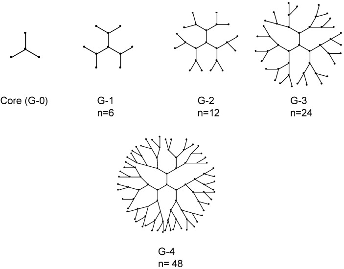
Dendrimers have attracted a great deal of interest in areas ranging from drug and nucleic acid delivery applications to processing, diagnostics and nanoengineering due to their uniform, well-defined, three dimensional structures [34,36,37,40,86]. The name "dendrimer" originated from the Greek work “Dendron” meaning tree and “meros” meaning part, which depicts a structure that consists of a central core molecule that acts as a root, from which a number of highly branched, tree-like arms originates in a symmetrical manner (Figure 1).
Figure 1.
Representation of dendrimers of generations 1–4. The n denotes number of terminal functional groups.
Dendrimers differ from classical monomers or oligomers by their defined architecture, high branching from the center/core of the molecule and terminal functional group density. Key properties such as molecular symmetry and a high ratio of multivalent surface moieties to molecular volume make these nano-sized materials of interest for the development of synthetic non-viral vectors for therapeutic nucleic acids [36]. Reactive end-groups of dendrimers allow the addition of repetitive units or branching in a controllable manner and versatility by modification of the end groups for multiple copies of various ligands including therapeutics and imaging agents for biomedical applications [38,87].
Synthesis of dendrimer-type molecules was first reported as 'cascade molecules' [88]. Some years later, Tomalia and co-workers pioneered the synthesis of this new class of polymer and refer to them as starburst “polymers” or “dendrimers” [89,90]. Dendrimers containing poly(amidoamine) (PAMAM) as the branching unit have been studied extensively compared to other classes of dendrimers. Dendrimers differ from classical random coil macromolecules due to three distinct characteristics: a central core, layers of repetitive units (termed generations) radically attached to the central core, and the presence of terminal functional groups amenable to conjugation for ligand attachments.
7. Synthesis
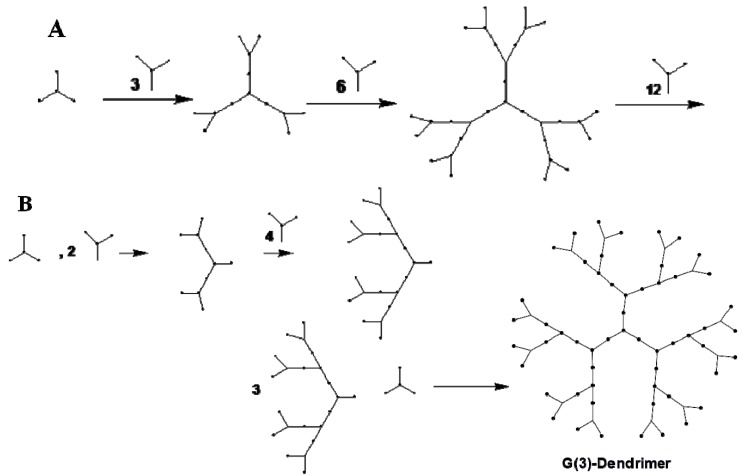
Dendrimers can be synthesized by two different approaches: divergent and convergent synthesis (Figure 2). Divergent synthesis involves outward, repeated addition of monomers or branching, starting from a multifunctional core by a series of stepwise polymerization reactions that attach layers of repetitive units/monomers to form dendrimers of various generations [89,91].
Figure 2.
Two common synthetic approaches of dendrimer assembly. Divergent (A) amd convergent (B) growth method.
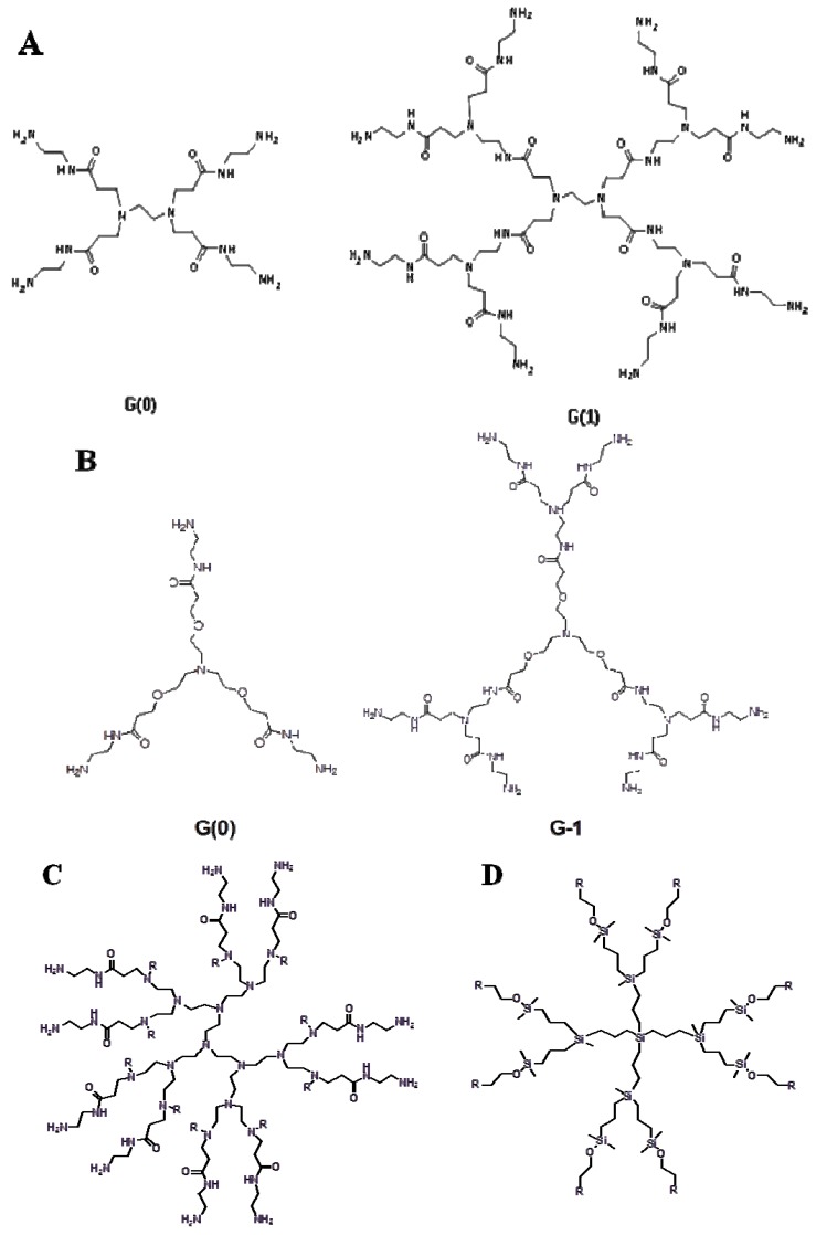
The valency of the core determines the starting number of branching points. For example, the ethylenediamine core in poly(amidoamine) dendrimers has four branching points (Figure 3A). The addition of four branchings of a repeating unit (-CH2-CH2-CONH-CH2-CH2-N-) generates a PAMAM dendrimer with four primary amine groups on the surface and two tertiary amines in the interior (referred to as generation 0). Addition of repeated branching makes PAMAM dendrimers of G=1, 2, 3, 4, 5 and so on with the number of exterior primary amine groups equal to 8, 16, 32, 64, 128 and so on. A schematic representation of various dendrimers is depicted in Figure 3. The convergent synthetic approach involves inward branching from the dendrimer surface to the inner core by formation of individual dendrons [92]. The dendrons are produced by repeated coupling (depending on the generation) of the branching units which are finally anchored to the core (Figure 2B). Both synthetic procedures pose a challenge for large scale production. Various alternative methods have been proposed [80,93,94,95]. Attempts have been made to reduce the reaction steps involved in the synthesis of dendrimers by the two procedures outlined above [94,95].
Figure 3.
Schematic representation of PAMAM dendrimers with-A, ethylenediamine; B, triethanolamine; C, PEI-PAMAM as the core; D, a carbosilane dendrimer of second generation.
Non-symmetrical dendrimers containing both a mannose binding and coumarin fluorescent unit have been prepared by high yield click chemistry of copper(I)-catalyzed azide-alkyne cycloadditions [95]. Maraval et al. developed a new straight forward synthesis method of dendrimers using two branched monomers where each generation was obtained in a single quantitative step [94]. PAMAM dendrimers are generally synthesized by the divergent approach. The initiator core is based on ethylene diamine or ammonia (Figure 3A, B). Addition of each new layer increases the molecular weight exponentially; the number or surface-active groups doubles and the diameter increases about 10 Å [37]. PAMAM dendrimers with high generation numbers have a high density of primary amine groups on the surface which make them efficient for binding nucleic acid molecules. Dendrimers possess low polydispersity. An increase in the generation number affects the shape of a dendrimer. Dendrimers of lower generations have a planar or elliptical shape, whereas dendrimers of higher generations typically have a spherical structure with a hydrophobic interior core, useful for the encapsulation of bioactive molecules [37].
Since their discovery, PAMAM dendrimers have gained interest as non-viral nucleic acid delivery vectors due to their high aqueous solubility, high transfection efficiency and minimal cytotoxicity. The positively charged primary amine groups on the surface of these dendrimers allows electrostatic interaction with negatively charged DNA molecules. The PAMAM dendrimer-nucleic acid complex can also be stable over a broad pH range [96].
8. Dendrimers as siRNA Delivery Vectors
Dendrimers are among the most widely used cationic polymers for nucleic acid delivery. However, as was said earlier, delivery of siRNA in particular is relatively unexplored. Several different formulations of dendrimer-DNA polyplexes were investigated including PAMAM-DNA, polyethylene glycol-modified PAMAM-DNA, PAMAM-PEG-PAMAM-DNA, PPI-DNA and PEI-DNA [39,97,98,99,100]. The relatively inflexible siRNA experiences a less efficient interaction with cationic polymers compared to DNA, indicating a cationic polymer efficient for plasmid DNA delivery may not be an effective siRNA carrier.
To elucidate the molecular mechanism of PAMAM-siRNA dendriplex self-assembly, Jensen et al. studied different generation PMAM dendrimers [101]. G4 and G7 displayed an equal efficiency for dendriplex formation. However, G1 with less charge density lacked the siRNA condensation ability. Reduced average size and increased polydispersity at a higher dendrimer concentration indicated a thermodynamically favorable electrostatic attraction. A spontaneous exothermic binding for G1 and a biphasic, initial exothermic and secondary endothermic binding with siRNA for G4 and G7 was observed for the formation of dendrimer-siRNA aggregates. Flexible G1 and rigid G7 displayed an entropic penalty, making G4 the most suitable for dendriplex formation with a favorable charge density for siRNA binding.
9. Dendrimers with Varied Structures for siRNA Delivery
Dendrimers with varied core/branching structures have been utilized for siRNA delivery [20,63,79,102,103]. Polymerized PEG-based dendrimeric core-shell structures including polyglycerolamine (PG-Amine), polyglyceryl pentaethylenehexamine carbamate, PEI-PAMAM and PEI-gluconolactone were synthesized and tested for their efficacy as siRNA delivery vectors. A representative structure of a PEI-PAMAM dendrimer is shown in Figure 3C. The study indicated that these cationic dendrimers exhibited low toxicity, strongly improved the stability of the siRNA and its intracellular trafficking and had both in vitro and in vivo gene silencing efficacy. These dendritic polymers demonstrated high efficacy in silencing the luciferase gene, ectopically over-expressed in human glioblastoma and murine mammary adenocarcinoma cells. The data demonstrated that the PG-amine exhibited the best ratio of silencing efficacy vs. toxicity in vitro. Intratumoral and intravenous administration of luciferase targeting siRNA-PG-amine polyplexes to tumor-bearing mice resulted in a significant luciferase gene silencing effect within 24 h of treatment. High luciferase gene silencing (68% and 69%) was accomplished in vivo within 24 h of treatment with 2.5 mg/kg of luciferase siRNA in subcutaneously implanted U87-Luc human glioblastoma cells in SKID mice and DA3-mCherry-Luc cells in BALB/C mice with both i.v. and i.t. dosing respectively.
A family of triazine dendrimers, differing in their core flexibility, generation number, and surface functionality was developed using a divergent synthesis approach and evaluated for its ability to condense and effectively deliver a luciferase targeting siRNA for target-specific knockdown of the reporter gene [63]. The triazine groups were introduced in the periphery, which was linked by specific diamine groups. The triazine dendrimers, which was the most effective DNA delivery vector, was not able to mediate gene silencing, whereas the moderately effective gene delivery vector delivered significant amount of siRNA [102,104]. A molecular modeling and in vivo imaging study were performed to identify a useful flexible triazine dendrimer for siRNA delivery. In comparison with PEI, 25 KDa, flexible G2-4 triazine dendrimers formed thermodynamically more stable complexes with siRNA and demonstrated less toxicity. Triazine dendrimer-based siRNA delivery systems were more efficiently charge-neutralized than PEI complexes, which reduced agglomeration. The hydrodynamic diameters ranged from 72.0 to 153.5 nm for dendriplexes compared to 312.8 to 480.0 nm for PEI-siRNA complexes. All dendriplexes were efficiently endocytosed and demonstrated significantly higher luciferase knockdown. However, dendriplexes of higher generation were captured by reticuloendothelial system due to their increased surface charge.
Another new class, carbosilane dendrimer, has been utilized for siRNA delivery (Figure 3D) [103,105]. Amino terminated carbosilane dendrimers were developed to protect and transport siRNA to HIV-infected lymphocytes [103]. The dendrimers were able to effectively bound siRNA via electrostatic interactions, and the dendrimer-bound siRNA was resistant to degradation by RNAse. The dendriplexes with a N/P ratio of 2 displayed the highest transfection efficiency with no cytotoxicity in hard-to-transfect HIV-infected peripheral blood mononuclear cells. The dendrimer-siRNA complex down-regulated GAPDH expression and reduced HIV-replication in the tested lymphocytic cell lines. Carbosilane dendrimers were also utilized to deliver siRNA to postmitotic neurons to study the function of hypoxia-inducible factor-1 alpha (HIF1-alpha) during chemical hypoxia-mediated neurotoxicity [105]. Carbosilane dendrimers were as effective as viral vectors in terms of their siRNA delivery and transfection efficiency. Carbosilane dendrimers with sixteen positive charges/molecule caused strong repression of various interleukins in macrophages involved in autoimmune diseases suggesting a potential pharmacological application of this dendrimer [106].
Polypropyleneimine (PPI) dendrimers appear to be an attractive non-viral vector for siRNA delivery [68]. A PPI dendrimer-siRNA nanoparticle was formulated using a layer-by-layer surface modification strategy, where the nanoparticle was caged with a dithiol containing cross linker molecule followed by coating with a poly(ethylene glycol) polymer to provide the stability to the nanoparticles needed to withstand the neutralizing environment in the blood stream. Specific cancer targeting of the nanoparticles was achieved by conjugating a cancer-homing peptide, luteinizing hormone-releasing hormone (LHRH) peptide to the distal end of the PEG chain. The disulfide bonds of the coated nanoparticles get reduced after cell uptake and the nucleic acid is released in to the cytoplasm [107]. The coating with a dithiol linkage on the dendriplex provides stability and leads to less agglomeration of the nanoparticles during their circulation [108]. However, this could also cause decreased transfection efficiency due to over-stabilization of the product and require utilization of less stable disulfide linkages [109]. The surface functionalization with a thiol linkage was useful in stabilizing the nanoparticles in terms of dissociation in presence of competing polyanions. The modified nanoparticles were stable in human serum for at least 48 h. PEGylation imparted stability against aggregation by decreasing the particle-particle and particle-protein interactions. The functionalization of the nanoparticle surface with LHRH peptide for cancer targeting should be an effective strategy. LHRH receptors are over-expressed in several types of cancer including ovarian, prostate, lung, breast, and colon cancer with undetectable expression in healthy visceral organs [110]. The results obtained from this in vivo biodistribution study indicated a high degree of nanoparticle accumulation in tumor compared to other organs.
Hayashi et al. used a lactose group-modified, α-cyclodextrin conjugated G3-dendrimer (Lac-α-CDE) for the delivery of hepatocyte-specific siRNA carriers for the treatment of transthyretin-related familial amyloidotic polyneuropathy [111,112]. Lac-α-CDE was condensed with a siRNA that targets transthyretin (TTR) gene expression. The dendrimer-siRNA complex had a potent RNAi effect against the TTR gene expression, efficient endosomal escape and delivery of the siRNA complex to the cytoplasm with negligible cytotoxicity. After intravenous administration, The Lac-α-CDE/siRNA complex demonstrated a potent in vivo gene silencing effect.
A flexible PAMAM dendrimer with triethanolamine (TEA) as the core, in which the branching units start at a distance of 10 successive bonds from the center amine has also been studied for siRNA delivery and gene silencing [29,113,114,115]. A TEA core offers flexibility compared to the commercially available PAMAM with an ammonia or ethylenediamine core, in which the branching starts at the central amine of the core. Higher generation dendrimers of this family can efficiently deliver siRNA and induce gene silencing. Dendrimers with amine end groups almost completely retarded siRNA in the agarose gel electrophoresis at N/P ratios above 2.5. However, dendrimers with terminal ester groups demonstrated no gel retardation, indicating no condensation with siRNA. TEA core PAMAM dendrimers efficiently delivered siRNA and inhibited the catalytic activity of Candida ribozymes [115]. A TEA core PAMAM dendrimer delivered HSP27-targeted siRNA into prostate cancer cells [113]. This dendrimer, which protects siRNA from enzymatic degradation, enhanced cellular uptake of siRNA. The siRNA also exhibited potent and specific gene silencing of heat shock protein 27, which is an attractive therapeutic target in castration-resistant prostate cancer.
10. Surface Modification for Improved Efficacy and Multifunctionality
Surface functionalization of nanocarriers with a wide variety of polymers and targeting ligands is a promising approach to achieve specific functions [116]. Surface modification of nanocarriers with a biocompatible and hydrophilic polymer, poly(ethylene glycol) (PEG), has found wide application as pegylation shields the nanocarrier from exposure to enzymes or opsonizing proteins in the systemic circulation [74,117,118,119,120,121]. Evading capture by the reticuloendothelial system provides prolonged systemic circulation, which is particularly essential for nanocarriers that eventually accumulate in an infarcted region or tumor via the enhanced permeability and retention (EPR) effect [74,122,123]. PEG-conjugation decreased the cytotoxicity of cationic polymers for nucleic acid delivery such as PLL and PEI by reducing or partially shielding the positive charge on the surface of these polycations [74,118,119,121,124]. PAMAM dendrimers of higher generations such as G4 and G5 are highly efficient as nucleic acid delivery vectors, However their high cytotoxicity, liver toxicity and hemolysis limit their in vivo application [91,125,126].
PEG-modified PAMAM dendrimers have demonstrated low toxicity and mediated efficient drug and gene delivery [127,128,129]. The amount of PEG on the surface affects its transfection efficacy and cytotoxicity. The G(5) PAMAM, conjugated to 10% PEG-3.4 KDa had a 20-fold increase in the efficacy of in vitro gene transfection compared to the unconjugated G(5) PAMAM dendrimer [130]. Qi et al. demonstrated that 8 mol % PEG-conjugated G5 and G6 dendrimers were the most efficient at gene silencing, when compared to three pegylated systems of G5 and G6 PAMAM dendrimers at a 4, 8, or 15% molar ratio of PEG on the surface [127].
PAMAM dendrimers have been surface functionalized for selective siRNA delivery. G(5)-PAMAM dendrimers were conjugated to cell penetrating TAT peptide for the purpose of intracellular delivery [131]. MDR1 gene silencing siRNA-dendrimer polyplexes weakly inhibited the gene expression. In this study, conjugation with TAT-peptide did not improve the delivery efficiency of the G(5)-PAMAM dendrimer. However, TAT-peptide functionalized dendrimer-oligonucleotide complexes were moderately effective for delivery of antisense compared to plain dendrimer. The effect of acetylation of primary amines of G5 on dendriplex formation was also studied [132]. The results indicated that acetylation reduced the cytotoxicity and that approximately 20% of the primary amines of G5-PAMAM could be modified while maintaining the siRNA delivery efficiency seen with unmodified PAMAM. A higher degree of amine neutralization reduced cellular delivery, caused entrapment in the endosome due to the reduction in buffering capacity and reduced gene silencing efficiency.
A novel approach to reducing the cytotoxicity of the dendrimers was outlined by the Minko group [133,134,135,136]. They evaluated the internally cationic, surface neutral dendrimers as nanocarriers for the targeted delivery of siRNA. An internally quaternized and surface acetylated PAMAM-G4 dendrimer was synthesized. The neutral surface of these dendrimers elicited low cytotoxicity compared to parent dendrimer with amino groups on the surface. Interaction of the siRNA with the cationic charge inside the dendrimer resulted in the formation of a compact nanoparticle, that potentially protects siRNA from environmental degradation. The shape of internally quaternized, surface neutral PAMAM/siRNA polyplexes at a charge N/P ratio of 3 was spherical, whereas condensation of siRNA with PAMAM-NH2 at the same N/P ratio resulted in the formation of ribbon-like nanofibers [134]. PAMAM-OH dendrimers were internally positively charged by the reaction of tertiary amines with methyl iodide, and this charge was utilized for siRNA condensation. Internal condensation of siRNA played a significant role in controlling the morphology of the complexes by encapsulating the siRNA in the core. Quaternized G4-PAMAM-OH dendrimers (QPAMAM-OH) were functionalized with a synthetic analog of the natural LHRH peptide for active cancer targeting [136]. LHRH peptide conjugated QPAMAM-OH-LHRH successfully targeted cancer cells and facilitated cellular uptake of dendrimer-siRNA complex via interaction with over-expressed LHRH receptors through receptor-mediated endocytosis and accumulated in the tumors, with minimal invasion of the healthy tissues to potentially limit adverse side-effects.
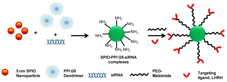
Functionalized inorganic nanomaterials including gold, iron oxide nanoparticles, quantum dots and carbon nanotubes provide a promising platform for siRNA delivery since these materials produce a high molecular weight polymer structure with functional groups on the surface [137,138]. The inorganic core functions as a space-filling material that presents surface-associated active functional groups. In a recent study, gold nanoparticles were functionalized with biodegradable glutamic acid scaffolds and cationic triethylenetetramine terminated dendron ligands for effective electrostatic interaction with siRNA [137]. In another study, poly(propyleneimine) generation 5 dendrimers (PPI G5) were cooperatively complexed with super paramagnetic iron oxide nanoparticles (SPION) and siRNA to develop a complex tumor-targeted drug delivery system for simultaneous delivery of siRNA with this MRI contrast agent specifically to cancer cells [138]. A schematic representation of the concept is shown in Figure 4. The dendrimer-siRNA-SPION particles were stabilized with PEG, the distal end of which was coupled with the tumor homing peptide, LHRH. This multi-functional system delivered siRNA specifically to the cancer cells and allowed monitoring of the therapeutic outcome using simultaneous imaging techniques.
Figure 4.
Schematic representation of the development of a cancer cell-targeted dendrimer-siRNA-SPION complex.
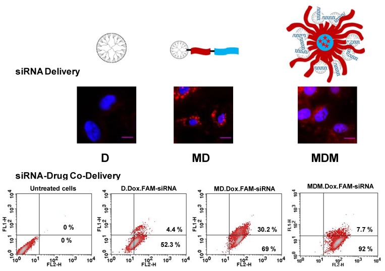
In our recent study, PAMAM-G4 dendrimer was lipid-modified for siRNA-drug co-delivery [139]. We synthesized a triblock co-polymeric system by conjugating G(4)-PAMAM dendrimer with poly(ethyleneglycol)-1,2-dioleoyl-sn-glycero-3-phosphoethanolamine (PEG-PE). The lipid block in the polymer provided optimum hydrophobicity and compatible cellular interaction for enhanced cell penetration. A mixed micellar system was developed using G(4)-D-PEG-PE and PEG-PE polymer at a 1:1 molar ratio. The lipid-modified, PEGylated dendrimer, G(4)-D-PEG-PE and the mixed micellar system form stable complexes with siRNA, showed excellent serum stability and significantly higher cellular uptake of siRNA that resulted in better targeted green fluorescence protein (GFP) downregulation compared to G(4)-PAMAM dendrimer. The core of the mixed micellar system loaded a chemotherapeutic drug, doxorubicin, efficiently, while the PEG-PE anchored G(4)-D condensed siRNA. The modified dendrimer demonstrated higher efficiency for siRNA delivery compared to G(4)-D and the mixed micellar system. The mixed micellar system appeared to be a promising career for siRNA/drug co-delivery.
Figure 5 showed confocal microscopy images of the dendrimer (D), lipid-modified dendrimer (MD), and mixed micellar system (MDM)-dosed cells, used to assess the intracellular siRNA delivery efficiency.
Figure 5.
Lipid modification of G(4)-PAMAM dendrimers for enhanced siRNA delivery and siRNA-drug co-delivery. Green fluorescence of FAM-labeled siRNA and red fluorescence of Doxorubicin were analyzed by flow cytometry to assess siRNA/drug co-delivery.
The flow cytometry data representing the efficiency of the polymer for siRNA-drug co-delivery are also shown. This study clearly demonstrated that lipid modification enhances the cellular association of the nanocarrier without compromising the siRNA condensation ability of the dendrimers. Moreover, a mixed micellar system is advantageous for its ability to simultaneously carry and effectively deliver both drug and siRNA.
11. Conclusions
In summary, dendrimer-mediated delivery of therapeutics including siRNA clearly should be considered a promising approach. Significant research is being carried out to develop dendrimer-based nanomedicine for the delivery of drugs and nucleic acids. The unique properties of this polymeric system, including ease of surface functionalization, enables engineering of truly multifunctional nanodevices for drug delivery applications. However, further addressing of the current limitations including non-specific cytotoxicity associated with higher generation-dendrimers, release kinetics of the associated bio-actives and rapid clearance issues would open up new perspectives for dendrimers as therapeutic nanoparticles.
Acknowledgments
The work was supported in part by NIH grants RO1 CA121838 and RO1 CA128486 to Vladimir P. Torchilin. We thank William C. Hartner for his helpful manuscript editing.
Conflict of Interest
The authors declare no conflict of interest.
References
- 1.Sifuentes-Romero I., Milton S.L., Garcia-Gasca A. Post-transcriptional gene silencing by rna interference in non-mammalian vertebrate systems: Where do we stand? Mutat. Res. 2011;728:158–171. doi: 10.1016/j.mrrev.2011.09.001. [DOI] [PubMed] [Google Scholar]
- 2.Mello C.C., Conte D., Jr. Revealing the world of rna interference. Nature. 2004;431:338–342. doi: 10.1038/nature02872. [DOI] [PubMed] [Google Scholar]
- 3.Monaghan M., Pandit A. Rna interference therapy via functionalized scaffolds. Adv. Drug Deliver. Rev. 2011;63:197–208. doi: 10.1016/j.addr.2011.01.006. [DOI] [PubMed] [Google Scholar]
- 4.Denli A.M., Hannon G.J. Rnai: An ever-growing puzzle. Trends Biochem. Sci. 2003;28:196–201. doi: 10.1016/S0968-0004(03)00058-6. [DOI] [PubMed] [Google Scholar]
- 5.Cerutti H. Rna interference: Traveling in the cell and gaining functions? Trends Genet. 2003;19:39–46. doi: 10.1016/S0168-9525(02)00010-0. [DOI] [PubMed] [Google Scholar]
- 6.Capecchi M.R. Altering the genome by homologous recombination. Science. 1989;244:1288–1292. doi: 10.1126/science.2660260. [DOI] [PubMed] [Google Scholar]
- 7.Aagaard L., Rossi J.J. Rnai therapeutics: Principles, prospects and challenges. Adv. Drug Deliver. Rev. 2007;59:75–86. doi: 10.1016/j.addr.2007.03.005. [DOI] [PMC free article] [PubMed] [Google Scholar]
- 8.Bumcrot D., Manoharan M., Koteliansky V., Sah D.W. Rnai therapeutics: A potential new class of pharmaceutical drugs. Nat. Chem. Biol. 2006;2:711–719. doi: 10.1038/nchembio839. [DOI] [PMC free article] [PubMed] [Google Scholar]
- 9.Daka A., Peer D. Rnai-based nanomedicines for targeted personalized therapy. Adv. Drug Deliver Rev. 2012;64:1508–1521. doi: 10.1016/j.addr.2012.08.014. [DOI] [PubMed] [Google Scholar]
- 10.Scherer L.J., Rossi J.J. Approaches for the sequence-specific knockdown of mrna. Nat. Biotechnol. 2003;21:1457–1465. doi: 10.1038/nbt915. [DOI] [PubMed] [Google Scholar]
- 11.Bertrand J.R., Pottier M., Vekris A., Opolon P., Maksimenko A., Malvy C. Comparison of antisense oligonucleotides and sirnas in cell culture and in vivo. Biochem. Biophys. Res. Commun. 2002;296:1000–1004. doi: 10.1016/S0006-291X(02)02013-2. [DOI] [PubMed] [Google Scholar]
- 12.Sontheimer E.J. Assembly and function of rna silencing complexes. Nat. Rev. Mol. Cell Biol. 2005;6:127–138. doi: 10.1038/nrm1568. [DOI] [PubMed] [Google Scholar]
- 13.Braasch D.A., Paroo Z., Constantinescu A., Ren G., Oz O.K., Mason R.P., Corey D.R. Biodistribution of phosphodiester and phosphorothioate sirna. Bioorg. Med. Chem. Lett. 2004;14:1139–1143. doi: 10.1016/j.bmcl.2003.12.074. [DOI] [PubMed] [Google Scholar]
- 14.Urban-Klein B., Werth S., Abuharbeid S., Czubayko F., Aigner A. Rnai-mediated gene-targeting through systemic application of polyethylenimine (pei)-complexed sirna in vivo. Gene Ther. 2005;12:461–466. doi: 10.1038/sj.gt.3302425. [DOI] [PubMed] [Google Scholar]
- 15.Higuchi Y., Kawakami S., Hashida M. Strategies for in vivo delivery of sirnas: Recent progress. BioDrugs. 2010;24:195–205. doi: 10.2165/11534450-000000000-00000. [DOI] [PubMed] [Google Scholar]
- 16.Nguyen J., Szoka F.C. Nucleic acid delivery: The missing pieces of the puzzle? Accounts Chem. Res. 2012;45:1153–1162. doi: 10.1021/ar3000162. [DOI] [PMC free article] [PubMed] [Google Scholar]
- 17.Shen H., Sun T., Ferrari M. Nanovector delivery of sirna for cancer therapy. Cancer Gene Ther. 2012;19:367–373. doi: 10.1038/cgt.2012.22. [DOI] [PMC free article] [PubMed] [Google Scholar]
- 18.Nimesh S. Polyethylenimine as a promising vector for targeted sirna delivery. Curr. Clin. Pharmacol. 2012;7:121–130. doi: 10.2174/157488412800228857. [DOI] [PubMed] [Google Scholar]
- 19.Nimesh S., Gupta N., Chandra R. Cationic polymer based nanocarriers for delivery of therapeutic nucleic acids. J. Biomed. Nanotechnol. 2011;7:504–520. doi: 10.1166/jbn.2011.1313. [DOI] [PubMed] [Google Scholar]
- 20.Posadas I., Guerra F.J., Cena V. Nonviral vectors for the delivery of small interfering RNAs to the CNS. Nanomedicine. 2010;5:1219–1236. doi: 10.2217/nnm.10.105. [DOI] [PubMed] [Google Scholar]
- 21.Gao Y., Liu X.L., Li X.R. Research progress on sirna delivery with nonviral carriers. Int. J. Nanomed. 2011;6:1017–1025. doi: 10.2147/IJN.S17040. [DOI] [PMC free article] [PubMed] [Google Scholar]
- 22.Akhtar S. Cationic nanosystems for the delivery of small interfering ribonucleic acid therapeutics: A focus on toxicogenomics. Expert Opin. Drug Metab. Toxicol. 2010;6:1347–1362. doi: 10.1517/17425255.2010.518611. [DOI] [PubMed] [Google Scholar]
- 23.Shuai L., Wang S., Zhang L., Fu B., Zhou X. Cationic porphyrins and analogues as new DNA topoisomerase i and ii inhibitors. Chem. Biodivers. 2009;6:827–837. doi: 10.1002/cbdv.200800083. [DOI] [PubMed] [Google Scholar]
- 24.Gopalakrishnan B., Wolff J. Sirna and DNA transfer to cultured cells. Methods Mol. Biol. 2009;480:31–52. doi: 10.1007/978-1-59745-429-2_3. [DOI] [PubMed] [Google Scholar]
- 25.Lungwitz U., Breunig M., Blunk T., Gopferich A. Polyethylenimine-based non-viral gene delivery systems. Eur. J. Pharm. Biopharm. 2005;60:247–266. doi: 10.1016/j.ejpb.2004.11.011. [DOI] [PubMed] [Google Scholar]
- 26.Zintchenko A., Philipp A., Dehshahri A., Wagner E. Simple modifications of branched pei lead to highly efficient sirna carriers with low toxicity. Bioconjug. Chem. 2008;19:1448–1455. doi: 10.1021/bc800065f. [DOI] [PubMed] [Google Scholar]
- 27.Tseng Y.C., Mozumdar S., Huang L. Lipid-based systemic delivery of sirna. Adv. Drug Deliv. Rev. 2009;61:721–731. doi: 10.1016/j.addr.2009.03.003. [DOI] [PMC free article] [PubMed] [Google Scholar]
- 28.Wu Z.W., Chien C.T., Liu C.Y., Yan J.Y., Lin S.Y. Recent progress in copolymer-mediated sirna delivery. J. Drug Target. 2012;20:551–560. doi: 10.3109/1061186X.2012.699057. [DOI] [PubMed] [Google Scholar]
- 29.Zhou J., Wu J., Hafdi N., Behr J.P., Erbacher P., Peng L. Pamam dendrimers for efficient sirna delivery and potent gene silencing. Chem. Commun. 2006;22:2362–2364. doi: 10.1039/b601381c. [DOI] [PubMed] [Google Scholar]
- 30.Jafari M., Soltani M., Naahidi S., Karunaratne D.N., Chen P. Nonviral approach for targeted nucleic acid delivery. Curr. Med. Chem. 2012;19:197–208. doi: 10.2174/092986712803414141. [DOI] [PubMed] [Google Scholar]
- 31.Tros de Ilarduya C., Sun Y., Düzgüneş N. Gene delivery by lipoplexes and polyplexes. Eur. J. Pharm. Sci. 2010;40:159–170. doi: 10.1016/j.ejps.2010.03.019. [DOI] [PubMed] [Google Scholar]
- 32.Zhang X.X., McIntosh T.J., Grinstaff M.W. Functional lipids and lipoplexes for improved gene delivery. Biochimie. 2012;94:42–58. doi: 10.1016/j.biochi.2011.05.005. [DOI] [PMC free article] [PubMed] [Google Scholar]
- 33.Lu J.J., Langer R., Chen J. A novel mechanism is involved in cationic lipid-mediated functional sirna delivery. Mol. Pharm. 2009;6:763–771. doi: 10.1021/mp900023v. [DOI] [PMC free article] [PubMed] [Google Scholar]
- 34.Boas U., Heegaard P.M. Dendrimers in drug research. Chem. Soc. Rev. 2004;33:43–63. doi: 10.1039/b309043b. [DOI] [PubMed] [Google Scholar]
- 35.Cheng Y., Wang J., Rao T., He X., Xu T. Pharmaceutical applications of dendrimers: Promising nanocarriers for drug delivery. Front. Biosci. 2008;13:1447–1471. doi: 10.2741/2774. [DOI] [PubMed] [Google Scholar]
- 36.Dufes C., Uchegbu I.F., Schatzlein A.G. Dendrimers in gene delivery. Adv. Drug Deliv. Rev. 2005;57:2177–2202. doi: 10.1016/j.addr.2005.09.017. [DOI] [PubMed] [Google Scholar]
- 37.Eichman J.D., Bielinska A.U., Kukowska-Latallo J.F., Baker J.R., Jr. The use of pamam dendrimers in the efficient transfer of genetic material into cells. Pharm. Sci. Technolo. Today. 2000;3:232–245. doi: 10.1016/S1461-5347(00)00273-X. [DOI] [PubMed] [Google Scholar]
- 38.Gao Y., Gao G., He Y., Liu T., Qi R. Recent advances of dendrimers in delivery of genes and drugs. Mini Rev. Med. Chem. 2008;8:889–900. doi: 10.2174/138955708785132729. [DOI] [PubMed] [Google Scholar]
- 39.Haensler J., Szoka F.C., Jr. Polyamidoamine cascade polymers mediate efficient transfection of cells in culture. Bioconjug. Chem. 1993;4:372–379. doi: 10.1021/bc00023a012. [DOI] [PubMed] [Google Scholar]
- 40.Gillies E.R., Frechet J.M. Dendrimers and dendritic polymers in drug delivery. Drug Discov. Today. 2005;10:35–43. doi: 10.1016/S1359-6446(04)03276-3. [DOI] [PubMed] [Google Scholar]
- 41.Lee Y., Kim M., Han J., Yeom K.H., Lee S., Baek S.H., Kim V.N. Microrna genes are transcribed by rna polymerase ii. EMBO J. 2004;23:4051–4060. doi: 10.1038/sj.emboj.7600385. [DOI] [PMC free article] [PubMed] [Google Scholar]
- 42.Preall J.B., Sontheimer E.J. Rnai: Risc gets loaded. Cell. 2005;123:543–545. doi: 10.1016/j.cell.2005.11.006. [DOI] [PubMed] [Google Scholar]
- 43.Provost P., Dishart D., Doucet J., Frendewey D., Samuelsson B., Radmark O. Ribonuclease activity and rna binding of recombinant human dicer. EMBO J. 2002;21:5864–5874. doi: 10.1093/emboj/cdf578. [DOI] [PMC free article] [PubMed] [Google Scholar]
- 44.Macrae I.J., Zhou K., Li F., Repic A., Brooks A.N., Cande W.Z., Adams P.D., Doudna J.A. Structural basis for double-stranded rna processing by dicer. Science. 2006;311:195–198. doi: 10.1126/science.1121638. [DOI] [PubMed] [Google Scholar]
- 45.Elbashir S.M., Harborth J., Lendeckel W., Yalcin A., Weber K., Tuschl T. Duplexes of 21-nucleotide rnas mediate rna interference in cultured mammalian cells. Nature. 2001;411:494–498. doi: 10.1038/35078107. [DOI] [PubMed] [Google Scholar]
- 46.Potti A., Schilsky R.L., Nevins J.R. Refocusing the war on cancer: The critical role of personalized treatment. Sci. Transl. Med. 2010;2:28cm13. doi: 10.1126/scitranslmed.3000643. [DOI] [PubMed] [Google Scholar]
- 47.Zimmermann T.S., Lee A.C., Akinc A., Bramlage B., Bumcrot D., Fedoruk M.N., Harborth J., Heyes J.A., Jeffs L.B., John M., et al. Rnai-mediated gene silencing in non-human primates. Nature. 2006;441:111–114. doi: 10.1038/nature04688. [DOI] [PubMed] [Google Scholar]
- 48.Dorn G., Patel S., Wotherspoon G., Hemmings-Mieszczak M., Barclay J., Natt F.J., Martin P., Bevan S., Fox A., Ganju P., et al. Sirna relieves chronic neuropathic pain. Nucleic Acids Res. 2004;32:e49. doi: 10.1093/nar/gnh044. [DOI] [PMC free article] [PubMed] [Google Scholar]
- 49.Shen J., Samul R., Silva R.L., Akiyama H., Liu H., Saishin Y., Hackett S.F., Zinnen S., Kossen K., Fosnaugh K., et al. Suppression of ocular neovascularization with sirna targeting vegf receptor 1. Gene Ther. 2006;13:225–234. doi: 10.1038/sj.gt.3302641. [DOI] [PubMed] [Google Scholar]
- 50.Behlke M.A. Chemical modification of sirnas for in vivo use. Oligonucleotides. 2008;18:305–319. doi: 10.1089/oli.2008.0164. [DOI] [PubMed] [Google Scholar]
- 51.Chiu Y.L., Rana T.M. Sirna function in rnai: A chemical modification analysis. RNA. 2003;9:1034–1048. doi: 10.1261/rna.5103703. [DOI] [PMC free article] [PubMed] [Google Scholar]
- 52.Elmen J., Thonberg H., Ljungberg K., Frieden M., Westergaard M., Xu Y., Wahren B., Liang Z., Orum H., Koch T., et al. Locked nucleic acid (LNA) mediated improvements in siRNA stability and functionality. Nucleic Acids Res. 2005;33:439–447. doi: 10.1093/nar/gki193. [DOI] [PMC free article] [PubMed] [Google Scholar]
- 53.Allerson C.R., Sioufi N., Jarres R., Prakash T.P., Naik N., Berdeja A., Wanders L., Griffey R.H., Swayze E.E., Bhat B. Fully 2'-modified oligonucleotide duplexes with improved in vitro potency and stability compared to unmodified small interfering rna. J. Med. Chem. 2005;48:901–904. doi: 10.1021/jm049167j. [DOI] [PubMed] [Google Scholar]
- 54.Braasch D.A., Jensen S., Liu Y., Kaur K., Arar K., White M.A., Corey D.R. Rna interference in mammalian cells by chemically-modified rna. Biochemistry. 2003;42:7967–7975. doi: 10.1021/bi0343774. [DOI] [PubMed] [Google Scholar]
- 55.Morrissey D.V., Blanchard K., Shaw L., Jensen K., Lockridge J.A., Dickinson B., McSwiggen J.A., Vargeese C., Bowman K., Shaffer C.S., et al. Activity of stabilized short interfering rna in a mouse model of hepatitis b virus replication. Hepatology. 2005;41:1349–1356. doi: 10.1002/hep.20702. [DOI] [PubMed] [Google Scholar]
- 56.Prakash T.P., Allerson C.R., Dande P., Vickers T.A., Sioufi N., Jarres R., Baker B.F., Swayze E.E., Griffey R.H., Bhat B. Positional effect of chemical modifications on short interference rna activity in mammalian cells. J. Med. Chem. 2005;48:4247–4253. doi: 10.1021/jm050044o. [DOI] [PubMed] [Google Scholar]
- 57.Choi H.S., Liu W., Misra P., Tanaka E., Zimmer J.P., Itty Ipe B., Bawendi M.G., Frangioni J.V. Renal clearance of quantum dots. Nat. Biotechnol. 2007;25:1165–1170. doi: 10.1038/nbt1340. [DOI] [PMC free article] [PubMed] [Google Scholar]
- 58.Van de Water F.M., Boerman O.C., Wouterse A.C., Peters J.G., Russel F.G., Masereeuw R. Intravenously administered short interfering rna accumulates in the kidney and selectively suppresses gene function in renal proximal tubules. Drug Metab. Dispos. 2006;34:1393–1397. doi: 10.1124/dmd.106.009555. [DOI] [PubMed] [Google Scholar]
- 59.Sledz C.A., Williams B.R. Rna interference in biology and disease. Blood. 2005;106:787–794. doi: 10.1182/blood-2004-12-4643. [DOI] [PMC free article] [PubMed] [Google Scholar]
- 60.Kariko K., Bhuyan P., Capodici J., Weissman D. Small interfering rnas mediate sequence-independent gene suppression and induce immune activation by signaling through toll-like receptor 3. J. Immunol. 2004;172:6545–6549. doi: 10.4049/jimmunol.172.11.6545. [DOI] [PubMed] [Google Scholar]
- 61.Jackson A.L., Bartz S.R., Schelter J., Kobayashi S.V., Burchard J., Mao M., Li B., Cavet G., Linsley P.S. Expression profiling reveals off-target gene regulation by rnai. Nat. Biotechnol. 2003;21:635–637. doi: 10.1038/nbt831. [DOI] [PubMed] [Google Scholar]
- 62.Jeong J.H., Park T.G., Kim S.H. Self-assembled and nanostructured sirna delivery systems. Pharm. Res. 2011;28:2072–2085. doi: 10.1007/s11095-011-0412-y. [DOI] [PubMed] [Google Scholar]
- 63.Merkel O.M., Mintzer M.A., Librizzi D., Samsonova O., Dicke T., Sproat B., Garn H., Barth P.J., Simanek E.E., Kissel T. Triazine dendrimers as nonviral vectors for in vitro and in vivo rnai: The effects of peripheral groups and core structure on biological activity. Mol. Pharm. 2010;7:969–983. doi: 10.1021/mp100101s. [DOI] [PMC free article] [PubMed] [Google Scholar]
- 64.Kebbekus P., Draper D.E., Hagerman P. Persistence length of rna. Biochemistry. 1995;34:4354–4357. doi: 10.1021/bi00013a026. [DOI] [PubMed] [Google Scholar]
- 65.Hagerman P.J. Flexibility of DNA. Annu Rev. Biophys. Biophys. Chem. 1988;17:265–286. doi: 10.1146/annurev.bb.17.060188.001405. [DOI] [PubMed] [Google Scholar]
- 66.Hagerman P.J. Investigation of the flexibility of DNA using transient electric birefringence. Biopolymers. 1981;20:1503–1535. doi: 10.1002/bip.1981.360200710. [DOI] [PubMed] [Google Scholar]
- 67.Shah S.A., Brunger A.T. The 1.8 a crystal structure of a statically disordered 17 base-pair rna duplex: Principles of rna crystal packing and its effect on nucleic acid structure. J. Mol. Biol. 1999;285:1577–1588. doi: 10.1006/jmbi.1998.2385. [DOI] [PubMed] [Google Scholar]
- 68.Taratula O., Garbuzenko O.B., Kirkpatrick P., Pandya I., Savla R., Pozharov V.P., He H., Minko T. Surface-engineered targeted ppi dendrimer for efficient intracellular and intratumoral sirna delivery. J. Control. Release. 2009;140:284–293. doi: 10.1016/j.jconrel.2009.06.019. [DOI] [PMC free article] [PubMed] [Google Scholar]
- 69.Spagnou S., Miller A.D., Keller M. Lipidic carriers of sirna: Differences in the formulation, cellular uptake, and delivery with plasmid DNA. Biochemistry. 2004;43:13348–13356. doi: 10.1021/bi048950a. [DOI] [PubMed] [Google Scholar]
- 70.Gary D.J., Puri N., Won Y.Y. Polymer-based sirna delivery: Perspectives on the fundamental and phenomenological distinctions from polymer-based DNA delivery. J. Control. Release. 2007;121:64–73. doi: 10.1016/j.jconrel.2007.05.021. [DOI] [PubMed] [Google Scholar]
- 71.Felgner J.H., Kumar R., Sridhar C.N., Wheeler C.J., Tsai Y.J., Border R., Ramsey P., Martin M., Felgner P.L. Enhanced gene delivery and mechanism studies with a novel series of cationic lipid formulations. J. Biol. Chem. 1994;269:2550–2561. [PubMed] [Google Scholar]
- 72.De Wolf H.K., Snel C.J., Verbaan F.J., Schiffelers R.M., Hennink W.E., Storm G. Effect of cationic carriers on the pharmacokinetics and tumor localization of nucleic acids after intravenous administration. Int. J. Pharm. 2007;331:167–175. doi: 10.1016/j.ijpharm.2006.10.029. [DOI] [PubMed] [Google Scholar]
- 73.Jere D., Jiang H.L., Arote R., Kim Y.K., Choi Y.J., Cho M.H., Akaike T., Cho C.S. Degradable polyethylenimines as DNA and small interfering rna carriers. Expert Opin. Drug Deliver. 2009;6:827–834. doi: 10.1517/17425240903029183. [DOI] [PubMed] [Google Scholar]
- 74.Lee M., Kim S.W. Polyethylene glycol-conjugated copolymers for plasmid DNA delivery. Pharm. Res. 2005;22:1–10. doi: 10.1007/s11095-004-9003-5. [DOI] [PubMed] [Google Scholar]
- 75.Wang J., Lu Z., Wientjes M.G., Au J.L. Delivery of sirna therapeutics: Barriers and carriers. AAPS J. 2010;12:492–503. doi: 10.1208/s12248-010-9210-4. [DOI] [PMC free article] [PubMed] [Google Scholar]
- 76.Lau C., Soriano H.E., Ledley F.D., Finegold M.J., Wolfe J.H., Birkenmeier E.H., Henning S.J. Retroviral gene transfer into the intestinal epithelium. Hum. Gene Ther. 1995;6:1145–1151. doi: 10.1089/hum.1995.6.9-1145. [DOI] [PubMed] [Google Scholar]
- 77.Howard K.A. Delivery of rna interference therapeutics using polycation-based nanoparticles. Adv. Drug Deliver. Rev. 2009;61:710–720. doi: 10.1016/j.addr.2009.04.001. [DOI] [PubMed] [Google Scholar]
- 78.Singha K., Namgung R., Kim W.J. Polymers in small-interfering rna delivery. Nucleic Acid Ther. 2011;21:133–147. doi: 10.1089/nat.2011.0293. [DOI] [PMC free article] [PubMed] [Google Scholar]
- 79.Ofek P., Fischer W., Calderon M., Haag R., Satchi-Fainaro R. In vivo delivery of small interfering rna to tumors and their vasculature by novel dendritic nanocarriers. FASEB J. 2010;24:3122–3134. doi: 10.1096/fj.09-149641. [DOI] [PubMed] [Google Scholar]
- 80.Svenson S., Tomalia D.A. Dendrimers in biomedical applications--reflections on the field. Adv. Drug Deliv. Rev. 2005;57:2106–2129. doi: 10.1016/j.addr.2005.09.018. [DOI] [PubMed] [Google Scholar]
- 81.Grzelinski M., Urban-Klein B., Martens T., Lamszus K., Bakowsky U., Hobel S., Czubayko F., Aigner A. Rna interference-mediated gene silencing of pleiotrophin through polyethylenimine-complexed small interfering rnas in vivo exerts antitumoral effects in glioblastoma xenografts. Hum. Gene Ther. 2006;17:751–766. doi: 10.1089/hum.2006.17.751. [DOI] [PubMed] [Google Scholar]
- 82.Thomas M., Lu J.J., Ge Q., Zhang C., Chen J., Klibanov A.M. Full deacylation of polyethylenimine dramatically boosts its gene delivery efficiency and specificity to mouse lung. Proc. Natl. Acad. Sci. USA. 2005;102:5679–5684. doi: 10.1073/pnas.0502067102. [DOI] [PMC free article] [PubMed] [Google Scholar]
- 83.Hobel S., Koburger I., John M., Czubayko F., Hadwiger P., Vornlocher H.P., Aigner A. Polyethylenimine/small interfering rna-mediated knockdown of vascular endothelial growth factor in vivo exerts anti-tumor effects synergistically with bevacizumab. J. Gene Med. 2010;12:287–300. doi: 10.1002/jgm.1431. [DOI] [PubMed] [Google Scholar]
- 84.Tan P.H., Yang L.C., Shih H.C., Lan K.C., Cheng J.T. Gene knockdown with intrathecal sirna of nmda receptor nr2b subunit reduces formalin-induced nociception in the rat. Gene Ther. 2005;12:59–66. doi: 10.1038/sj.gt.3302376. [DOI] [PubMed] [Google Scholar]
- 85.Navarro G., Sawant R.R., Biswas S., Essex S., Tros de Ilarduya C., Torchilin V.P. P-glycoprotein silencing with sirna delivered by dope-modified pei overcomes doxorubicin resistance in breast cancer cells. Nanomedicine. 2012;7:65–78. doi: 10.2217/nnm.11.93. [DOI] [PMC free article] [PubMed] [Google Scholar]
- 86.Twyman L.J., King A.S., Martin I.K. Catalysis inside dendrimers. Chem. Soc. Rev. 2002;31:69–82. doi: 10.1039/b107812g. [DOI] [PubMed] [Google Scholar]
- 87.Patri A.K., Majoros I.J., Baker J.R. Dendritic polymer macromolecular carriers for drug delivery. Curr. Opin. Chem. Biol. 2002;6:466–471. doi: 10.1016/S1367-5931(02)00347-2. [DOI] [PubMed] [Google Scholar]
- 88.Buhleier E., Wehner W., Vogtle F. Cascade-chain-like and nonskid-chain-like syntheses of molecular cavity topologies. Synthesis-Stuttgart. 1978:155–158. [Google Scholar]
- 89.Tomalia D.A., Baker H., Dewald J., Hall M., Kallos G., Martin S., Roeck J., Ryder J., Smith P. A new class of polymers: Starburst-dendritic macromolecules. Polymer J. 1985;17:117–132. doi: 10.1295/polymj.17.117. [DOI] [Google Scholar]
- 90.Tomalia D.A., Baker H., Dewald J., Hall M., Kallos G., Martin S., Roeck J., Ryder J., Smith P. Dendritic macromolecules: Synthesis of starburst dendrimers. Macromolecules. 1986;19:2466–2468. doi: 10.1021/ma00163a029. [DOI] [Google Scholar]
- 91.Roberts J.C., Bhalgat M.K., Zera R.T. Preliminary biological evaluation of polyamidoamine (pamam) starburst dendrimers. J. Biomed. Mater. Res. 1996;30:53–65. doi: 10.1002/(SICI)1097-4636(199601)30:1<53::AID-JBM8>3.0.CO;2-Q. [DOI] [PubMed] [Google Scholar]
- 92.Hawker C.J., Frechet J.M.J. Preparation of polymers with controlled molecular architecture. A new convergent approach to dendritic macromolecules. J. Am. Chem. Soc. 1990;112:7638–7647. doi: 10.1021/ja00177a027. [DOI] [Google Scholar]
- 93.Brauge L., Magro G., Caminade A.M., Majoral J.P. First divergent strategy using two ab (2) unprotected monomers for the rapid synthesis of dendrimers. J. Am. Chem Soc. 2001;123:6698–6699. doi: 10.1021/ja0029228. [DOI] [PubMed] [Google Scholar]
- 94.Maraval V., Pyzowski J., Caminade A.M., Majoral J.P. "Lego" chemistry for the straightforward synthesis of dendrimers. J. Org. Chem. 2003;68:6043–6046. doi: 10.1021/jo0344438. [DOI] [PubMed] [Google Scholar]
- 95.Wu P., Malkoch M., Hunt J.N., Vestberg R., Kaltgrad E., Finn M.G., Fokin V.V., Sharpless K.B., Hawker C.J. Multivalent, bifunctional dendrimers prepared by click chemistry. Chem. Commun. 2005;0:5775–5777. doi: 10.1039/b512021g. [DOI] [PubMed] [Google Scholar]
- 96.Hui Z., He Z.G., Zheng L., Li G.Y., Shen S.R., Li X.L. Studies on polyamidoamine dendrimers as efficient gene delivery vector. J. Biomater. Appl. 2008;22:527–544. doi: 10.1177/0885328207080005. [DOI] [PubMed] [Google Scholar]
- 97.Kim T.I., Seo H.J., Choi J.S., Jang H.S., Baek J.U., Kim K., Park J.S. Pamam-peg-pamam: Novel triblock copolymer as a biocompatible and efficient gene delivery carrier. Biomacromolecules. 2004;5:2487–2492. doi: 10.1021/bm049563j. [DOI] [PubMed] [Google Scholar]
- 98.Schatzlein A.G., Zinselmeyer B.H., Elouzi A., Dufes C., Chim Y.T., Roberts C.J., Davies M.C., Munro A., Gray A.I., Uchegbu I.F. Preferential liver gene expression with polypropylenimine dendrimers. J. Control. Release. 2005;101:247–258. doi: 10.1016/j.jconrel.2004.08.024. [DOI] [PubMed] [Google Scholar]
- 99.Forrest M.L., Gabrielson N., Pack D.W. Cyclodextrin-polyethylenimine conjugates for targeted in vitro gene delivery. Biotechnol. Bioeng. 2005;89:416–423. doi: 10.1002/bit.20356. [DOI] [PubMed] [Google Scholar]
- 100.Richardson S.C., Pattrick N.G., Man Y.K., Ferruti P., Duncan R. Poly(amidoamine)s as potential nonviral vectors: Ability to form interpolyelectrolyte complexes and to mediate transfection in vitro. Biomacromolecules. 2001;2:1023–1028. doi: 10.1021/bm010079f. [DOI] [PubMed] [Google Scholar]
- 101.Jensen L.B., Pavan G.M., Kasimova M.R., Rutherford S., Danani A., Nielsen H.M., Foged C. Elucidating the molecular mechanism of pamam-sirna dendriplex self-assembly: Effect of dendrimer charge density. Int. J. Pharm. 2011;416:410–418. doi: 10.1016/j.ijpharm.2011.03.015. [DOI] [PubMed] [Google Scholar]
- 102.Mintzer M.A., Merkel O.M., Kissel T., Simanek E.E. Polycationic triazine-based dendrimers: Effect of peripheral groups on transfection efficiency. New J. Chem. 2009;33:1918–1925. doi: 10.1039/b908735d. [DOI] [PMC free article] [PubMed] [Google Scholar]
- 103.Weber N., Ortega P., Clemente M.I., Shcharbin D., Bryszewska M., de la Mata F.J., Gomez R., Munoz-Fernandez M.A. Characterization of carbosilane dendrimers as effective carriers of sirna to hiv-infected lymphocytes. J. Control. Release. 2008;132:55–64. doi: 10.1016/j.jconrel.2008.07.035. [DOI] [PubMed] [Google Scholar]
- 104.Merkel O.M., Mintzer M.A., Sitterberg J., Bakowsky U., Simanek E.E., Kissel T. Triazine dendrimers as nonviral gene delivery systems: Effects of molecular structure on biological activity. Bioconjug. Chem. 2009;20:1799–1806. doi: 10.1021/bc900243r. [DOI] [PMC free article] [PubMed] [Google Scholar]
- 105.Posadas I., Lopez-Hernandez B., Clemente M.I., Jimenez J.L., Ortega P., de la Mata J., Gomez R., Munoz-Fernandez M.A., Cena V. Highly efficient transfection of rat cortical neurons using carbosilane dendrimers unveils a neuroprotective role for hif-1alpha in early chemical hypoxia-mediated neurotoxicity. Pharm. Res. 2009;26:1181–1191. doi: 10.1007/s11095-009-9839-9. [DOI] [PubMed] [Google Scholar]
- 106.Gras R., Almonacid L., Ortega P., Serramia M.J., Gomez R., de la Mata F.J., Lopez-Fernandez L.A., Munoz-Fernandez M.A. Changes in gene expression pattern of human primary macrophages induced by carbosilane dendrimer 2g-nn16. Pharm. Res. 2009;26:577–586. doi: 10.1007/s11095-008-9776-z. [DOI] [PubMed] [Google Scholar]
- 107.Ooya T., Lee J., Park K. Hydrotropic dendrimers of generations 4 and 5: Synthesis, characterization, and hydrotropic solubilization of paclitaxel. Bioconjug. Chem. 2004;15:1221–1229. doi: 10.1021/bc049814l. [DOI] [PubMed] [Google Scholar]
- 108.Trubetskoy V.S., Loomis A., Slattum P.M., Hagstrom J.E., Budker V.G., Wolff J.A. Caged DNA does not aggregate in high ionic strength solutions. Bioconjugate Chem. 1999;10:624–628. doi: 10.1021/bc9801530. [DOI] [PubMed] [Google Scholar]
- 109.Miyata K., Kakizawa Y., Nishiyama N., Harada A., Yamasaki Y., Koyama H., Kataoka K. Block catiomer polyplexes with regulated densities of charge and disulfide cross-linking directed to enhance gene expression. J. Am. Chem. Soc. 2004;126:2355–2361. doi: 10.1021/ja0379666. [DOI] [PubMed] [Google Scholar]
- 110.Dharap S.S., Wang Y., Chandna P., Khandare J.J., Qiu B., Gunaseelan S., Sinko P.J., Stein S., Farmanfarmaian A., Minko T. Tumor-specific targeting of an anticancer drug delivery system by lhrh peptide. Proc. Natl. Acad. Sci. USA. 2005;102:12962–12967. doi: 10.1073/pnas.0504274102. [DOI] [PMC free article] [PubMed] [Google Scholar]
- 111.Hayashi Y., Mori Y., Yamashita S., Motoyama K., Higashi T., Jono H., Ando Y., Arima H. Potential use of lactosylated dendrimer (G3)/α-cyclodextrin conjugates as hepatocyte-specific sirna carriers for the treatment of familial amyloidotic polyneuropathy. Mol. Pharm. 2012;9:1645–1653. doi: 10.1021/mp200654g. [DOI] [PubMed] [Google Scholar]
- 112.Hayashi Y., Mori Y., Higashi T., Motoyama K., Jono H., Sah D.W., Ando Y., Arima H. Systemic delivery of transthyretin sirna mediated by lactosylated dendrimer/α-cyclodextrin conjugates into hepatocyte for familial amyloidotic polyneuropathy therapy. Amyloid. 2012;19(Suppl 1):47–49. doi: 10.3109/13506129.2012.674581. [DOI] [PubMed] [Google Scholar]
- 113.Liu X.X., Rocchi P., Qu F.Q., Zheng S.Q., Liang Z.C., Gleave M., Iovanna J., Peng L. Pamam dendrimers mediate sirna delivery to target hsp27 and produce potent antiproliferative effects on prostate cancer cells. Chem. Med. Chem. 2009;4:1302–1310. doi: 10.1002/cmdc.200900076. [DOI] [PubMed] [Google Scholar]
- 114.Shen X.C., Zhou J., Liu X., Wu J., Qu F., Zhang Z.L., Pang D.W., Quelever G., Zhang C.C., Peng L. Importance of size-to-charge ratio in construction of stable and uniform nanoscale RNA/dendrimer complexes. Org. Biomol. Chem. 2007;5:3674–3681. doi: 10.1039/b711242d. [DOI] [PubMed] [Google Scholar]
- 115.Wu J., Zhou J., Qu F., Bao P., Zhang Y., Peng L. Polycationic dendrimers interact with rna molecules: Polyamine dendrimers inhibit the catalytic activity of candida ribozymes. Chem. Commun. 2005;41:313–315. doi: 10.1039/b414241a. [DOI] [PubMed] [Google Scholar]
- 116.Torchilin V.P. Multifunctional nanocarriers. Adv. Drug Deliver. Rev. 2006;58:1532–1555. doi: 10.1016/j.addr.2006.09.009. [DOI] [PubMed] [Google Scholar]
- 117.Lee M., Kim S.W. Polyethylene glycol-conjugated copolymers for plasmid DNA delivery. Pharm. Res. 2005;22:1–10. doi: 10.1007/s11095-004-9003-5. [DOI] [PubMed] [Google Scholar]
- 118.Bhadra D., Bhadra S., Jain N.K. Pegylated lysine based copolymeric dendritic micelles for solubilization and delivery of artemether. J. Pharm. Sci. 2005;8:467–482. [PubMed] [Google Scholar]
- 119.Choi Y.H., Liu F., Kim J.S., Choi Y.K., Park J.S., Kim S.W. Polyethylene glycol-grafted poly-l-lysine as polymeric gene carrier. J. Control. Release. 1998;54:39–48. doi: 10.1016/S0168-3659(97)00174-0. [DOI] [PubMed] [Google Scholar]
- 120.Morato R.G., Bueno M.G., Malmheister P., Verreschi I.T., Barnabe R.C. Changes in the fecal concentrations of cortisol and androgen metabolites in captive male jaguars (panthera onca) in response to stress. Braz. J. Med. Biol. Res. 2004;37:1903–1907. doi: 10.1590/s0100-879x2004001200017. [DOI] [PubMed] [Google Scholar]
- 121.Kursa M., Walker G.F., Roessler V., Ogris M., Roedl W., Kircheis R., Wagner E. Novel shielded transferrin-polyethylene glycol-polyethylenimine/DNA complexes for systemic tumor-targeted gene transfer. Bioconjug. Chem. 2003;14:222–231. doi: 10.1021/bc0256087. [DOI] [PubMed] [Google Scholar]
- 122.Torchilin V.P., Omelyanenko V.G., Papisov M.I., Bogdanov A.A., Jr., Trubetskoy V.S., Herron J.N., Gentry C.A. Poly(ethylene glycol) on the liposome surface: On the mechanism of polymer-coated liposome longevity. Biochem. Biophys. Acta. 1994;1195:11–20. doi: 10.1016/0005-2736(94)90003-5. [DOI] [PubMed] [Google Scholar]
- 123.Maeda H., Sawa T., Konno T. Mechanism of tumor-targeted delivery of macromolecular drugs, including the epr effect in solid tumor and clinical overview of the prototype polymeric drug smancs. J. Control. Release. 2001;74:47–61. doi: 10.1016/S0168-3659(01)00309-1. [DOI] [PubMed] [Google Scholar]
- 124.Bikram M., Ahn C.-H., Chae S.Y., Lee M., Yockman J.W., Kim S.W. Biodegradable poly (ethylene glycol)-co-poly(l-lysine)-g-histidine multiblock copolymers for nonviral gene delivery. Macromolecules. 2004;37:1903–1916. doi: 10.1021/ma035650c. [DOI] [Google Scholar]
- 125.Malik N., Wiwattanapatapee R., Klopsch R., Lorenz K., Frey H., Weener J.W., Meijer E.W., Paulus W., Duncan R. Dendrimers: Relationship between structure and biocompatibility in vitro, and preliminary studies on the biodistribution of 125i-labelled polyamidoamine dendrimers in vivo. J. Control. Release. 2000;65:133–148. doi: 10.1016/S0168-3659(99)00246-1. [DOI] [PubMed] [Google Scholar]
- 126.Chen H.T., Neerman M.F., Parrish A.R., Simanek E.E. Cytotoxicity, hemolysis, and acute in vivo toxicity of dendrimers based on melamine, candidate vehicles for drug delivery. J. Am. Chem. Soc. 2004;126:10044–10048. doi: 10.1021/ja048548j. [DOI] [PubMed] [Google Scholar]
- 127.Qi R., Gao Y., Tang Y., He R.R., Liu T.L., He Y., Sun S., Li B.Y., Li Y.B., Liu G. Peg-conjugated pamam dendrimers mediate efficient intramuscular gene expression. AAPS J. 2009;11:395–405. doi: 10.1208/s12248-009-9116-1. [DOI] [PMC free article] [PubMed] [Google Scholar]
- 128.Liu M., Kono K., Fréchet J.M.J. Water-soluble dendrimer-poly(ethylene glycol) starlike conjugates as potential drug carriers. J. Polymer Sci. Part A. 1999;37:3492–3503. doi: 10.1002/(SICI)1099-0518(19990901)37:17<3492::AID-POLA7>3.0.CO;2-0. [DOI] [Google Scholar]
- 129.Tang Y., Li Y.B., Wang B., Lin R.Y., van Dongen M., Zurcher D.M., Gu X.Y., Banaszak Holl M.M., Liu G., Qi R. Efficient in vitro sirna delivery and intramuscular gene silencing using peg-modified pamam dendrimers. Mol. Pharm. 2012;9:1812–1821. doi: 10.1021/mp3001364. [DOI] [PMC free article] [PubMed] [Google Scholar]
- 130.Luo D., Haverstick K., Belcheva N., Han E., Saltzman W.M. Poly(ethylene glycol)-conjugated pamam dendrimer for biocompatible, high-efficiency DNA delivery. Macromolecules. 2002;35:3456–3462. doi: 10.1021/ma0106346. [DOI] [Google Scholar]
- 131.Kang H., DeLong R., Fisher M.H., Juliano R.L. Tat-conjugated pamam dendrimers as delivery agents for antisense and sirna oligonucleotides. Pharm. Res. 2005;22:2099–2106. doi: 10.1007/s11095-005-8330-5. [DOI] [PubMed] [Google Scholar]
- 132.Waite C.L., Sparks S.M., Uhrich K.E., Roth C.M. Acetylation of pamam dendrimers for cellular delivery of sirna. BMC Biotechnol. 2009;9:38. doi: 10.1186/1472-6750-9-38. [DOI] [PMC free article] [PubMed] [Google Scholar]
- 133.Minko T., Patil M.L., Zhang M., Khandare J.J., Saad M., Chandna P., Taratula O. Lhrh-targeted nanoparticles for cancer therapeutics. Methods Mol. Biol. 2010;624:281–294. doi: 10.1007/978-1-60761-609-2_19. [DOI] [PubMed] [Google Scholar]
- 134.Patil M.L., Zhang M., Betigeri S., Taratula O., He H., Minko T. Surface-modified and internally cationic polyamidoamine dendrimers for efficient sirna delivery. Bioconjugate Chem. 2008;19:1396–1403. doi: 10.1021/bc8000722. [DOI] [PubMed] [Google Scholar]
- 135.Patil M.L., Zhang M., Minko T. Multifunctional triblock nanocarrier (pamam-peg-pll) for the efficient intracellular sirna delivery and gene silencing. ACS Nano. 2011;5:1877–1887. doi: 10.1021/nn102711d. [DOI] [PMC free article] [PubMed] [Google Scholar]
- 136.Patil M.L., Zhang M., Taratula O., Garbuzenko O.B., He H., Minko T. Internally cationic polyamidoamine pamam-oh dendrimers for sirna delivery: Effect of the degree of quaternization and cancer targeting. Biomacromolecules. 2009;10:258–266. doi: 10.1021/bm8009973. [DOI] [PMC free article] [PubMed] [Google Scholar]
- 137.Kim S.T., Chompoosor A., Yeh Y.-C., Agasti S.S., Solfiell D.J., Rotello V.M. Dendronized gold nanoparticles for sirna delivery. Small. 2012;8:3253–3256. doi: 10.1002/smll.201201141. [DOI] [PMC free article] [PubMed] [Google Scholar]
- 138.Taratula O., Garbuzenko O., Savla R., Wang Y.A., He H., Minko T. Multifunctional nanomedicine platform for cancer specific delivery of sirna by superparamagnetic iron oxide nanoparticles-dendrimer complexes. Curr. Drug Deliver. 2011;8:59–69. doi: 10.2174/156720111793663642. [DOI] [PubMed] [Google Scholar]
- 139.Biswas S., Deshpande P.P., Navarro G., Dodwadkar N.S., Torchilin V.P. Lipid modified triblock pamam-based nanocarriers for sirna drug co-delivery. Biomaterials. 2013;34:1289–1301. doi: 10.1016/j.biomaterials.2012.10.024. [DOI] [PMC free article] [PubMed] [Google Scholar]


