Abstract
The key role of Cyclin-dependent kinase 5 (Cdk5) in neuronal function has been well established but understanding of its importance in sensory pathways is in its infancy. Recently we described the important role of Cdk5 in pain signaling. Our studies indicated that conditional deletion of Cdk5 in small sensory neurons causes hypoalgesia. In current study, we identified development of atypical non-healing skin lesions in these mutant mice during the general colony maintenance. Detailed examination of these lesions clearly distinguishes them from ulcerative dermatitis. Here we hypothesize that these skin lesions are due to general sensation loss in these mice as evident from deep skin scratches that turn into unhealed wounds.
INTRODUCTION
Cyclin-dependent kinase 5 (Cdk5) is a member of the small proline directed serine/threonine kinase family. In a normal cell, Cdk5 activity remains under tight control due to its binding with its activators,p35 and/or p39. This kinase is an essential component for neuronal function; however, deregulated activity of Cdk5 results in improper cell functioning including disrupted cell migration, uncontrolled cell growth, apoptosis and improper cell signaling. Therefore, a balance of Cdk5 activity is required for normal maintenance of cell health. Deregulated Cdk5 activity has also been reported to be a prime cause of various disease processes including neurodegenerative diseases (Alzheimer's disease, Parkinson's disease, Niemen Pick's disease), addictions, Type 2 Diabetes, etc. This unique functionality of Cdk5 has made it a prime target for drug development for treatment of various disorders. Recently, improper sensing behavior of Cdk5 and p35 germ line mutant mice has stimulated interest in identifying the role of Cdk5/p35 activity in sensory neurons. Cdk5 knockout mice die in utero or just after birth and show abnormal responses to pinching stimuli whereas p35 knockout mice with significant reduction in Cdk5 activity demonstrate hypoalgesia. In contrast overexpression of p35 results in hyperalgesia.1, 2 These results indicate the prerequisite of normal Cdk5 activity in pain signaling. However, these mouse models cannot address the specificity of Cdk5 requirement in either primary or secondary order sensory neurons during pain signaling. To overcome this problem, we recently developed a transgenic mouse where Cdk5 was specifically deleted in small sensory neurons, without altering its activity in other tissues3. These mice developed hypoalgesia due to non-functionality of Transient receptor potential vanaloid-1 (TRPV1); caused by a lack of Cdk5 mediated phosphorylation.3 Interestingly, here we observed that with age these mice depict a loss of general sensation resulting in atypical non-healing skin lesions. Since ulcerative dermatitis is reported to occur in C57Bl/6 mice, we compared our non-healing lesions with typical ulcerative dermatitis and found no significant similarities. The skin lesions observed in these mice were quite distinct and severe from general ulcerative dermatitis symptoms and demonstrated no evidence of healing. Although we do not have enough compelling evidence showing the involvement of Cdk5 in wound healing, our preliminary observations indicate that a loss of Cdk5 activity in the primary sensory neurons cause these mice to loose the general sensation and then develop unhealed skin lesions as a result of trauma. These results provide new insights into the concealed role of Cdk5 in the sensory pathway.
RESULTS
Incidence of disease
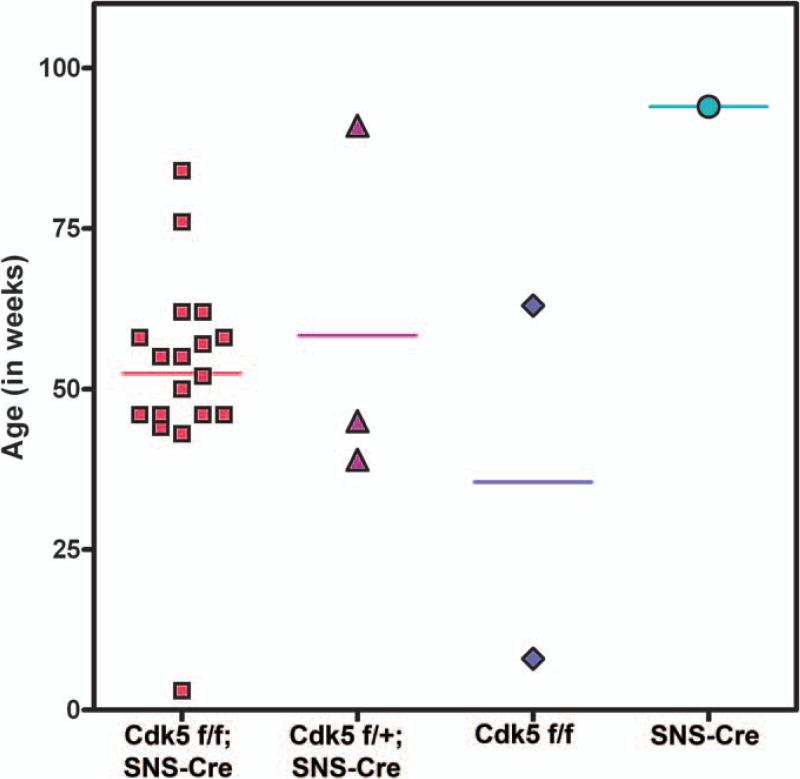
The main transgenic line that was affected by atypical lesions was the Cdk5 conditional knockout mouse (Cdk5-SNS-CoKO) generated by crossing Cdk5 flox/- and SNS-Cre mice. These mice were of mixed strains of C57BL6/129SvJ. As described earlier, these mice showed delayed responses against basal thermal heat stimuli, suggesting the altered pain signaling due to lack of Cdk5 activity in primary sensory neurons3. These lesions arose in the affected Cdk5-SNS-CoKo mice when they were on average 53 weeks old and either singly housed or in groups up to five. (Fig 1).
Figure 1.
A comparative analysis of the Incidence of occurrence of atypical lesions in Cdk5 conditional knockout mice and their littermate controls: Mice were monitored routinely for signs of atypical skin lesions and analyzed by described histological techniques. The numbers of positively affected mice from each group (15 to 25 mice) were plotted on the day these lesions were observed. Cdk5-SNS-CoKO mice affected in significantly higher number (18) on mean age of 53 weeks, compared to control groups i.e. Cdk5 f/+;SNS-Cre (3), Cdk5 f/f (2) and SNS-Cre (1).
Type of skin lesions
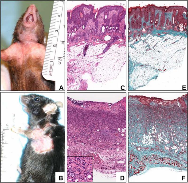
After further analysis, skin lesions due to fighting, barbering, parasitic disease and chronic ulcerative dermatitis were ruled out. Fighting was ruled out based on the appearance and location of the lesions, lack of scab formation, and other signs expected of traumatic lesions. Skin disease secondary to parasitic involvement was ruled out based on lack of organisms observed at gross necropsy and on histological examination. Chronic ulcerative dermatitis was excluded due to the location of the lesions and absence of any signs indicative of healing. The non-healing lesions had very distinguishing characteristics. All of the atypical skin lesions in Cdk5-SNS-CoKO mice were located on the ventrocervical or pectoral areas with subsequent spread distally along the forelimbs. (Fig.2)
Figure 2.
A: In the earlier stage the lesions are hyperplastic and exudative. The animal shows a thickened hairless with focal superficial ulcerations (A). As the lesions progress the ulcers become deeper and extensive and do not heal (B). Microscopically, the earlier stages are characterized by a hyperplastic epidermis skin with fibrotic dermis, which is associated with atrophic hair follicles that may develop cystic structures, and relatively hypertrophic sebaceous glands (C). The fibrosis reaches the skeletal muscle layer and the hypodermis, as shown in green by the trichrome staining (E). The absence of an inflammatory reaction is conspicuous at this stage. As the lesion becomes ulcerated and chronifies it develops a superficial layer of debris and fibrin (D), which lies on a more cellular fibrous tissue. Even at this stage the infiltration is mainly composed of histiocytes and neutrophils with scattered mast cells. Only a few lymphocytes and plasma cells are observed (Inset). The whole process is now embedded in a dense fibrotic tissue involving the whole thickness of the skin (F).
Disease progression
The atypical lesions all progressed in similar patterns. The edges of the lesions were irregular but well demarcated. The initial lesions were most commonly seen at the base of the neck. Three to three and a half weeks after the initial observation the lesions became exudative and started spreading caudally over the pectoral region and thorax and distally along the forelimbs. Between week five and eight there were no major changes in the appearance of the skin lesions. The cases that survived longer than eight weeks developed secondary lesions on the opposite side of the body near the front limbs. Severe pruritus is one of the common characteristics of chronic ulcerative dermatitis.4 However, only six of the 26 animals with the atypical skin lesions exhibited pruritus.
Lack of response to treatment
Treatment was given to all but six of the 26 atypical lesions cases. Treatment included bacitracin, neomycin, and polymixin ointment (BNP), BNP with hydrocortisone, povidone-iodine topical antiseptic, the injectable antibiotic trimethoprimsulfa, or dexamethasone, an anti-inflammatory steroid. Treatment was started at the initial sign of the lesions. In all of the treated non-healing cases, the lesions were not responsive to the treatment. June was found to have the most frequent number of atypical lesions cages (7 of 26). The remaining 19 cases were spread throughout the year.
Pathology of skin lesions
Several interesting characteristics were observed on the H & E slides. Foremost, there was no sign of healing in any of the skin biopsies from the lesions samples. The epithelium was seen to be proliferating, but it was not healing the wound. It also ended abruptly just before the lesions. All of the lesions showed signs of necrosis, inflammation, and fibrosis. Some were so deep they compromised the whole thickness of the skin, reaching to the dermis. An interesting finding was the fact that even in chronic-looking lesions there were not many lymphocytes or plasma cells. While most of the normal skin biopsies appeared normal, a couple of the slides showed signs of mild dermal fibrosis. There were no signs of bacteria, parasites, or fungus.
DISCUSSIONS
Although ulcerative dermatitis has been reported to occur in C57BL/6 mice there have not been many reports of this syndrome in transgenic and knockout mouse lines of different strains. Since more and more transgenic and knockout mouse lines are being generated in different strains, it is imperative to identify ulcerative dermatitis as a distinct phenotype from the skin conditions resulting from aging, fighting, scratching, and from the background strain of the animal itself. There are several different mechanisms that we can speculate as to the cause of the non-healing skin lesions in Cdk5-SNS-CoKO mice. These mice have significant reduction in Cdk5 activity in small diameter Sensory neurons (C-fibers). We have reported earlier that loss of Cdk5 activity in these neurons result in complete abrogation of phosphorylation of heat receptor, TRPV1, affecting function of this receptor and eventually resulting in hypoalgesia in these mice. It is quite possible that due to impaired perception of pain these mice do not respond to general sensation and thus results in unhealed wounds. Previous studies, as early as 1971, have shown chronic ulcerated dermatitis to occur in C57 and B6 mice.5 As these were the background strains of our mice; one could argue that the phenotype of these transgenic mice simply predisposes this dermatitis. This could explain why the unhealed lesions in this study occurred in mice with C57 or B6 strains and initially started near the base of the neck. However, as compared with normal C57 chronic ulcerative dermatitis, the cases observed in our study did not respond to treatment, spread under the neck, down the front legs and lacked lymphocytes. A retrospective study of ulcerative dermatitis in C57Bl/6 background mice was performed to identify the disease trends and predisposed lines.5 The histopathological findings in that study correlated with the classic ulcerative dermatitis, including fibrosis, neutrophils, and the presence of lymphocytes. The location of the lesions, at the base of the neck, also followed the classic cases. However, in our Cdk5-SNS-CoKO mice the location of the lesions differs from the classic ulcerative dermatitis and also lacks lymphocytes. The typical ulcerative dermatitis lesions occurred in mice with either black or agouti coat colors whereas in our study we did not observe any correlation in the occurrence of lesions and the coat color. As mentioned earlier, p35−/− mice also exhibit hypoalgesia, but we did not observe any incidence of such atypical lesions in these mice. There can be two possible explanations for these discrepancy among p35−/− and Cdk5-SNS-CoKO mice: first these lesions are apparent around 53 weeks of age in Cdk5-SNS-CoKO and p35−/− mice die early before that and second in Cdk5-SNS-CoKO mice there is complete loss of Cdk5 activity in peripheral neurons whereas we cannot rule out possibility of residual Cdk5 activity in p35−/− mice contributed from the other Cdk5 activator p39. In conclusion, our results provide a new dimension to the functional role of Cdk5. If Cdk5 activity is require for the maintenance of normal sensation and the wound healing process, it would be interesting to check if restoration of Cdk5 activity in these mice (genetically or by topical Cdk5 application) can help in healing these lesions.
MATERIALS AND METHODS
Mice
The different strains (Cdk5 flox/flox-C57Bl6/129SVJ; SNS-Cre-C57BL6; WT-C57Bl6 and 129SVJ) of transgenic, knockout and wild-type mice were housed individually in ventilated polycarbonate microisolator cages (Lab Products, Seaford, DE) on paper bedding (Tek-fresh, Harlan, Frederick, MD), and each single cage was provided with a Sheppard Shack, (Shepherd Specialty Paper, Baltimore, MD). The room was maintained on a 14/10-hour light dark cycle with a temperature of 68-74 degrees Fahrenheit. Mice were fed NIH rodent formula #07 22.5-5 (Zeigler Bros, Gardens, PA) and chlorinated tap water. The animal study protocols were approved by the NIDCR Animal Care and Use Committee.
Clinical records
In order to characterize the epidemiology of the skin lesions in the transgenic mouse lines, we kept detailed records of the clinical cases observed during January to December 2006. The age, gender, strain, genotype, and type of social housing were recorded for each case. They were then divided into one of the two categories, lesions with a likely cause and not unexpected in a mouse colony versus lesions that were considered atypical based on history, appearance and histopathological analysis. Typical or expected lesions were determined to be due to fighting, strain related ulcerative dermatitis, or expected phenotype. Atypical lesions discussed here were characterized by a lack of response to treatment, no grossly visible signs of healing, bacteriological, fungal or ectoparasitic infections, and frequently in locations not associated with lesions due to fighting, chronic ulcerative or parasitic disease.
Histological analysis
Mice were euthanized by CO2 inhalation in a standard CO2 chamber. Skin samples were obtained from areas of the lesions and normal skin of the affected mice. The skin sections were fixed in formalin. Paraffin sections were obtained and stained with hematoxylin and eosin (H&E). Additional slides were stained for connective tissue and nerve cells and fibers. Ayoub-Shklar (keratin and prekeratin), Fraser-Lendrum (fibrin), Gomori's Trichrome (collagen and muscle), Jone's PAMS (basement membrane), and Movat Pentachrome (elastic, muscle, collagen, and muccosaccharides) stains were used to view the connective tissues. Nerve cells and fibers were stained with Bielschowsky (axons and dendrites), Bodian (nerve fibers), Vogt's Method (nissl substance), and Woelcke (myelin).
REFERENCES
- 1.Pareek TK, Keller J, Kesavapany S, Pant HC, Iadarola MJ, Brady RO, Kulkarni AB. Cyclin-dependent kinase 5 activity regulates pain signaling. Proceedings of the National Academy of Sciences of the United States of America. 2006;103:791–6. doi: 10.1073/pnas.0510405103. [DOI] [PMC free article] [PubMed] [Google Scholar]
- 2.Pareek TK, Kulkarni AB. Cdk5: a new player in pain signaling. Cell cycle. 2006;5:585–8. doi: 10.4161/cc.5.6.2578. [DOI] [PubMed] [Google Scholar]
- 3.Pareek TK, Keller J, Kesavapany S, Agarwal N, Kuner R, Pant HC, Iadarola MJ, Brady RO, Kulkarni AB. Cyclin-dependent kinase 5 modulates nociceptive signaling through direct phosphorylation of transient receptor potential vanilloid 1. Proceedings of the National Academy of Sciences of the United States of America. 2007;104:660–5. doi: 10.1073/pnas.0609916104. [DOI] [PMC free article] [PubMed] [Google Scholar]
- 4.Kastenmayer RJ, Fain MA, Perdue KA. A retrospective study of idiopathic ulcerative dermatitis in mice with a C57BL/6 background. J Am Assoc Lab Anim Sci. 2006;45:8–12. [PubMed] [Google Scholar]
- 5.Lawson GW, Sato A, Fairbanks LA, Lawson PT. Vitamin E as a treatment for ulcerative dermatitis in C57BL/6 mice and strains with a C57BL/6 background. Contemporary topics in laboratory animal science / American Association for Laboratory Animal Science. 2005;44:18–21. [PubMed] [Google Scholar]


