Abstract
[Purpose] The purpose of this study was to investigate the effects of isokinetic passive exercise and motion velocity on passive stiffness. In addition, we also discuss the effects of the contraction of agonist and antagonist muscles on passive stiffness. [Subjects] The subjects were 20 healthy men with no bone or joint disease. [Methods] Isokinetic passive exercise and isometric muscle contraction were performed on an isokinetic dynamometer. The angular acceleration measured by the accelerometer was compared before and after each task. [Results] After the passive exercise, the angular acceleration increased in the phase of small damped oscillation. Moreover, the effect was higher at high-speed movement. The angular acceleration was decreased by the contraction of the agonist muscle. Conversely, the angular acceleration was increased by the contraction of the antagonist muscle. [Conclusion] Isokinetic passive exercise reduced passive stiffness. Our results suggest the possibility that passive stiffness is increased by agonist muscle contraction and decreased by antagonist muscle contraction.
Key words: Passive stiffness, Angular acceleration, Isokinetic passive exercise
INTRODUCTION
In the field of engineering, stiffness is defined as the amount of displacement on application of force. The term “stiffness” is also used in the medical field to describe the hardness of the skin caused by scarring, decreased joint movement, and spasticity because of increased muscle tone1). Changes in stiffness affect the course of clinical rehabilitation. When a patient has joint contractures caused by spasticity or limitation of the range of motion (ROM) associated with immobility (disuse or fixation), joint stiffness is increased. These impairments cause activity disturbance and decline in the efficiency of activity2, 3). In the field of sports medicine, decreased joint stiffness reportedly diminishes performance because of the loss of power transmission efficiency4). The state of joint stiffness is important in deciding the strategy for rehabilitation and the therapeutic effect. Joint stiffness is classified into dynamic stiffness and passive stiffness. Passive stiffness is joint stiffness during passive exercise and represents the characteristics of the elastic element of the soft tissue5). Dynamic stiffness is the stiffness of joints during activity, and includes not only structural hardness of the tissue but also the element of muscle activity; therefore, it is calculated as the slope of the regression line between the joint moment and the joint angle during joint movement6). Passive stiffness varies with lifestyle and reportedly decreases with certain stretching methods5, 7). There are few reports on the influence on passive stiffness of passive ROM exercises, which have been widely adopted in clinical practice8, 9). Furthermore, the influence of the motion speed of the passive ROM exercise on passive stiffness has not been investigated.
Muscle hardness increases after strength training10), and affects the cross-linked structure of actin and myosin in resting muscle11). Thus, the potential for muscle contraction can change passive stiffness. Therefore, the intensity, frequency, or the practice order of muscle strengthening exercises may have a positive or negative effect on ROM. This study used the angular acceleration of a leg in the pendulum drop test (PDT)12) as an indicator of passive stiffness. We investigated the effects of isokinetic passive exercise on passive stiffness and determined the difference between the effects of high-speed and low-speed tasks. In addition, we examined the effect of the contraction of agonist and antagonist muscles on the passive stiffness of the knee extension element.
SUBJECTS AND METHODS
The subjects were 20 healthy men (age, 26.8 ± 3.8 years) with no bone or joint disease. Thigh length, leg length, leg circumference, height, weight, body fat percentage, and body mass index were measured as the physical characteristics of the subjects. The subjects provided their consent to participation in this study, and the study protocol was approved by the Medical Ethics Committee of Kanazawa University (certificate no. 354).
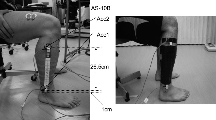
The acceleration of a freely falling leg in the PDT was measured using 2 accelerometers in the resting state, and after the tasks of isometric muscle contraction and constant-velocity passive exercise. The accelerometer used was a small acceleration transducer (AS-10B; Kyowa Electronic Instruments Co., Ltd., Japan). The accelerometers were fixed to a plastic rod (length, 31 cm; width, 2.5 cm; and thickness, 0.5 mm) with surgical tape and double-sided tape, so that the sensitive direction of the accelerometer would correspond to the long axis of the leg. Accelerometer 1 (Acc1) was attached 1 cm above the bottom of the bar, and accelerometer 2 (Acc2) was affixed 26.5 cm above the top of Acc1. The center of the head of the fibula and the lateral malleolus were used as landmarks to determine the position of the plastic rod, and the plastic rod was attached 1 cm above the center of the lateral malleolus (Fig. 1).
Fig. 1.
Mounting of accelerometer
Electromyographic data were collected from the vastus lateralis (VL) and biceps femoris (BF) muscles to detect the stretch reflex during the isokinetic passive exercise, and to measure the muscle activity during isometric contraction. The myoelectric signal was recorded using a 16-bit A/D converter (MQ8; Kissei Comtec Co., Ltd., Japan). The sampling frequency was 1,024 Hz, and a band-pass filter of 10–500 Hz was used. Skin preparation consisted of shaving and abrading with a skin preparation gel (NuPrep; DO Weaver & Co., Aurora, Co., USA) (4 × 8 cm) to achieve a skin resistance of ≤5 kΩ. Disposable silver–silver chloride (Blue Sensor) electrodes were attached at an interelectrode distance of 2.5 cm. The electrode for the VL recording was located on the outer femur surface 5 finger breadths proximal to the upper edge of the patella; the electrode for the BF recording was located at the midpoint of the line segment connecting the ischial tuberosity and head of the fibula; and the ground electrode was located on the tibial tuberosity. Electromyography (EMG) radio transmitters were fixed to the waist of the subject by a belt after affixing the electrodes.
Isometric muscle contraction and isokinetic passive exercise tasks were performed using an isokinetic dynamometer (Cybex 770-Norm; Cybex International Inc., USA). Subjects were positioned on the seat after the recording devices were attached, and the upper trunk and pelvis were secured with belts. The seat was adjusted so that the subject's knee-joint axis matched the rotational axis of the lever arm. During this time, the subject was instructed to rest.
Measurements were taken using the following procedure.
-
1)
PDT before the task of isokinetic passive exercise
After practicing several times, the free-fall acceleration of the leg was measured in the PDT. The starting position was 30°of knee joint flexion. The accelerometer was reset after checking the position with a goniometer. We also monitored the displacement of the zero position at the start angle with the accelerometer. All these checks were performed by the same person.
-
2)
Isokinetic passive exercise at 500°/s and 30°/s
Motor tasks were performed on the left and right sides, and the 2-speed tasks were allocated randomly to either the left or the right leg. The tasks were performed using the Cybex CPM mode; the slow speed was 30°/s and the high speed was 500°/s. The tasks consisted of flexing and extending the knee and were repeated 40 times. The ROM was set from 0° to 120° of the knee joint. The examiner also confirmed the presence or absence of the muscle stretch reflex on EMG.
-
3)
PDT after the task of isokinetic passive exercise
The PDT was carried out immediately after the completion of the isokinetic passive exercise task. The distal part of the leg cuff was removed, and the lever arm was quickly rotated upward. The resultant acceleration of the free fall of the lower leg was measured.
-
4)
Isometric contraction of the knee extensor
Subjects performed isometric contraction of the knee extensor muscles at maximum effort, with the knee flexed at 60°. The contraction time was 5 s with a resting time of 10 s, and the task was repeated 3 times. The examiner provided verbal encouragement during the task. EMG data were recorded from the start until the end of the measurement.
-
5)
PDT after the task of isometric contraction of the knee extensor
-
6)
Isometric contraction of the knee flexor
Subjects performed isometric contraction of the knee flexor muscles at maximum effort, with the knee flexed at 60°. The contraction time was 5 s with a resting time of 10 s, and the task was repeated 3 times. The examiner provided verbal encouragement during the task. EMG data were recorded from the start until the end of the measurement.
-
7)
PDT after the task of isometric contraction of the knee flexor
The measurement was repeated 3 times to confirm the reproducibility of the PDT data in 5 subjects (10 limbs).
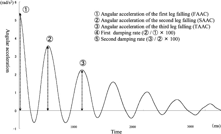
The accelerometer data were processed using analysis software (BIMUTAS II; Kissei Comtec Co., Japan), which applied a low-pass filter of 6 Hz after the calibration process. The acceleration data from the 2 sites were converted to angular acceleration according to the method of Kusuhara et al.13) (Acc1 − Acc2)/0.265. The following 5 parameters were used as indicators of passive stiffness (Fig. 2): first angular acceleration (FAAC), second angular acceleration (SAAC), third angular acceleration (TAAC), first attenuation factor (SAAC/FAAC × 100), and second attenuation factor (TAAC/SAAC × 100). Further, the percentage decrease of angular acceleration was calculated (after the task/before the task × 100), to determine the effect of the isokinetic passive exercise on movement velocity, and compared between the angular accelerations of 500°/s and 30°/s.
Fig. 2.
Angular acceleration as the passive stiffness index in the PDT
The angular acceleration was analyzed only in the direction of flexion from the extended position, because it would be expected to be slow in the direction of extension with a wide range of antigravity.
The EMG data of BF and VL were analyzed using BIMUTAS II. The root mean square (RMS) of the EMG was calculated on the basis of a 5-s isometric contraction task, with the initial and the final seconds excluded. The RMS values were calculated at intervals of 30-ms during the 3-s analysis period, and the mean RMS was calculated from the RMS values of the BF and VL. The mean RMS of the BF or VL during the task of knee extension or flexion was normalized to the mean RMS of the maximum isometric contraction of the corresponding muscle (BF mean RMS during the extension task/BF mean RMS during the maximum isometric contraction × 100 ; %RMS).
Statistical analysis was performed using SPSS for Windows 11.0 J (SPSS Inc., 1989–2001). The Bonferroni multiple comparison test was used for the comparison of angular acceleration data before and after the isokinetic passive exercise and isometric knee extension tasks. The paired t-test was used to compare the angular acceleration data before and after the isometric knee flexion task and the percentage increase of angular acceleration. The intraclass correlation coefficient (ICC) was used to test the reproducibility of the PDT data.
RESULTS
The ICCs for 3 repeats of the PDT data were 0.936, 0.949, and 0.956 for the FAAC, SAAC, and AAC, respectively, indicating good reproducibility.
FAAC: At 30°/s, the angular acceleration was 7.39 ± 2.12 rad/s2 before the task, 7.79 ± 2.51 rad/s2 after isokinetic passive exercise, and 6.11 ± 2.59 rad/s2 after isometric muscle contraction of knee joint extension. A significant difference was observed in the angular acceleration between before and after isometric muscle contraction of knee joint extension (p<0.05). The angular acceleration after isokinetic passive exercise significantly differed from that after isometric muscle contraction of knee joint extension (p<0.01). At 500°/s, the angular acceleration was 7.10 ± 1.99 rad/s2 before the task, 7.68 ± 1.71 rad/s2 after isokinetic passive exercise, and 6.50 ± 1.84 rad/s2 after isometric muscle contraction of knee joint extension. A significant difference was observed between the angular acceleration after isokinetic passive exercise and that after isometric muscle contraction of knee joint extension (p<0.01) (Table 1).
Table 1. Changes of FAAC after each motor task.
| Angular velocity | Angular acceleration (rad/s2) | ||
| Before task | After isokinetic passive exercise |
After isometric contraction |
|
| 30°/sec | 7.39 ± 2.12 | 7.79 ± 2.51 | 6.11 ± 2.59*, ** |
| 500°/sec | 7.10 ± 1.99 | 7.68 ± 1.71 | 6.50 ± 1.84** |
Values are presented as the average ± SD. *; between before and after isometric contraction, p<0.05. **; between after isokinetic passive exercise and after isometric contraction, p<0.01
SAAC: At 30°/s, the angular acceleration was 5.45 ± 1.79 rad/s2 before the task, 5.89 ± 1.79 rad/s2 after isokinetic passive exercise, and 4.61 ± 2.02 rad/s2 after isometric muscle contraction of knee joint extension. A significant difference was observed between the angular acceleration values before and after isometric muscle contraction of knee joint extension (p<0.05). Furthermore, the angular acceleration after isokinetic passive exercise significantly differed from that after isometric muscle contraction of knee joint extension (p<0.01). At 500°/s, the angular acceleration was 5.31 ± 2.03 rad/s2 before the task, 5.90 ± 1.44 rad/s2 after isokinetic passive exercise, and 4.87 ± 1.76 rad/s2 after isometric muscle contraction of knee joint extension. A significant difference was observed between the angular acceleration after isokinetic passive exercise and that after isometric muscle contraction of knee joint extension (p<0.01) (Table 2).
Table 2. Changes of SAAC after each motor task.
| Angular velocity | Angular acceleration (rad/s2) | ||
| Before task | After isokinetic passive exercise |
After isometric contraction |
|
| 30°/sec | 5.45 ± 1.79 | 5.89 ± 1.79 | 4.61 ± 2.02*,** |
| 500°/sec | 5.31 ± 2.03 | 5.90 ± 1.44 | 4.87 ± 1.76** |
Values are presented as the average ± SD. *; between before and after isometric contraction, p<0.05. **; between after isokinetic passive exercise and after isometric contraction, p<0.01
TAAC: At 30°/s, the angular acceleration was 4.14 ± 1.36 rad/s2 before the task, 4.27 ± 1.51 rad/s2 after isokinetic passive exercise, and 3.28 ± 1.72 rad/s2 after isometric muscle contraction of knee joint extension. The angular acceleration differed significantly before and after the isometric muscle contraction of knee joint extension (p<0.05). A significant difference was observed between the angular acceleration after isokinetic passive exercise and that after isometric muscle contraction of knee joint extension (p<0.01). At 500°/s, the angular acceleration was 3.85 ± 1.66 rad/s2 before the task, 4.44 ± 1.42 rad/s2 after the isokinetic passive exercise, and 3.58 ± 1.44 rad/s2 after the isometric muscle contraction of knee joint extension. A significant difference was observed in the angular acceleration before and after isokinetic passive exercise (p<0.05). In addition, the angular acceleration after the isokinetic passive exercise significantly differed from those after isometric muscle contraction of knee joint extension (p<0.01) (Table 3).
Table 3. Changes of TAAC after each motor task.
| Angular velocity | Angular acceleration (rad/s2) | ||
| Before task | After isokinetic passive exercise |
After isometric contraction |
|
| 30°/sec | 4.14 ± 1.36 | 4.27 ± 1.51 | 3.28 ± 1.72*,** |
| 500°/sec | 3.85 ± 1.66 | 4.44 ± 1.42 | 3.58 ± 1.44†,** |
Values are presented as the average ± SD. *; between before and after isometric contraction, p<0.05. **; between after isokinetic passive exercise and after isometric contraction, p<0.01. †; between before and after isokinetic passive exercise, p<0.05
The first damping extinction ratio was 74.4 ± 6.9% before the task, 76.1 ± 5.2% after isokinetic passive exercise, and 74.8 ± 8.8% after isometric muscle contraction of the knee joint extension at 30°/s, and 76.2 ± 10.6% before the task, 78.5 ± 8.0% after isokinetic passive exercise, and 75.2 ± 10.6% after isometric muscle contraction of knee joint extension at 500°/s. No significant differences were observed between groups.
The second damping ratio was 73.4 ± 6.2% before the task, 73.0 ± 7.8% after isokinetic passive exercise, and 70.8 ± 12.7% after isometric muscle contraction of knee joint extension at 30°/s, with no significant differences between groups. At 500°/s, the second damping ratio was 70.0 ± 9.2% before the task, 73.8 ± 8.8% after isokinetic passive exercise, and 71.0 ± 10.4% after isometric muscle contraction of knee joint extension, with a significant difference between the values before and after isokinetic passive exercise (p<0.05).
The percentage increases in angular acceleration after isokinetic passive exercise were 106.0 ± 17.1% (FAAC), 110.0 ± 17.8% (SAAC), and 105.0 ± 24.1% (TAAC). At 500°/s, the percentage increases were 111.5 ± 22.5% (FAAC), 120.0 ± 32.6% (SAAC), and 124.0 ± 32.6% (TAAC). The percentage increase was significantly greater at 500°/s in TAAC (P=0.019).
The angular acceleration was significantly greater than the angular acceleration after the isometric knee extension in FAAC and SAAC at 30°/s (Table 4). The degree of muscle activity of BF was 7.18 ± 2.75% during isometric knee extension, whereas that of the VL was 9.49 ± 6.69% during isometric knee flexion.
Table 4. Change of the angular acceleration after the flexor contraction.
| Isometric contraction task (rad / s2) | ||||
| Extensor | Flexor | |||
| FAAC | 30°/sec | 6.11 ± 2.59 | 6.72 ± 2.55 | * |
| 500°/sec | 6.50 ± 1.84 | 6.67 ± 2.03 | ||
| SAAC | 30°/sec | 4.61 ± 2.02 | 5.09 ± 2.42 | * |
| 500°/sec | 4.87 ± 1.76 | 5.11 ± 1.94 | ||
| TAAC | 30°/sec | 3.28 ± 1.72 | 3.57 ± 2.18 | |
| 500°/sec | 3.58 ± 1.44 | 3.77 ± 1.73 | ||
Values are presented as the average ± SD. *; significantly difference, p<0.05
DISCUSSION
In the present study, changes in passive stiffness before and after isokinetic passive exercise and the influence of the motion speed of passive ROM exercise were investigated. After isokinetic passive exercise, the angular acceleration increased by 6–10% at 30°/s and by 11–24% at 500°/s. The percentage increase in the angular acceleration at 500°/s tended to be higher than that at 30°/s in FAAC and SAAC, and was significantly higher in TAAC. This result suggests that passive stiffness is reduced by isokinetic passive exercise, and that this effect is greater in high-speed motion.
Muscles exhibit thixotropy14, 15), a phenomenon in which agitation and shear stress causes a decrease in viscosity. Agitation and shear stress increases with an increase in the motion speed in passive exercise. As a result, the effect of thixotropy is greater at fast motion, which might explain the increased angular acceleration observed in the present study. ROM exercises are usually performed over the entire ROM at slow speeds in consideration of pain development and the stretch reflex. Joint movement at slow speeds is appropriate for inhibiting the stretch reflex16, 17). The multiple limiting factors in ROM include muscle, connective tissue, and skin18, 19). Therefore, if the main limiting factor is the fascia with the characteristics of glide and connective tissue, ROM exercises may be more effectively performed at high speed, thereby increasing the agitation power and shearing stress per unit time. In FAAC and SAAC at both 30°/s and 500°/s, the angular acceleration tended to increase; however, no significant difference was observed between before and after each task. In the initial phases of the lower leg fall, the angular acceleration in the PDT did not reflect the changes in passive stiffness because of increases in both the angular acceleration and the amount of displacement. The damped oscillation of the falling lower leg is affected by the lower leg shape, length, weight, and femoral muscle mass. In the present study, individual differences were not considered in the investigation of the rate of change before and after the task. We consider that the variation in angular acceleration was increased by the influence of individual differences such as lower leg shape in the FAAC and SAAC, particularly in large angular accelerations. In recent years, the mitigation model of individual differences has been devised for the PDT20). Future studies should incorporate modifications using this technique.
In the FAAC, SAAC, and TAAC at both 30°/s and 500°/s, the angular acceleration significantly decreased after the task of isometric muscle contraction of knee joint extension compared with the task of isokinetic passive exercise. This result suggests that passive stiffness in the direction of knee flexion increases after isometric muscle contraction of knee joint extension. Noda et al.21) reported that the viscous constant of the muscle is significantly greater before than after isometric muscle contraction. On the other hand, a study of the hardness of muscle reported that muscle hardness was increased after exercise10). In an isometric contraction task, the fascia and connective tissue are not stretched because there is no articulation. It seems that the influence of these factors on angular acceleration is small. Therefore, we consider the decrease in angular acceleration after isometric contraction is caused by changes in the muscle contraction element. The results of the present study revealed increased passive stiffness after agonist muscle contraction. Thus, we propose that when treatment is intended to increase ROM, exercises generating strong muscle contractions may inhibit ROM. ROM exercises preceding muscle strength training are considered optimal for effective rehabilitation. In contrast, higher joint rigidity may aid stability, because the transmission efficiency of the joint torque in muscle contraction is excellent.
In patients with hemiplegia, rigid ankles caused by abnormal muscle tone resulting from spasticity may contribute to the stabilization of walking. Based on these considerations, increased joint stiffness caused by muscle contraction before surgery may improve performance and surgical outcome of patients with joint instability.
However, the effect of increasing the duration of passive stiffness is unknown. Future research on this topic is warranted.
In FAAC and SAAC at 30°/s, the angular acceleration after the isometric muscle contraction of knee joint flexion significantly increased compared with that in the task of isometric knee extension. During the motor task, the %RMS was 9.4% on average for VL, but the angular acceleration was increased and the passive stiffness in the direction of knee flexion was decreased. We consider that the increased involvement of the contractile element resulted in an articulation different from that in isometric knee extension, and a significant difference in the phase of large angular acceleration in the initial free-fall motion of the lower leg. Afferent nerves from the muscle spindle inhibit the motor neurons of the antagonist muscle through the interneurons, in addition to the excitation from the firing of the same neurons, when the agonist muscle contracts22). We consider that the extensor muscle is inhibited by the contraction of the flexor muscle; therefore, angular acceleration is increased. Our results, confirm the theory of the relaxation technique using Ia-inhibition, and the effect of previous isometric contraction of the antagonist muscle improving the agonist ROM exercise was verified.
Acknowledgments
The authors are grateful to T. Nakagawa, M. Yokogawa, as well as to all subjects who were involved in this study.
REFERENCES
- 1.Muraki T: Assessment of skeletal muscle and joint with stiffness measurement. Phys Ther Jpn, 2010, 37: 654–657 [Google Scholar]
- 2.Lamontagne A, Malouin F, Richards C: Contribution of passive stiffness to ankle plantar flexor moment during gait after stroke. Arch Phys Med Rehabil, 2000, 81: 351–358 [DOI] [PubMed] [Google Scholar]
- 3.Sinkjaer T, Magnussen I: Passive intrinsic and reflex-mediated stiffness in ankle extensors of hemiparetic patients. Brain, 1994, 117: 355–363 [DOI] [PubMed] [Google Scholar]
- 4.Fowles JR, Sale DG, MacDougall JD: Reduced strength after passive stretch of the human plantarflexors. J Appl Physiol, 2000, 89: 1179–1188 [DOI] [PubMed] [Google Scholar]
- 5.Nordez A, Comu C, McNair P: Acute effects of stretching on passive stiffness of hamstring muscle calculated using different mathematical models. Clin Biomech (Bristol, Avon), 2006, 21: 755–760 [DOI] [PubMed] [Google Scholar]
- 6.Davis RB, DeLuca P: Gait characterization via dynamic joint stiffness. Gait Posture, 1996, 4: 224–231 [Google Scholar]
- 7.Nordez A, McNair PJ, Casari P, et al. : Static and cyclic stretching: their different effects on the passive torque-angle curve. J Sci Med Sport, 2010, 13: 156–160 [DOI] [PubMed] [Google Scholar]
- 8.Tanaka N, Okajima Y, Taki M, et al. : Effect of continuous range of motion exercise on passive resistive joint torque. Jpn J Rehabil Med, 1998, 35: 491–495 [Google Scholar]
- 9.Lakie M, Walsh EG, Wright GW: Resonance at the wrist demonstrated by the use of a torque motor: an instrumental analysis of the muscle tone in man. J Physiol, 1984, 353: 265–285 [DOI] [PMC free article] [PubMed] [Google Scholar]
- 10.Matsubara Y, Awai H, Kimura G, et al. : Effect of vibration stimulation on recovery of muscle stiffness after isometric muscle contraction had achieved fatigue. Rigakuryoho Kagaku, 2004, 19: 341–345 [Google Scholar]
- 11.Seki M: Biomechanical properties during passive ankle movement in spastic hemiplegic patients. Jpn J Rehabil Med, 2001, 38: 259–267 [Google Scholar]
- 12.Wartenberg R: Pendulousness of the leg as a diagnostic test. Neurology, 1951, 1: 18–24 [DOI] [PubMed] [Google Scholar]
- 13.Kusuhara T, Jikuya K, Nakamura T, et al. : Accuracy of knee-joint angular acceleration detector by using two linear accelerometers for pendulum test. IEICE Tech Rep MBE, 2010, 19: 17–20 [Google Scholar]
- 14.Zhang Q, Noryati I, Cheng LH: Effects of pH on the functional properties of chicken muscle proteins-modified waxy cornstarch blends. J Food Sci, 2008, 73: 82–87 [DOI] [PubMed] [Google Scholar]
- 15.Sekihara C, Izumizaki M, Yasuda T, et al. : Effect of cooling on thixotropic position-sense error in human biceps muscle. Muscle Nerve, 2007, 35: 781–787 [DOI] [PubMed] [Google Scholar]
- 16.Williams PE, Goldspink G: Changes in sarcomere length and physiological properties in immobilized muscle. J Anat, 1978, 127: 459–468 [PMC free article] [PubMed] [Google Scholar]
- 17.Rowe RW: Morphology of perimysial and endomysial connective tissue in skeletal muscle. Tissue Cell, 1981, 13: 681–690 [DOI] [PubMed] [Google Scholar]
- 18.Chino N: Modern Textbook of Rehabilitation Medicine. Tokyo: Kanehara and Co, Ltd., 1999, pp 217–219. [Google Scholar]
- 19.Bang MD, Deyle GD: Comparison of supervised exercise with and without manual physical therapy for patients with shoulder impingement syndrome. J Orthop Sports Phys Ther, 2000, 30: 126–137 [DOI] [PubMed] [Google Scholar]
- 20.Jikuya K, Michinishi H, Okamoto T, et al. : A method for decreasing influence of individual differences on pendulum test –In case of normal subjects–. IEICE Tech Rep MBE, 2006, 22: 1–4 [Google Scholar]
- 21.Noda M, Shibayama A, Ishige Y, et al. : Change of viscoelastic properties in human triceps surae after isometric endurance exercise. JJBSE, 2000, 4: 232–243 [Google Scholar]
- 22.FitzGerald MJ: Neuroanatomy Basic and Clinical. Galway: W.B.Saunders, 1996, pp 78–81. [Google Scholar]


