Abstract
The technical advancement in the anaesthesia workstations has made the peri-operative anaesthesia more safer. Apart from other monitoring options, respiratory gas analysis has become an integral part of the modern anaesthesia workstations. Monitoring devices, such as an oxygen analyser with an audible alarm, carbon dioxide analyser, a vapour analyser, whenever a volatile anaesthetic is delivered have also been recommended by various anaesthesia societies. This review article discusses various techniques for analysis of flow, volumes and concentration of various anaesthetic agents including oxygen, nitrous oxide and volatile anaesthetic agents.
Keywords: Anaesthesia machine, gas analyser, gas flows, gas volumes, oxygen analyser, volatile agent analyser
INTRODUCTION
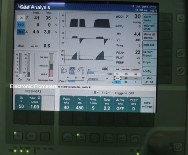
A well-equipped anaesthesia workstation is a boon for better anaesthesia practice. The technical advancement in anaesthesia workstations has made the intra-operative anaesthesia better and more safer. Apart from other monitoring options, respiratory gas analysis has become an integral part of the modern anaesthesia workstation. This has been incorporated not only for operating room anaesthesia machines but also for that used in intensive care units, transfer of ventilated patients and at other places outside the operating room where anaesthesia services are required. Monitoring devices, such as an oxygen analyser with an audible alarm, carbon dioxide analyser, a vapour analyser, whenever a volatile anaesthetic is delivered have also been recommended by various anaesthesia societies.[1,2,3,4] Nowadays, anaesthesia workstations are being configured by manufacturers in either a modular way or as a preconfigured approach. In the modular approach, the machine is equipped with basic monitors while in the preconfigured approach, it is more integrated with multiple monitoring units and includes gas flow monitors and analysers as well [Figure 1]. Some units also have the option of integrating, analyzing, displaying and recording the data obtained from monitoring sensors.
Figure 1.

Monitor showing gas analysis and electronic flowmeters on anaesthesia workstation
This review is based on a thorough search of published articles in English, across Pubmed, Medline, Ovid and Embase databases. The literature search was performed using keywords in various combinations: ‘anaesthesia machine, gas analyser, oxygen analyser, volatile agent analyser, nitrous oxide analyser, flow and volume measurement, sampling techniques’. The articles were further searched from the cross references. The manufactures information was also searched but were verified from the published articles or cross checked from the mentioned references.
HISTORICAL PERSPECTIVE
Gas analysis has evolved a lot over time.[4,5,6,7] The earliest gas monitor was based on the effect of gas on a silicone rubber band. The relaxation of a lightly tensioned silicone rubber band by anaesthetic gases was transmitted mechanically to a measuring scale via a lever. The scale was calibrated with various anaesthetic agents and thus provided the gas concentration with an acceptable accuracy within the range of 0-3%.[5] This monitoring was limited by the certain factors affecting monitoring like the presence of water vapour and nitrous oxide. The sampling could be done either from inspiratory or expiratory limbs.
The use of the mass spectrometer was introduced in 1981 for monitoring anaesthetic gases on breath-by-breath basis.[7] This technique involved side-stream sampling (250 mL/min) and transfering it to a centrally located mass spectrometer for analysis by a multiplexing valve.[8] This device could measure and quantify up to eight unique gases.[6] Few years later, PPG (Pittsburgh Plate Glass) SARA (System for Anaesthetic and Respiratory Analysis), with sampling flow of 330 mL/min was introduced.[5,8] Gas analyses in these devices were influenced by gases like aerosol propellants, helium and anaesthetic agents for which these were not designed. Later, stand-alone mass spectrometers, like the Ohmeda 6000, were introduced which provided continuous readings as compared to intermittent analysis in centralised devices.[9,10] These devices required low sampling gas flow and thus were considered worth for paediatric anaesthesia cases. Also, these devices could be easily calibrated with any gas or a new agent.[10] Subsequently, various stand-alone sidestream gas analysers were introduced which were based on infrared spectrometry, Raman spectrometry, infrared photoacoustic spectrometry and piezoelectric crystal agent analysis technology.[11,12] These devices could identify the specific agents and thus were of help in case any agent was misfed in the vapouriser mistakenly. Some of these technologies such as Raman spectroscopy, infrared photoacoustic spectrometry and piezoelectric crystal agent could not gain regular use in clinical practice because of various limitations like requirement of high sampling gas flow, analysis affected by the presence of other gases or contaminants and slow response time. The technique of infrared photoacoustic and piezoelectric devices was not agent-specific and thus limited in its clinical use.[13] The technique of infrared photospectrometer became popular because of its lower cost and agent identification functionality could be easily incorporated in these analysers.[13]
OXYGEN ANALYSIS
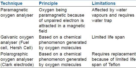
Oxygen analysers are recommended for all anaesthesia cases as measuring oxygen is of utmost importance to prevent any untoward event related to hypoxia.[14] These have been incorporated in anaesthesia workstations and monitors oxygen concentration either at the common gas outlet or in the inspiratory limb of the circle system.[1,15] The oxygen is also monitored at the patient end using paramagnetic analysers. The three commonly reported techniques for measurement of the inspired oxygen concentration include galvanic, polarographic and paramagnetic techniques [Table 1]. The newer anaesthesia machines more commonly have paramagnetic method for oxygen analysis.
Table 1.
Technique, principle and limitations of oxygen analysers in anaesthesia machines

Paramagnetic oxygen analyser
An oxygen molecule has unpaired electrons in its outer electron ring which makes it paramagnetic and thus is attracted in the magnetic field. This forms the working principle of paramagnetic oxygen analysers.[1,15,16] This device samples gas from breathing circuit which should be close to the patient. Older paramagnetic analysers used dumb-bell and torsion wire system but the newer analysers use switched electromagnetic field which is generated at approximately 110 Hz.[1] The analysers comprise of two chambers (sampling and reference chamber) with a sensitive pressure transducer in between. The sampling chamber receives sample gas via sampling tube while a reference chamber receives room air. The changing magnetic field is created by electromagnet which rapidly switches on and off which causes oxygen molecules to be attracted and agitated. This in turn results in pressure on either side of the pressure transducer and the pressure difference across the transducer is proportional to the oxygen partial pressure difference between the sample gas and the reference gas (room air, containing 21% oxygen). The pressure fluctuations of approximately 20–50 mbar is sensed by the transducer and is converted to a DC voltage, which is directly proportional to the concentration of oxygen.[1] Though the measurement is in partial pressure, it is programmed to display in percentage. Such devices require calibrations which are scheduled in anaesthesia workstations. They are accurate and highly sensitive as well with a rapid response allowing measurement of oxygen on a breath-to-breath basis. These devices are incorporated with water trap from the sampled gas as water vapours could affect oxygen analysis.
Galvanic oxygen analyser (Fuel cell, Hersh cell)
These analysers work on the basis of chemical phenomenon generated by oxygen molecules. Oxygen diffuses across the membrane and an electrolyte solution to a cathode (gold or silver).[16,17] This is connected to a lead anode via the electrolyte solution. This generates an electrical current which is proportional to partial pressure of oxygen in the sampled gas. The chemical reaction involves:
Pb + 2OH- → PbO + H2O + 2e-
They are usually connected to inspiratory limb. These require calibration with 100% oxygen and room air (21% oxygen) and have a response time of around 20 seconds with an accuracy of 3%.[15] These do not require a water trap as water vapour does not affect their performance. These analysers have a limited life span because of the depletion of the battery due to continuous exposure to oxygen.
Polarographic oxygen analyser (Clark electrode)
These analysers have a similar principle like that of a galvanic oxygen analyser.[15] Here the oxygen molecules move across a Teflon membrane and current flows between a silver cathode and a platinum anode. The current so generated is proportional to oxygen concentration of the samples gas. It is usually attached to inspiratory limb of the circuit. It has a life span of around 3 years because of deterioration of the Teflon membrane.[15]
OTHER GASEOUS (CARBON DIOXIDE, NITROUS OXIDE AND VOLATILE AGENTS) ANALYSIS
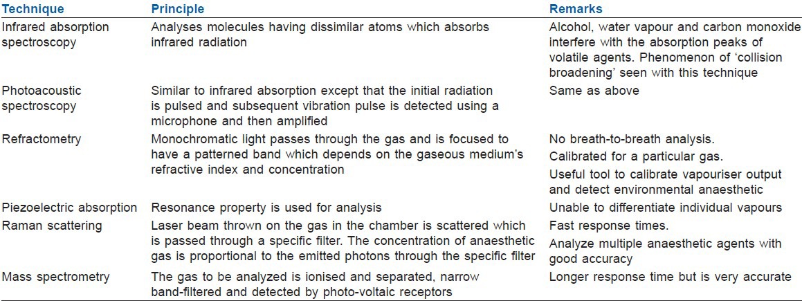
In anaesthesia practice, the measurements of other anaesthetic gases like nitrous oxide, volatile anaesthetic agents and carbon dioxide are also required. These could be analysed by using various techniques like infrared absorption spectroscopy, photoacoustic spectroscopy, silicone rubber and piezoelectric absorption, refractometry, Raman scattering and mass spectrometry [Table 2].[1,15] The infrared analysers are commonly used for measuring anaesthetic agents concentration by side-sampling method on the anaesthesia machines.
Table 2.
Technique, principle and limitations of anaesthetic gases analysers in anaesthesia machines

Infrared absorption spectroscopy
Most commonly, either a dispersive infrared (DIR) method or a non-dispersive infrared (NDIR) method is used to isolate the absorbance characteristics of the gas sample.[1,15] The dispersive method uses a single optical filter and either a prism or a diffraction grating to separate the component wavelengths for each agent. The non-dispersive technique incorporates multiple narrow-band optical filters through which the infrared emission is passed to determine the gas present in the mixture.
The infrared absorption spectroscopy analyses molecules having dissimilar atoms which absorb infrared radiation. The energy so generated causes molecular vibration. The frequency of vibration in turn depends on molecular mass and atomic bonding within the molecule being analysed. The absorption of infrared by a specific molecule occurs at a specific wavelength and according to ‘Beer-Lambert law’ (logarithmic dependence between the transmission of light through a substance and the concentration of that substance).[2,18] This forms the principle of identifying and analysing the concentration of the gas molecule like the anaesthetic agent. The absorbance peaks for carbon dioxide and nitrous oxide are located at the 4-5 μm range, and the anaesthetic agents are found at the 8-13 μm range. The specific infrared wavelengths are selected by narrow band filters in a chopper wheel assembly through which infrared radiation is focused. Such devices have a reference channel and sample channel which are aligned side by side. They have means of detecting the transmitted infrared (photocells or thermopiles), amplifying and processing the signal. Pressure and temperature are integrated with the data. At times, photoacoustic spectroscopy is applied when the initial radiation is pulsed and subsequent vibration pulse is detected using a microphone and then amplified. Certain molecules like carbon dioxide, nitrous oxide, alcohol, water vapour and carbon monoxide interfere with the absorption peaks of volatile agents as these molecules absorb infrared between 3 and 12 μm. The recent analysers avoid such problems by analysing series of absorption peaks and thus correct identification of the volatile agent. Another phenomenon of ‘collision broadening’ is seen with carbon dioxide and nitrous oxide which broadens each other's peaks and thus those of volatile agents. Here the energy absorbed by one molecule is transferred to another which allows further energy absorption of the previous molecule and thus discrepancy in overall energy absorption. This discrepancy is again taken care by analysing various absorption peaks which appears predictable and is done by electronic programming.[18,19]
The infrared absorption spectroscopy may be used with both mainstream and sidestream sampling method. The mainstream analysers are attached to patient end but add bulk to the circuit. They are encased in plastic housing having a source of infrared and adetector as well. Presently, these are available for analysing carbon dioxide. They do not require sampling of gas from the breathing circuit. Sidestream analysers are more commonly used in modern anaesthesia workstation and require sampling of gas. In modern gas analysers, the gas sampling rate from the breathing circuit is in the range of 50 to 250 mL/min. The response time is based on sampling speed and also on length of sampling tube which may be 2.5 s for 3 m tubing approximately. These require water trap as water vapour hinders analysis.
Refractometry
The monochromatic light source emits beams through a gaseous medium and are focused on a screen.[1,20] This creates a typical pattern of bands which depends on the light waves arriving in or out of phase of each other, which in turn will depend on the gaseous medium's refractive index and concentration. Thus, the concentration of agent is analysed and displayed. Rayleigh refractometers have series of prisms which split the light source through sampling and control tubes. These devices are calibrated for a particular gas. The fringe patterns are created by each sample; a scale can be made to give the concentration of that particular anaesthetic agent. They are calibrated for halothane but can be used for other anaesthetic agents by reference to conversion tables of different gases. These analysers do not give breath-to-breath analysis. They are a useful tool to calibrate vaporiser output and detect environmental anaesthetic gas exposure.[20]
Piezoelectric absorption
The analysers use a piezoelectric compound such as quartz and resonance property is used for analysis of an anaesthetic agent.[1] In such devices, two quartz crystals (one coated with silicone based oil and other remains uncoated) are mounted between electrodes. The oil-coated quartz crystal absorbs the halogenated vapours and changes the resonant frequency in proportion to the concentration of vapour present. These devices have a fast response time. They are limited by inability to differentiate individual vapours of anaesthetic gases.
Raman scattering
This technique uses intense, coherent and monochromatic light (i.e., from a laser).[21] When this light interacts with an object i.e. anaesthetic gas molecule, the light will be scattered, usually elastically, with no change in energy state (Rayleigh scattering).[1] Also, some of the light's energy will be absorbed leading to transformational shift, either by absorption of the energy or by release of the energy as a photon, with a different wavelength (Raman scattering).[1] This principle is used for anaesthetic gas analysis. The availability of argon lasers and smaller cooling systems has improved the gas analysis. In such devices, laser beam is concentrated in the analysis chamber containing the gas to be analyzed. The scattered light is passed through specialised optics and a series of narrowband filters, to a photo-detector and signal processing unit. The filters are specific for the gas to be analyzed. The concentration of anaesthetic gas is proportional to the emitted photons through the specific filter. These analysers have fast response times. They can analyze multiple anaesthetic agents with good accuracy (greater than infrared spectroscopy, approaching mass spectrometry).
Mass spectrometry
For such analysers, a sample gas is drawn or injected into a low-pressure sample chamber which is connected to another chamber, at a pressure nearing that of a vacuum created by vacuum pumps.[1] The sample gas molecules are ionised in the second chamber and accelerated by a cathode plate. The ions are separated based on the ion's mass and charge by fixed magnets or electromagnets. The ions are narrowband-filtered and detected by photo-voltaic receptors, and the signal amplified and processed. Thus, the identity and concentration of the molecule is displayed. These have a longer response time but are very accurate.
PRACTICAL CONCERNS WITH ANAESTHESIA GAS ANALYSERS
The conventional anaesthesia sidestream gas analyser uses high sample flow rate (which can exceed 200 mL/min). This may be of concern in paediatric anaesthesia. If the patient exhales at a flow rate less than the sampling rate, then inspired gas will contaminate the sample. This is also an issue with the use of low flow anaesthesia where very low flows are used where the possibility exists for more gas to be removed from the patient circuit than is added to it by the anaesthesia delivery system.[22,23] The sidestream analysers are also affected with water vapour, liquid water and patient secretions. So the design of the sensor should be such that these are controlled and prevented from reaching and damaging the analysers or influencing the accuracy of the measurements. The water trap and Nafion® tubing is a simple solution being used to alleviate this concern. Nafion® removes gases based on their chemical affinity for sulfuric acid.[22,23] Certain agents and degradation products in the circuit may hinder the accurate anaesthetic gas analysis. Compounds like terafluoroethane which are replacing choloroflurocarbons, as medical aerosol propellants, interfere with infrared spectroscopy identification of anaesthetic gases because of sharing of 8-12 μm wavelength.[24,25,26]
SITE OF SAMPLING FOR GAS ANALYSIS
The two ways for sampling gas for analysis may be by either a sidestream or a mainstream analyser.[15]
Sidestream sampling
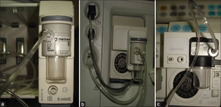
The sampling tube is used for sampling gas for analysis [Figure 2a-c]. It is usually of 1.2 mm internal diameter. The tube is connected to a lightweight adapter near the patient's end of the breathing system. It delivers the gas to the sample chamber. It is made of Teflon so it is impermeable to carbon dioxide and does not react with anaesthetic agents. Typical infrared instruments sample at a flow rate between 50 and 150 ml/min. The sampled gas may be returned to breathing circuit and is of concern when low flows are used. Also, the point of sampling should always be as near as possible to the patient's airway and the sampled gas mixture must not be contaminated by inspired gas during the expiratory phase.
Figure 2.

(a-c) Sidestream gas analysers with water trap – different manufacturers
Mainstream sampling
In mainstream type of analysers, the sample chamber is positioned within the gas stream near the patient's end of the breathing system. It does not remove any gas and no issue of water condensation from humidity from expired air happens.
MEASUREMENT OF VOLUME AND FLOW OF GASES
The measurement of flows and volumes are essential for anaesthetists.[27,28] For the understanding of the working principles of flow and volume in anaesthesia workstation, review of basic science is needed.
Types of flow
Many physical variables influence whether the flow is laminar or turbulent and this may be depicted by Reynolds’ number, Re = vρd/η (where v is linear velocity, ρ is density, d is diameter and η is viscosity).[3,29] Laminar flow exists when Re is less than 2000 and Re over 2000 indicates flow is likely to be turbulent. Laminar flow is efficient, with layers passing smoothly over each other producing a parabolic flow profile, with the greatest velocity centrally. It is determined by the Hagen-Poiseuille formula, Q = P πr4/8 ηl (where P is pressure drop, r is the radius of the tube and l is the length of the tube). This implies that flow is directly proportional to the pressure drop, proportional to the fourth power of the radius and related to the viscosity but not the density of the gas. Turbulent flow is less efficient, with multiple eddy currents occurring in the overall direction of flow. Because of the variable nature of turbulence, there is no precise and comprehensive equation to calculate flow, but turbulent flow is related to the square root of the pressure drop and density of the gas rather than its viscosity.
Measurement principles
The gases flow and volume could be measured directly or indirectly. The technique of direct measurement of gases may be done using bulk filling of a container of known volume. Certain gadgets like vitalograph, gas meter and water-displacement spirometer may be used for direct measurement of the gases. Such devices are in limited use in clinical practice. For clinical use, the measurement of gases is usually done indirectly, using a property of the gas that changes in parallel to flow or volume.
Pressure drop across a resistance technique utilises the phenomenon of measuring drop in pressure when a gas flows across. This effect can be used to calculate flow either by keeping the resistance constant and measuring the pressure change as the flow varies (e.g. pneumotachograph) or keeping a constant pressure drop and varying the resistance in a measurable way (e.g. bobbin rotameter).[28,29] The mechanical movement technique utilises kinetic energy of the moving gas molecules for rotation of a vane or bending a flexible obstruction. These are measurable events and may be transduced into an electrical signal. In the heat transfer technique, the heated element is cooled by a flowing gas which is calibrated and programmed to depict amount of gas flowing past a heated element e.g. hot-wire anemometer. In ultrasound interference, when a gas flows across the ultrasound signal, the velocity of the ultrasound signal either increases if gas is flowing alongside it in the same direction or decreases if the gas is flowing against it. This change in velocity is measurable and gives the flow measurement.
DEVICES FOR FLOW AND VOLUME MEASUREMENT
The various devices available for measurement of flow and volume may also give flow characteristics.[30,31,32] The methods of measuring the flow and volume may be based on total or partial volumes. This means some devices measure the whole gas and give the reading. However, some devices split a known quantity of gas and measure the split volume. This is extrapolated to depict the total flow or volume of the gas. The former technique is more commonly used. Some of the devices are described below [Table 3].
Table 3.
Devices for gas flow and volume measurement

Pneumotachograph
The technique of pneumotachograph measures flow by pressure drop across a resistance in the gas flow pathway. The pressure transducer measures rapidly and accurately the pressure drop which is extrapolated to find the flow rate and volume of the gas. For better accuracy, laminar flow is maintained by means of series of small-bore tubes arranged in parallel through which the gas flow must pass. The water vapour and its condensation may affect the accuracy of flow measurements. These sensors are accompanied with heating element to prevent water condensation. It can be used for both the inspired and expired flow by incorporating the sensors in both the inspiratory and expiratory limbs. The values may also be extrapolated to measure airway pressures and compliance can be calculated and displayed in real time.[30]
Rotameters
This is one of the common methods of measurement of continuous flow volume of gases in an anaesthesia machine.[31] The rotameters are specific for a particular gas. It comprises of special tube called ‘thorpe tube’.[33] Thorpe tube is a vertical tapered tube containing a bobbin or a ball. This moves up and down by the flow of the gas. The bobbin weight, i.e. the pressure drop required to maintain it, is balanced by the gas flow. The higher the bobbin rises in the thorpe tube, more is the area around it for gas flow and thus more is the flow. The resulting dimensions lead to laminar flow but at the top of the tube produce turbulent flow. Hence, the viscosity determines the flow at bottom of the thorpe tube and density at the top of the tube.[16] The readings are measured by a marker which is usually on the top of bobbin or centre of the ball. For accurate measurements over a large range, two tubes are added, one for low and one for high flow rates.[33] The measurement could be affected by static electricity on inner side of the tube that may lead to sticking of bobbin to the tube. The bobbin top is fluted for easy viewing of its rotation. The inner tube is also painted with antistatic spray. The other components of flow meter assembly are needle valve, indicator float, knobs, valve stops assembly.[33]
Mechanical flow transducers
Such equipments have a small metal disc attached to a flexible pin. The gas flow is proportionately split and passed into small chamber with the disc attached, which is perpendicular to the direction of flow. The strain gauge is placed behind the pin which bends with the flow of the gas. The resulting electrical signal is processed to calculate the flow rate. This has a high accuracy.
Hot wire anemometer
Such equipments work on the principle of cooling of a heating wire usually of platinum by the flow of a gas which is extrapolated to display the flow.[34] Some devices use heated screen or film instead of a wire. The wire is incorporated into the balanced Wheatstone bridge circuit and any change in its temperature unbalances the bridge.[2] The temperature of the wire is maintained constant by application of correcting current. These anemometers are very accurate.
Ultrasonic flowmeters
These are based on change in velocity of an ultrasound signal with the change in gas flow.[35] The flowmeters comprise of a design so as to have a pair of ultrasound beams directed in opposite direction. So when a gas flow occurs, the time difference happens for the two beams and then this is used to calculate the gas velocity and flow rate.
Electronic flowmeters
These are most commonly used in modern day anaesthesia workstation.[36,37] The gas flow is indicated on the screen in the form of bar. Such flowmeters comprise of a needle valve by which gas is conducted to a chamber of known volume having solenoid. The gas is held here till the transduced pressure within the chamber reaches a preset limit. This measures the gas flow.
SUMMARY
The advancement in technology has been a boon for perioperative care. The respiratory gas analysis has progressed from mechanical crude techniques to more advanced, automated and programmed technique with better accuracy and reliability. The increasing use of low flow anaesthesia has made the need of gas analysers including oxygen analysers mandatory for anaesthesia management.
Footnotes
Source of Support: Nil
Conflict of Interest: None declared
REFERENCES
- 1.Langton JA, Hutton A. Respiratory gas analysis. Cont Educ Anaesth Crit Care Pain. 2009;9:19–23. [Google Scholar]
- 2.Szocik J, Barker SJ, Tremper KK. Fundamental principles of monitoring instrumentation. In: Miller RD, editor. Miller's Anaesthesia. 7th ed. Philadelphia: Churchill Livingstone Elsevier; 2010. pp. 197–1227. [Google Scholar]
- 3.Vallis CJ, Hutton P, Clutton-Brock TH, Stedman R. The anaesthetic machine and breathing system. In: Healy TE, Knight PR, editors. Wylie and Churchill-Davidson's A Practice of Anaesthesia. 7th ed. New York: Oxford University Press; 2013. pp. 465–86. [Google Scholar]
- 4.Singaravelu S, Barclay P. Automated control of end-tidal inhalation anaesthetic concentration using the GE Aisys Carestation. Br J Anaesth. 2013;110:561–66. doi: 10.1093/bja/aes464. [DOI] [PubMed] [Google Scholar]
- 5.Harry JL, Karl L. Clinical and laboratory evaluation of an expired anaesthetic gas monitor (Narko-Test) Anesthesiology. 1971;34:378–82. doi: 10.1097/00000542-197104000-00024. [DOI] [PubMed] [Google Scholar]
- 6.White DC, Wardley-Smith B. The narkotest anaesthetic meter. Br J Anaesth. 1972;44:1100–4. doi: 10.1093/bja/44.10.1100. [DOI] [PubMed] [Google Scholar]
- 7.Caplan RA, Vistica MF, Posner KL, Cheney FW. Adverse anaesthetic outcomes arising from gas delivery equipment: A closed claims analysis. Anesthesiology. 1997;87:741–8. doi: 10.1097/00000542-199710000-00006. [DOI] [PubMed] [Google Scholar]
- 8.Ozanne GM, Young WG, Mazzei WJ, Severinghaus JW. Multipatient anaesthetic mass spectrometry. Anesthesiology. 1981;55:62–7. [PubMed] [Google Scholar]
- 9.Able M, Eisenkraft JB. Erroneous Mass Spectrometer Readings Caused by Desflurane and Sevoflurane. J Clin Monit. 1995;11:152–8. doi: 10.1007/BF01617715. [DOI] [PubMed] [Google Scholar]
- 10.Schulte TG, Block FE. Evaluation of a Single-Room, Dedicated Mass Spectrometer. Int J Clin Monit Comput. 1991;8:179–81. doi: 10.1007/BF01738890. [DOI] [PubMed] [Google Scholar]
- 11.Block FE, Schulte GT. Observations on use of wrong agent in an anaesthesia agent vapourizer. J Clin Monit. 1999;15:57–61. doi: 10.1023/a:1009979531850. [DOI] [PubMed] [Google Scholar]
- 12.Morrison JE, McDonald C. Erroneous data from an infrared anaesthetic gas analyzer. J Clin Monit. 1993;9:293–4. doi: 10.1007/BF02886702. [DOI] [PubMed] [Google Scholar]
- 13.Walder B, Lauber R, Zbinden AM. Accuracy and cross sensitivity of 10 different anaesthetic gas monitors. J Clin Monit. 1993;9:364–73. doi: 10.1007/BF01618679. [DOI] [PubMed] [Google Scholar]
- 14.Moyle JT, Davey A. Physiological monitoring: Gases. In: Ward C, editor. Wards Anaesthetic Equipment. 4th Ed. London: WB Saunders Company; 1998. pp. 279–92. [Google Scholar]
- 15.McFayden G. Respiratory gas analysis: Updates in anaesthesia. [Last assessed on 2013 June 15]. Available from: http://update.anaesthesiologists.org/2008/12/01/respiratory-gas-analysis .
- 16.Hill RW. Determination of oxygen consumption byuse of the paramganetic oxygen analyzer. J Appl Physiol. 1972;33:261–3. doi: 10.1152/jappl.1972.33.2.261. [DOI] [PubMed] [Google Scholar]
- 17.Meyer RM. Oxygen analyzers: Failure rates and life spans of galvanic cells. J Clin Monit. 1990;6:196–202. doi: 10.1007/BF02832146. [DOI] [PubMed] [Google Scholar]
- 18.Mortier E, Rolly G, Verschelen L. Methane influences infrared technique anaesthetic agent monitors. J Clin Monit Comput. 1998;14:85–8. doi: 10.1023/a:1007417828768. [DOI] [PubMed] [Google Scholar]
- 19.Verkaaik AP, VanDijk G. High flow closed circuit anaesthesia. Anaesth Intensive Care. 1994;22:426–34. doi: 10.1177/0310057X9402200418. [DOI] [PubMed] [Google Scholar]
- 20.Allison J, Gregory R, Bich K, Crowder J. Determination of anaesthetic agent concentration by refractometry. Br J Anaesth. 1995;74:85–8. doi: 10.1093/bja/74.1.85. [DOI] [PubMed] [Google Scholar]
- 21.Nagashima Y, Suzuki T, Terada S, Tsuji S, Misawa K. In Vivo molecular labeling of halogenated volatile anaesthetics via intrinsic molecular vibrations using nonlinear Raman spectroscopy. J Chem Phys. 2011;134 doi: 10.1063/1.3526489. 024525. [DOI] [PubMed] [Google Scholar]
- 22.Mushlin PS, Mark JB, Elliot WR, Striz S, Goldman DB, Philip J. Inadvertent development of subatmospheric airway pressure during cardiopulmonary bypass. Anesthesiology. 1989;71:459–62. doi: 10.1097/00000542-198909000-00028. [DOI] [PubMed] [Google Scholar]
- 23.Huffman LM, Riddle RT. Mass spectrometer and/or capnograph use during low flow, closed circuit anaesthesia administration. Anesthesiology. 1987;66:439–40. doi: 10.1097/00000542-198703000-00040. [DOI] [PubMed] [Google Scholar]
- 24.Levin PD, Levin D, Avidan A. Medical aerosol propellent interference with infrared anaesthetic gas monitors. Br J Anaesth. 2004;92:865–9. doi: 10.1093/bja/aeh154. [DOI] [PubMed] [Google Scholar]
- 25.Nunn G. Low flow anaesthesia. Cont Educ Anaesth Crit Care Pain. 2008;8:1–4. [Google Scholar]
- 26.Hawkes CA. Factitious halothane detection during trigger free anaesthesia in a malignant hyperthermia susceptible patient. Can J Anaesth. 1999;46:567–70. doi: 10.1007/BF03013548. [DOI] [PubMed] [Google Scholar]
- 27.Mitchell V, Cheesman K. Gas, tubes and flow. Anaesth Intensive Care Med. 2010;11:32–5. [Google Scholar]
- 28.Mandal NG. Measurement of volume and flow in gases. Anaesth Intensive Care Med. 2009;10:52–6. [Google Scholar]
- 29.The Medicine Publishing Company Ltd; 2003. [Last assessed on 2013 Jun 15]. Anaesthesia UK. ©. created 31/7/2006. Available from: http://www.frca.co.uk/article.aspx?articleid=100390 . [Google Scholar]
- 30.Bachiller PR, McDonough JM, Feldman JM. Do new anaesthesia ventilators deliver small tidal volumes accurately during volume controlled ventilation? Anesth Analg. 2008;106:1392–400. doi: 10.1213/ane.0b013e31816a68c6. [DOI] [PubMed] [Google Scholar]
- 31.Otteni JC, Ancellin J, Cazalaa JB, Feiss P. Anaesthesia equipment: Fresh gas delivery systems: Mechanical systems with rotameters and calibrated vapourizers. Ann Fr Reanim. 1999;18:956–75. doi: 10.1016/s0750-7658(00)87945-9. [DOI] [PubMed] [Google Scholar]
- 32.Lawrenceville, New Jersey 08648-1057 USA: Published by ProMed Strategies, LLC under an educational grant from PHASEIN AB, Sweden. Copyright © 2009, ProMed Strategies, LLC. 5 Branchwood Court; [Last assessed on 2013 Jun 15]. Anaesthesia gas monitoring: Evolution of a de facto Standard of care. Available from: http://ebookbrowse.com/anaesthesia-gas-monitoring-evolution-of-a-de-facto-standard-ofcare-pdf-d385419618 . [Google Scholar]
- 33.Dorsch JA, Dorsch SE. The anaesthesia machine. In: Dorsch JA, Dorsch SE, editors. Understanding anaesthesia equipment. 5th ed. Walnut Street, Philadelphia: Lippincot Williams and Wilkins; 2008. pp. 75–121. [Google Scholar]
- 34.Kann T, Hald A, Jorgensen FE. A new transducer for respiratory monitoring. a description of hot wire anemometer and test procedure for general use. Acta Anaesthesiol Scand. 1979;23:349–58. doi: 10.1111/j.1399-6576.1979.tb01462.x. [DOI] [PubMed] [Google Scholar]
- 35.King R, Bretland M, Wilkes A, Dingley J. Xenon measurement in breathing systems: A comparison of ultrasonic and thermal conductivity methods. Anaesthesia. 2005;60:1226–30. doi: 10.1111/j.1365-2044.2005.04379.x. [DOI] [PubMed] [Google Scholar]
- 36.Odin L, Nalthan N. What are changes in pediatric anaesthesia practice afforded by new anaesthetic ventilators? Ann Fr Anesth Reanim. 2006;25:417–23. doi: 10.1016/j.annfar.2005.10.003. [DOI] [PubMed] [Google Scholar]
- 37.White DC. Electronic measurement and control of gas flow. [Last assessed on 2013 Jun 15];Anaesth Intensive Care. 1994 22:409–14. doi: 10.1177/0310057X9402200415. anaesthesia-gas-monitoring-evolution-of-a-de-facto-standard- of-care-pdf-d385419618. [DOI] [PubMed] [Google Scholar]


