Key Points
AAV delivery of ZFNs and corrective Donor vectors to adult mouse liver results in stable human factor IX levels, normalizing hemophilic clotting times.
Abstract
Monogenic diseases, including hemophilia, represent ideal targets for genome-editing approaches aimed at correcting a defective gene. Here we report that systemic adeno-associated virus (AAV) vector delivery of zinc finger nucleases (ZFNs) and corrective donor template to the predominantly quiescent livers of adult mice enables production of high levels of human factor IX in a murine model of hemophilia B. Further, we show that off-target cleavage can be substantially reduced while maintaining robust editing by using obligate heterodimeric ZFNs engineered to minimize unwanted cleavage attributable to homodimerization of the ZFNs. These results broaden the therapeutic potential of AAV/ZFN-mediated genome editing in the liver and could expand this strategy to other nonreplicating cell types.
Introduction
We previously reported successful in vivo gene targeting in a neonatal mouse model of hemophilia B (HB). Specifically, we demonstrated that systemic codelivery of adeno-associated virus (AAV) vectors, encoding a zinc finger nuclease (ZFN) pair targeting the human F9 gene (AAV-ZFN) and a gene-targeting vector with arms of homology flanking a corrective complementary DNA (cDNA) cassette (AAV-Donor), resulted in the correction of a defective hF9 gene engineered into the mouse genome (referred to as hF9mut locus) in the livers of neonatal HB mice. Importantly, we obtained stable levels of human factor IX (hF.IX) expression sufficient to normalize clotting times.1
However, the neonatal model differs from the anticipated clinical setting2 in the degree of hepatocyte proliferation. Although neonatal mouse livers undergo multiple rounds of cell division during development, the rate of turnover in adult hepatocytes is very low.3 In the quiescent or G0 phase of the cell cycle, DNA double-strand breaks such as those created by ZFNs are expected to be repaired mainly by nonhomologous end joining (NHEJ), 1 of 2 major repair pathways.4,5 Interestingly, despite NHEJ being the predominant repair choice in mammalian cells, few approaches have exploited this mechanism. Genome editing has traditionally been considered synonymous with the other repair pathway, homology-directed repair (HDR), which is mainly active in the S/G2 phases of the cell cycle. We therefore investigated whether ZFN-driven gene targeting could be generalized to adult animals where the impact of a largely quiescent liver could be assayed.
Study design
Animal experiments
Animal experiments were approved by the Institutional Animal Care and Use Committee at the Children’s Hospital of Philadelphia. Previously described heterozygous male hF9mut transgenic mice1 or wild-type (WT) littermate controls, 8 to 10 weeks of age, were used. hF9mut/HB male mice were obtained by crossing male homozygote hF9mut mice with previously described HB females.6 Partial hepatectomies were performed as previously described.7
ZFN reagents and targeting vectors
ZFNs targeting the hF9mut locus and F9-targeting vectors have been described previously,1 as well as the ELD:KKR mutations to the FokI domain to construct the obligate heterodimeric ZFNs.8 Experiments shown in Figure 1 used the WT FokI domain with the exception of Figure 1D, in which the obligate heterodimeric ZFNs were used. AAV serotype 8 vectors were produced and titered as previously described.9,10
Figure 1.
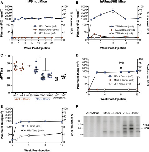
In vivo gene correction in adult mice results in stable circulating factor IX levels and correction of clotting times in hemophilic animals. (A-B) Levels of hF.IX in plasma of hF9mut (A) and hF9mut/HB (B) mice following intravenous injection at week 8 of life with 1 × 10¹¹ vg AAV8-ZFN alone, 1 × 10¹¹ vg AAV8-Mock and 5 × 10¹¹ vg AAV8-Donor, or 1 × 10¹¹ vg AAV8-ZFN and 5 × 10¹¹ vg AAV8-Donor. The previously described1 hF9mut mouse model of hF.IX deficiency contains a liver-specific, promoter-driven human coagulation factor IX cDNA devoid of necessary catalytic exons 7 to 8 knocked into the Rosa26 locus. (C) Measurement of clot formation by activated partial thromboplastin time (aPTT) prior to, 2 weeks, and 80 weeks after AAV administration. The aPTT of WT mice is shown for comparison. A 2-tailed Mann-Whitney test was used to compare 2 groups. (D) Levels of hF.IX in treated mice remain stable following two-thirds partial hepatectomy. (E) hF.IX expression is more than 10-fold greater following AAV8-ZFN and AAV8-Donor treatment in hF9mut mice harboring the ZFN target site (hF9mut) compared with littermate controls that do not (WT). (F) PCR analysis showing successful gene targeting by both HDR and NHEJ 5 months after intravenous coinjection of ZFN and Donor vectors. PCR products were resolved by 5% polyacrylamide gel electrophoresis and autoradiographed. See supplemental Figure 2 for maps. Mice treated with ZFN alone or with Mock and Donor showed no evidence of targeting. Identity of the PCR products was confirmed by sequencing. The lower band (*) appears in all donor treated samples, which is an artifact apparently generated by the reverse primer and the AAV–inverted terminal repeat resulting in amplification from nonintegrated AAV genomes. hF.IX plasma levels were assayed by enzyme-linked immunosorbent assay (ELISA) and represent repeated measurements, obtained by serial bleeding, on the same group of animals over the time of the study (n = number of mice in each cohort). Error bars denote standard error of the mean. Plasma factor IX data are representative of at least 2 independent experiments.
Factor IX levels and activity
The hF.IX ELISA kit (Affinity Biologicals, Ancaster, ON, Canada) was used to quantify plasma factor IX. All readings below the last value of the standard curve (15 ng/mL) were arbitrarily given the value of 15 ng/mL except where otherwise indicated. The aPTT assay was performed using 1:1:1 sample plasma, HB human plasma, and aPTT reagent (Trinity Biotech, Bray, Wicklow, Ireland).
Polymerase chain reaction (PCR)-based genotyping
Primers binding to a sequence upstream of exon 1 unique to the hF9mut minigene and within a sequence unique to the donor were used to amplify targeting events from genomic DNA.
Clustered integration site analysis (CLIS)
The mapping of in vivo ZFN cleavage sites was performed as previously described,4 using sequencing data from our previous study.1
Surveyor nuclease (Cel-I) assay
Liver function tests and immunohistochemical analysis
Quantification of plasma alanine aminotransferase was performed using an alanine aminotransferase colorimetric assay (Teco Diagnostics, Anaheim, CA). Immunohistochemical staining was performed using Ki-67 antibody (abcam ab16667) to stain formalin-fixed, paraffin-embedded tissue.
Reverse transcription–PCR
Tissues were homogenized in Qiazol, cellular RNA was isolated with miRNeasy Mini kits (Qiagen, Valencia, CA) and treated with DNaseI (Life Technologies, Carlsbad, CA), and 1 µg was reverse-transcribed using the High Capacity RNA-to-cDNA Kit (Life Technologies). PCR was performed using AccuPrime Pfx SuperMix (Life Technologies).
Additional details including CLIS parameters, primer sequences, and antibody dilutions are provided in the supplemental Methods (see the Blood Web site).
Results and discussion
Eight-week-old hF9mut mice (harboring the ZFN target site) were treated with 1 × 10¹¹ vg of AAV-ZFN, expressing the hF9-specific ZFN pair, and 5 × 10¹¹ vg of AAV-Donor, containing arms of homology flanking the corrective partial cDNA cassette (supplemental Figure 1). Treated animals exhibited long-term expression (>60 weeks) of hF.IX averaging 23% of normal (1146 ± 100 ng/mL) at week 60 (Figure 1A). In contrast, plasma hF.IX levels in mice coinjected with a luciferase-expressing AAV-Mock vector and AAV-Donor or mice injected with AAV-ZFN alone were below the limit of detection (15 ng/mL) (Figure 1A). Treatment of hF9mut/HB mice with AAV-ZFN and AAV-Donor produced similarly high hF.IX expression levels (Figure 1B) and reduced clotting times to those of WT mice (Figure 1C). Mice aged 7 to 8 months at initial injection exhibited lower but nonetheless therapeutic levels of hF.IX (supplemental Figure 2). hF.IX expression was derived predominantly from stably integrated, on-target gene correction based on 2 independent observations. First, following two-thirds partial hepatectomy, known to induce hepatocyte proliferation and subsequent loss of episomal AAV genomes,11 plasma hF.IX levels were not reduced (Figure 1D). Second, treatment of WT littermates lacking the hF9mut locus (and therefore the ZFN target site) supported just ∼50 ng/mL hF.IX (Figure 1E). As predicted by the use of a liver-specific promoter to drive ZFN expression and the hepatotropism of the AAV8 serotype, hF9 mRNA was exclusively detected in the liver and only when both the ZFN and Donor vectors were administered (supplemental Figure 3). Proliferation levels in adult mouse liver were confirmed to be low by Ki-67 staining shortly after treatment (supplemental Figure 4). Nonquantitative PCR-based molecular analyses 5 months post injection revealed gene insertion in the liver through both homology-directed and homology-independent processes precisely at the ZFN-specified locus, each of which is predicted to result in hF.IX expression (Figure 1F and supplemental Figure 5). We next sought to determine the impact of arms of homology on genome editing. We measured hF.IX levels resulting from treatment of hF9mut mice with ZFN and donors containing no substantial homology to the target site, establishing that homology arms are not required for clinically meaningful levels of correction (supplemental Figure 6). Together these data demonstrate that nuclease-driven gene targeting in the quiescent livers of adult mice via intravenous administration of AAV vectors results in substantial levels of hF.IX, sufficient to correct the phenotypic defect in HB mice.
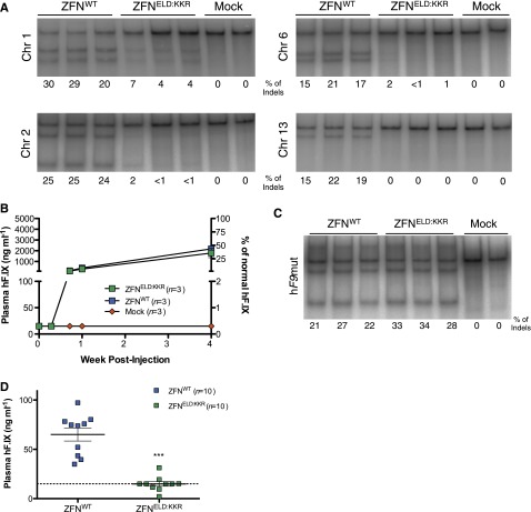
For therapeutic application, the risk of ZFN cleavage at off-target sites should be minimized. To assay off-target activity, we first identified potential cleavage sites using a genome-wide CLIS analysis12 of 454 deep-sequencing data from our previous in vivo ZFN study.1 To identify integration sites attributed to ZFN activity, we compared integration sites in ZFN- vs Mock-treated animals and excluded those common to both groups of mice, which generated a list of 9 potential off-target sites in the ZFN-treated mice. Next, using the Cel-I assay,13 we confirmed 4 of these sites as bona fide targets of ZFN cleavage in livers of mice treated with AAV-ZFN at week 4 post treatment (Figure 2A). Because all 4 cleavage sites mapped to a ZFN target sequence resulting from the homodimerization of 1 of the 2 ZFNs, we constructed an AAV-ZFN vector employing an obligate heterodimeric FokI architecture containing the ELD:KKR mutations in the dimerization interface between the 2 ZFNs to reduce cleavage.8 On-target activity was similar for the WT and ELD:KKR ZFNs as measured by the levels of hF.IX expression (Figure 2B) and cleavage at the hF9mut locus at week 4 post treatment (Figure 2C). In hF9mut mice treated with the obligate heterodimeric AAV-ZFNELD:KKR vector, the levels of off-target cleavage/repair were significantly reduced (relative to AAV-ZFNWT controls) at all sites analyzed (Figure 2A). Consistent with this reduction in off-target cleavage, the low background levels of hF.IX expression observed in AAV-ZFNWT–treated mice that lack the hF9mut locus, potentially arising from an integration event on chr2 (supplemental Figure 7), were reduced to below our limit of detection by the ELD:KKR variants (Figure 2D). Thus, although the use of WT ZFNs appears to be well tolerated, producing stable hF.IX expression over 60 weeks without change in circulating liver enzymes (Figure 1A and supplemental Figure 8), these data support the use of the obligate heterodimeric ZFNs, which provide an increase in specificity without reducing hF.IX expression.
Figure 2.
Obligate heterodimer ZFNs reduce off-target cleavage in vivo while preserving equivalent levels of hF.IX secretion. (A) Cel-I cleavage at the 4 validated off targets identified by clustered integration site analysis 4 weeks after treatment with 5 × 10¹¹ vg AAV8-Donor and either 1 × 10¹¹ vg AAV8-ZFNWT, 1 × 10¹¹ vg AAV8-ZFNELD:KKR, or 1 × 10¹¹ vg AAV8-Mock. (B) Plasma hF.IX levels following treatment. (C) On-target cleavage measured by Cel-I assay 4 weeks after AAV injection. (D) hF.IX expression in WT mice lacking ZFN target site 5 weeks after injection of 5 × 10¹¹ vg AAV8-Donor and either 1 × 10¹¹ vg AAV8-ZFNWT or 1 × 10¹¹ vg AAV8-ZFNELD:KKR. hF.IX plasma levels were assayed by ELISA. A two-tailed t test was used. ***P < .0001 compared with ZFNWT (n = number of mice in each cohort). Error bars denote standard error of the mean. Frequency of ZFN-induced insertions and deletions is indicated as “% of Indels” below each lane. Each lane represents an individual mouse. Plasma factor IX data are representative of at least 2 independent experiments.
In summary, these data provide the first demonstration of robust in vivo genome editing in the context of the predominantly quiescent hepatocytes of adult animals, resulting in complete amelioration of the extended clotting times in HB mice. We observe an ∼5-fold increase in hF.IX expression compared with our previous study in mice treated as neonates, which could be attributable to the loss of AAV vector genomes during neonatal liver development and/or differential promoter activity in each group. Alternatively, the difference could reflect increased genome-editing activity in adult mice. Importantly, we demonstrate substantial levels of genome editing in the absence of homology arms, which may further expand the utility of this approach by (1) allowing correction of cells unlikely to be amenable to HDR; (2) increasing the effective packaging capacity of donor vectors by obviating the need for homology arms; and (3) permitting efficient targeting without the need to design donors that incorporate patient-specific polymorphisms. Lastly, by using obligate heterodimer FokI nuclease domains, we show a marked enhancement in the specificity of ZFN action in vivo. Future directions include determining whether the levels of hF.IX observed in this model can be achieved by targeting endogenous loci, which may differ in chromatin status and promoter activity. The findings presented in this study serve to demonstrate the feasibility of genome editing in adult, noncycling tissues as a potential approach to the treatment of a wide range of monogenic diseases.
Supplementary Material
Acknowledgments
The authors thank Daniel Martinez and the CHOP Pathology Core for support with immunohistochemistry.
This work was supported by the National Institutes of Health: National Heart, Lung, and Blood Institute (HL64190 and HL078810, K.A.H.; T32-HL007971, R.S.), the Howard Hughes Medical Institute, and the Center for Cellular and Molecular Therapeutics at the Children's Hospital of Philadelphia.
Footnotes
The online version of this article contains a data supplement.
The publication costs of this article were defrayed in part by page charge payment. Therefore, and solely to indicate this fact, this article is hereby marked “advertisement” in accordance with 18 USC section 1734.
Authorship
Contribution: X.M.A., R.S., Y.D., P.D.G., M.C.H., and K.A.H. designed the experiments; X.M.A., R.S., Y.D., H.L., V.H., M.E.R., S.Y.W., R.J.D., S.Z., and M.C.H. generated reagents and performed the experiments; and X.M.A., R.S., Y.D., J.C.M., P.D.G., M.C.H., and K.A.H. analyzed the data and wrote the manuscript.
Conflict-of-interest disclosure: K.A.H. has consulted for companies developing AAV-based gene therapeutics and is an inventor on issued and pending patents on ZFN and AAV gene-transfer technologies. H.L. is an inventor on issued and pending patents on ZFN and AAV gene-transfer technologies. Y.D., J.C.M., M.E.R., S.Y.W., P.D.G., and M.C.H. are employees of Sangamo BioSciences, which designed and provided the ZFNs used in this study. The remaining authors declare no competing financial interests.
Correspondence: Dr Katherine A. High, Children’s Hospital of Philadelphia, 3501 Civic Center Blvd, Colket Translational Research Building, Room 5060, Philadelphia, PA 19104; e-mail: high@e-mail.chop.edu.
References
- 1.Li H, Haurigot V, Doyon Y, et al. In vivo genome editing restores haemostasis in a mouse model of haemophilia. Nature. 2011;475(7355):217–221. doi: 10.1038/nature10177. [DOI] [PMC free article] [PubMed] [Google Scholar]
- 2.Nathwani AC, Tuddenham EGD, Rangarajan S, et al. Adenovirus-associated virus vector-mediated gene transfer in hemophilia B. N Engl J Med. 2011;365(25):2357–2365. doi: 10.1056/NEJMoa1108046. [DOI] [PMC free article] [PubMed] [Google Scholar]
- 3.Magami Y, Azuma T, Inokuchi H, et al. Cell proliferation and renewal of normal hepatocytes and bile duct cells in adult mouse liver. Liver. 2002;22(5):419–425. doi: 10.1034/j.1600-0676.2002.01702.x. [DOI] [PubMed] [Google Scholar]
- 4.Branzei D, Foiani M. Regulation of DNA repair throughout the cell cycle. Nat Rev Mol Cell Biol. 2008;9(4):297–308. doi: 10.1038/nrm2351. [DOI] [PubMed] [Google Scholar]
- 5.Moynahan ME, Jasin M. Mitotic homologous recombination maintains genomic stability and suppresses tumorigenesis. Nat Rev Mol Cell Biol. 2010;11(3):196–207. doi: 10.1038/nrm2851. [DOI] [PMC free article] [PubMed] [Google Scholar]
- 6.Lin H-F, Maeda N, Smithies O, Straight DL, Stafford DW. A coagulation factor IX-deficient mouse model for human hemophilia B. 1997;90(10):3962–3966. [PubMed] [Google Scholar]
- 7.Mitchell C, Willenbring H. A reproducible and well-tolerated method for 2/3 partial hepatectomy in mice. Nat Protoc. 2008;3(7):1167–1170. doi: 10.1038/nprot.2008.80. [DOI] [PubMed] [Google Scholar]
- 8.Doyon Y, Vo TD, Mendel MC, et al. Enhancing zinc-finger-nuclease activity with improved obligate heterodimeric architectures. Nat Methods. 2011;8(1):74–79. doi: 10.1038/nmeth.1539. [DOI] [PubMed] [Google Scholar]
- 9.Ayuso E, Mingozzi F, Montane J, et al. High AAV vector purity results in serotype- and tissue-independent enhancement of transduction efficiency. Gene Ther. 2010;17(4):503–510. doi: 10.1038/gt.2009.157. [DOI] [PubMed] [Google Scholar]
- 10.Wright JF, Zelenaia O. Otto-Wilhelm M, Al-Rubeai M, editors. Vector characterization methods for quality control testing of recombinant adeno-associated viruses. Viral Vectors for Gene Therapy. Methods in Molecular Biology, vol. 737. New York, NY: Humana Press; 2011; 247-278. [DOI] [PubMed]
- 11.Song S, Lu Y, Choi Y-K, et al. DNA-dependent PK inhibits adeno-associated virus DNA integration. Proc Natl Acad Sci USA. 2004;101(7):2112–2116. doi: 10.1073/pnas.0307833100. [DOI] [PMC free article] [PubMed] [Google Scholar]
- 12.Gabriel R, Lombardo A, Arens A, et al. An unbiased genome-wide analysis of zinc-finger nuclease specificity. Nat Biotechnol. 2011;29(9):816–823. doi: 10.1038/nbt.1948. [DOI] [PubMed] [Google Scholar]
- 13.Perez EE, Wang J, Miller JC, et al. Establishment of HIV-1 resistance in CD4+ T cells by genome editing using zinc-finger nucleases. Nat Biotechnol. 2008;26(7):808–816. doi: 10.1038/nbt1410. [DOI] [PMC free article] [PubMed] [Google Scholar]
Associated Data
This section collects any data citations, data availability statements, or supplementary materials included in this article.


