Description
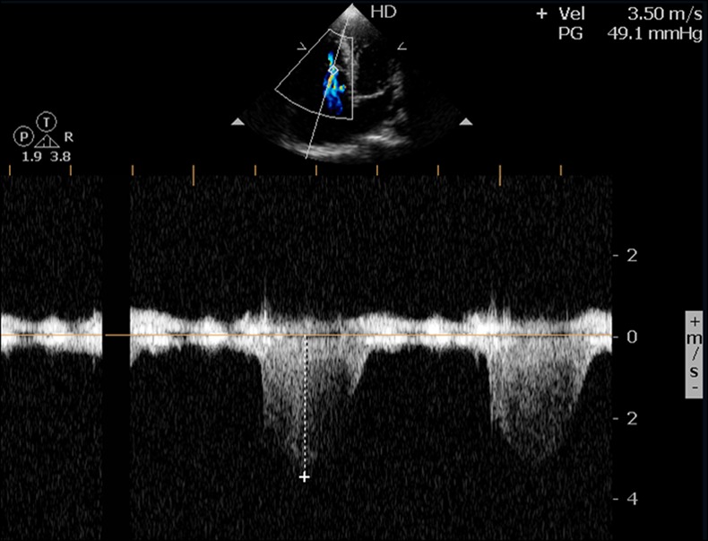
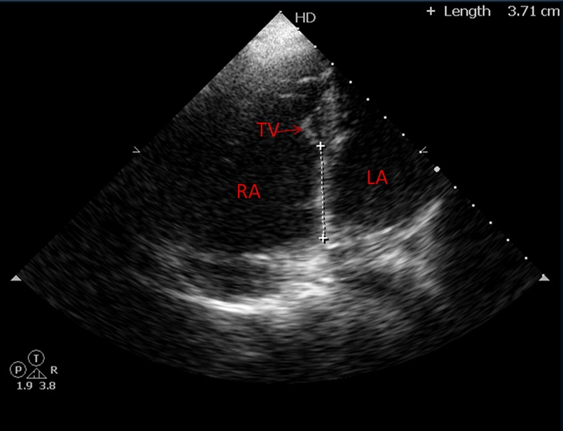
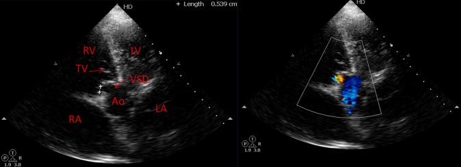
We report the case of a 42-year-old man who presented with effort intolerance of New York Heart Association (NYHA) class II for the past 1 year. On clinical examination, there was presence of cyanosis, clubbing, ejection systolic murmur of grade III/VI at left lower parasternal area, splitting of both first and second heart sound and fourth heart sound. An echocardiography revealed apical displacement of the septal leaflet of the tricuspid valve by about 37 mm (figure 1, video 1) and presence of tricuspid regurgitation (TR) with a TR jet of 49 mm Hg (figure 2). There was presence of Gerbode-like defect with perimembranous ventricular septal defect (VSD) of 5 mm connecting the left ventricle (LV) to the right atrium (RA) with left to right shunt (figure 3, video 2). A persistent foramen ovale of 2 mm with right to left shunt was also associated with Ebstein's anomaly in our case and it was also the cause for cyanosis and clubbing in our case (video 3). So, in our case Gerbode-like VSD was associated with Ebstein's anomaly. This kind of VSD is rarely associated with Ebstein's anomaly. So far only one case has been reported in the literature where Gerbode-type VSD was associated with Ebstein's anomaly along with Wolff–Parkinson–White syndrome.1 But, in our case this adult patient has cyanosis and significant effort intolerance without any arrhythmia. It is speculated that this rare association, allowing LV to RA flow will cause RA volume overload and will increase right to left shunt and that is why he had significant cyanosis and NYHA class II symptoms at presentation. In contrast, when there is a connection between the LV to the right ventricle, adequate forward pulmonary blood flow will be seen and these kinds of patients reported to have favourable prognosis.2
Figure 1.

Apical 4C view revealed apical displacement of the septal leaflet of the tricuspid valve by 37 mm.
Figure 2.

Colour Doppler showed a TR jet of 49 mm Hg originating from apically displaced tricuspid valve leaflets.
Figure 3.

Colour Doppler at apical 5C view showed presence of Gerbode-like communication with perimembranous ventricular septal defect connecting between left ventricle and right atrium.
Apical 4C view revealed apical displacement of septal leaflet of tricuspid valve with TR.
Apical 5C view showed presence of VSD between LV & RA with left to right shunt.
Subcostal view showed presence of PFO with right to left shunt.
Learning points.
Ebstein’s anomaly is a rare congenital heart defect associated with apical displacement of septal leaflet of tricuspid valve.
Patent foramen ovale and ostium secundum atrial septal defect are most commonly associated lesions with Ebstein's anomaly.
Gerbode's defect, which is a connection between left ventricle and right atrium, rarely associated with Ebstein's anomaly.
Footnotes
Contributors: All authors were involved in the management of this patient.
Competing interests: None.
Patient consent: Obtained.
Provenance and peer review: Not commissioned; externally peer reviewed.
References
- 1.Bayar N, Canbay A, Uçar O, et al. Association of Gerbode-type defect and Wolff-Parkinson-White syndrome with Ebstein's anomaly. Anadolu Kardiyol Derg 2010;2013:88–90 [DOI] [PubMed] [Google Scholar]
- 2.Del Pasqua A, de Zorzi A, Sanders SP, et al. Severe Ebstein's anomaly can benefit from a small ventricular septal defect: two cases. Pediatr Cardiol 2008;2013:217–19 [DOI] [PubMed] [Google Scholar]
Associated Data
This section collects any data citations, data availability statements, or supplementary materials included in this article.
Supplementary Materials
Apical 4C view revealed apical displacement of septal leaflet of tricuspid valve with TR.
Apical 5C view showed presence of VSD between LV & RA with left to right shunt.
Subcostal view showed presence of PFO with right to left shunt.


