Abstract
We previously described potent inhibition of Ebola virus entry by a ‘C-peptide’ based on the GP2 C-heptad repeat region (CHR) targeted to endosomes (‘Tat-Ebo’). Here, we report the synthesis and evaluation of C-peptides conjugated to cholesterol, and Tat-Ebo analogs containing covalent side chain–side chain crosslinks to promote α-helical conformation. We found that the cholesterol-conjugated C-peptides were potent inhibitors of Ebola virus glycoprotein (GP)-mediated cell entry (~103-fold reduction in infection at 40 µM). However, this mechanism of inhibition is somewhat non-specific because the cholesterol-conjugated peptides also inhibited cell entry mediated by vesicular stomatitis virus glycoprotein G. One side chain–side chain crosslinked peptide had moderately higher activity than the parent compound Tat-Ebo. Circular dichroism revealed that the cholesterol-conjugated peptides unexpectedly formed a strong α-helical conformation that was independent of concentration. Side chain–side chain crosslinking enhanced α-helical stability of the Tat-Ebo variants, but only at neutral pH. These result provide insight into mechanisms of C-peptide inhibiton of Ebola virus GP-mediated cell entry.
Keywords: C-peptide, Peptide design, Viral membrane fusion, Ebola virus, Hemorrhagic fever
Ebolaviruses and marburgviruses (the ‘filoviruses’, members of the family Filoviridae) are causative agents of a severe and rapidly progressing hemorrhagic fever.1,2 Although filovirus infections are rare in the United States and Europe, outbreaks have been occurring with increased frequency in endemic regions. In 2012 alone, the Centers for Disease Control (CDC) reported five filovirus outbreaks or isolated cases of infection in Africa: two caused by Sudan virus, SUDV, one by Bundibugyo virus, BDBV (both ebola virus species) and two caused by Marbug virus, MARV.3 In larger outbreaks, mortality rates range from 30% to 90%. Currently, there are no approved therapies or vaccines to treat filovirus infections in the United States. Due to their rapid proliferation and high mortality rates associated with infection, filoviruses are classified as Category A biodefense pathogens by the National Institutes of Allergy and Infectious Diseases (NIAID) and CDC.
Among the filoviruses, the Zaire Ebola virus species (EBOV) has been most extensively studied.2,4,5 EBOV particles are enveloped, filamentous and contain a negatively stranded RNA genome. Infection requires fusion of the host and EBOV membranes for delivery of the viral genomic material. This process is thought to occur in host endosomal compartments and is mediated by the transmembrane glycoprotein subunit GP2.6,7 In the prefusion state, the glycoprotein (GP) spike assembly consists of three copies each of the surface subunit (GP1) and the transmembrane subunit (GP2).2,4,5 Cell attachment involves binding of cell surface host factors by GP1, followed by uptake of the EBOV particle by macropinocytosis. Once in the endosome, host proteases Cathepsin L and Cathepsin B (CatL and CatB, respectively) process GP1 to relieve constraints on GP2 and expose a receptor-binding domain (RBD). Interaction between the RBD and at least one host receptor, Niemann Pick C1 (NPC-1), is required to trigger membrane fusion.8
The subsequent events leading to membrane fusion are analogous to mechanisms proposed for HIV-1 and influenza, and are based mostly on structural information.2,4,5,9,10 GP2 is thought to undergo a conformational transition that results in projection of its N-terminal fusion loop into the host endosomal membrane. This results in a transient intermediate known as the extended or prehairpin intermediate that spans the viral and host cell membranes. Two heptad repeat regions, N- and C-terminal (the NHR and CHR, respectively) next refold into a highly stable six-helix bundle.6,7 This refolding event brings the host and virus membranes into proximity and thus facilitates initial fusion events. Ultimately, a fusion pore is formed through which the viral contents are delivered into the host cytosol. Three-dimensional structures of the core domain from EBOV and MARV GP2 in the ‘post-fusion’ conformation have been described and are highly similar.6,7,11 The six-helix bundle contains a long, central NHR core (a triple-stranded coiled-coil) with the three shorter CHR segments packed in an antiparallel configuration. An intramolecular disulfide bond stabilizes a loop/helix-turn-helix motif between the NHR and CHR.
Evidence for the extended intermediate in most ‘class I’ fusion proteins (i.e., those that contain α-helical segments) is largely indirect.9,10 Two recent reports provide specific information about interbilayer spacing and glycoprotein clustering where the extended intermediate occurs.12,13 However, the major evidence for this conformation arises from the fact that synthetic peptides corresponding to the CHR (‘C-peptides’) can block infection.9,10 It is assumed that C-peptides function by binding the preformed NHR core trimer in the transiently exposed extended intermediate, thus sequestering these segments and preventing formation of the sixhelix bundle. One HIV-1 C-peptide is an approved drug (Enfurvitide/Fuzeon/T-20).
The fusion reaction for HIV-1 occurs primarily at the cell surface, but most other membrane viruses are taken up into endosomal compartments where low pH or other endosomal factors are required to trigger membrane fusion.9,10 Therefore, inhibition of viral entry within endosomes requires a C-peptide that localizes to the sites of membrane fusion. We previously reported potent and broad-spectrum inhibition of filovirus infection by an EBOV C-peptide consisting of the CHR segment appended to an arginine-rich cell-penetrating undecapeptide from HIV-1 transactivator protein (‘Tat-Ebo’).12 Here we explore the effect of two peptide modifications on the activity of EBOV C-peptides: conjugation to cholesterol and side chain–side chain crosslinking.
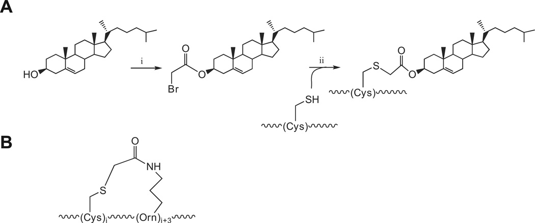
Other groups have found that HIV-1, paramyxoviruses, as well as endosomal viruses (e.g., influenza virus) can be potently inhibited by C-peptides conjugated to a cholesterol group.13–16 It is believed that the cholesterol group localizes the C-peptide to membrane surfaces such that it is primed to interact with the NHR of the transient extended intermediate when it forms.13–16 Presumably, this cholesterol-mediated localization effect is not specific to endosomal or plasma membranes, explaining why both HIV-1 and influenza virus can be targeted effectively in this way. For HIV-1 C-peptides, conjugation to cholesterol confers up to 400-fold improvement in IC50.13 For paramyxoviruses and influenza, cholesterol conjugation imparts activity on an otherwise inactive C-peptide.14–16 With these factors in mind, we designed and synthesized peptides 1 and 2 in which the EBOV GP2 CHR peptide segment contains either an N-terminal or C-terminal free cysteine as a reactive handle for conjugation. In both cases, a tetralysine segment was included to enhance solubility. Peptides 1 and 2 were produced by standard solid-phase peptide chemistry, and purified by HPLC. Cholesterol was bromoacetylated at the 3-C hydroxyl using bromoacetic acid and N-N’-diisopropylcarbodiimide; the bromoacetylated cholesterol was conjugated to the free sulfhydryls of 1 or 2 to form 1-Chol or 2-Chol (Scheme 1a). We found that the cholesterol ester was stable to hydrolysis for periods of up to 72 h at neutral pH (data not shown).
Scheme 1.
(A) Synthesis of cholesterol-conjugated peptides: (i) Bromoacetic acid/N,N’-diisopropylcarbodiimide/DMAP/DCM, (ii) DMSO/DIEA; (B) Thioetheramide side chain–side chain linkage.
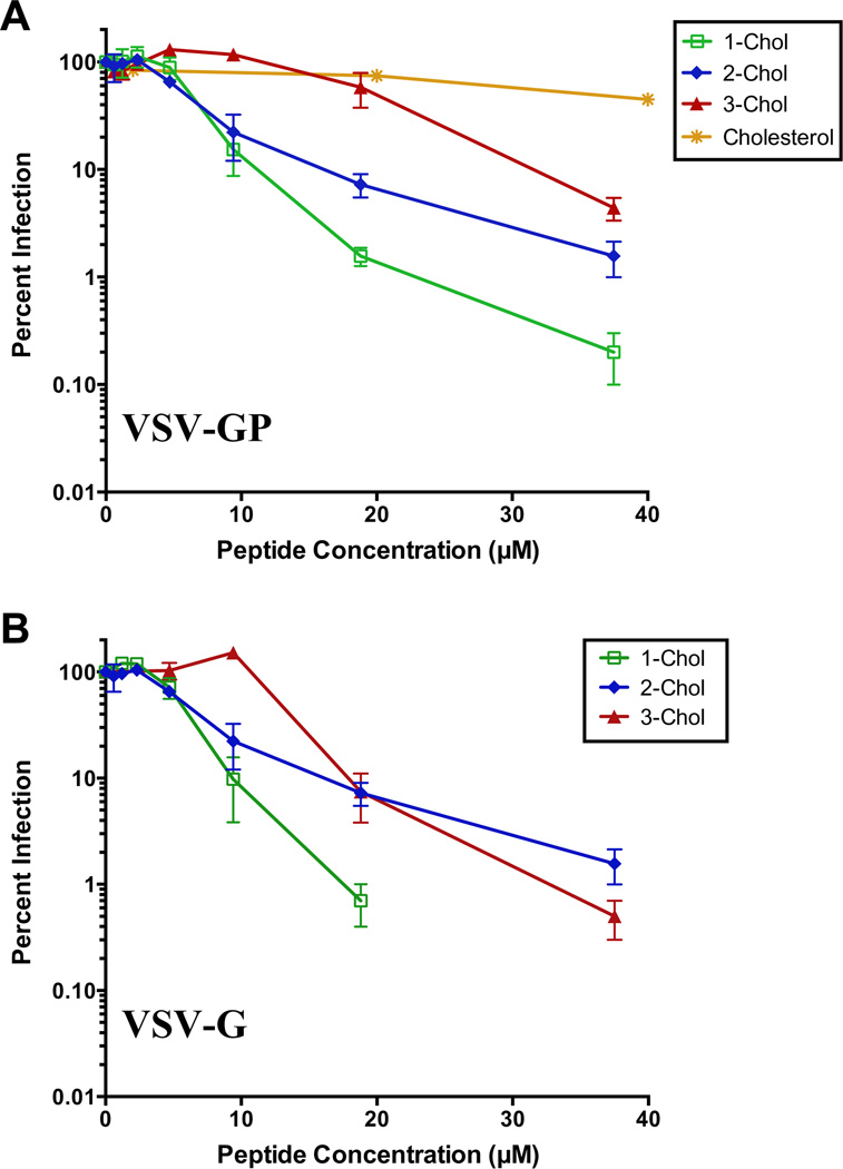
The capacity for 1-Chol or 2-Chol to inhibit GP-mediated viral entry was assessed with a vesicular stomatitis virus particle bearing the EBOV GP in place of the native glycoprotein G (‘VSV-GP’).12 The matrix protein contains an eGFP fusion allowing primary infection events to be quantified by fluorescence confocal microscopy. Vero cells were used as the host cell line. Peptides 1-Chol and 2-Chol resulted in potent inhibition of VSV-GP entry with ~103-fold reduction in infection at 40 µM peptide concentrations (Fig. 1). This level of inhibition far exceeds previously reported VSV-GP inhibition by Tat-Ebo, which was ~102-fold reduction in entry at higher peptide concentrations (75 µM).12 Notably, cholesterol alone had relatively modest effects on VSV-GP entry. However, we found that this activity was not specific to the EBOV glycoprotein. When 1-Chol and 2-Chol were tested for inhibition of VSV bearing its native envelope glycoprotein G (‘VSV-G’), both had potent activity for inhibition of VSV-G, with ~90% reduction at 10 µM peptide concentration and >99% reduction at higher concentrations. Importantly, both peptides exhibited only modest cell toxicity (~20%) under these conditions (see Supplementary data). The potent inhibition of VSV-G by 1-Chol and 2-Chol suggests that a major aspect of the neutralization mechanism is non-specific, although the fact that cholesterol had only modest effects on VSV-GP entry suggests this mechanism requires some combination of peptide and cholesterol moieties. This behavior contrasts with that of cholesterol-conjugated C-peptides derived from other viruses.13–16 For example, cholesterol-conjugated HIV-1 C-peptides had no activity against VSV-G in previous studies.13 Cholesterol-conjugated C-peptides based on influenza had activity against VSV pseudotyped with influenza HA but not against VSV pseudotyped with the GPC from Nipah virus.16 However, activity against the native VSV G protein was not explored in this case. Furthermore, this non-specific effect appears to be related to the cholesterol-conjugated peptide; we previously reported that Tat-Ebo has no activity against VSV-G at 75 µM.12
Figure 1.
Inhibition of VSV-GP (A) or VSV-G (B) entry by cholesterol-conjugated peptides. For VSV-GP, cholesterol alone was included as a control.
To further explore requirements for inhibition of viral entry by cholesterol-conjugated peptides, we produced 3-Chol, a peptide that consists of a tetralysine segment conjugated to cholesterol. We found 3-Chol was active against both VSV-GP and VSV-G (Fig. 1). Binding to the NHR should be dependent on inclusion of the CHR sequence, which is not present in 3-Chol. At 20 µM, 3-Chol is much less potent against VSV-GP then 1-Chol and 2-Chol, suggesting that some component of activity is dependent on the native CHR sequence. However, at higher concentrations, 3-Chol inhibits VSV-GP entry by >90%. These results indicate a non-specific component of VSV-GP inhibition by cholesterol-conjugated peptides since 3-Chol does not contain the GP CHR sequence. In contrast, we previously reported that inhibition of VSV-GP entry by Tat-Ebo was dependent on the native GP2 CHR sequence since a sequence isomer in which the CHR segment was scrambled had no effect on entry.12
The non-specific contribution to inhibitory activity against VSV-GP is further exemplified by the fact that 1-Chol is more potent than 2-Chol. The orientation of the NHR core trimer α-helices is thought to be directional in the extended intermediate (with the N-terminal end pointed toward the cell membrane). It was therefore predicted that 2-Chol would be more potent than 1-Chol because the C-terminal location of the cholesterol in 2-Chol should affix the C-peptide in the native antiparallel orientation relative to the NHR in the extended intermediate. For HIV-1 C-peptides containing N- or C-conjugated cholesterol, the C-terminal conjugated C-peptide has >2000-fold higher activity than the N-terminal conjugated peptide.13 However, 1-Chol is more potent than 2-Chol, indicating that the orientation of the C-terminal end of the C-peptide toward the membrane is not an important factor for potency. This result suggests either that the C-peptide is liberated from the cholesterol group during infection, or that inhibition involves a more complex mechanism.
We sought to evaluate a second strategy for improvement of antiviral properties of EBOV C-peptides by incorporation of structure-promoting modifications. The activity of the CHR segment in Tat-Ebo is likely dependent on α-helical conformation, since the native CHR segment participates in a six-helix bundle in the post-fusion core domain structure.6,7 Therefore, enforcing an α-helical structure by side chain–side chain cross-links is expected to enhance activity of Tat-Ebo. To test this hypothesis, we produced peptides 4 and 5, which contain Cys and Orn residues at i and i + 3 positions that are not expected to impact CHR binding according to the post-fusion GP2 structure. Dawson and coworkers demonstrated that a thioetheramide side chain–side chain crosslink between Cys and Orn residues at this spacing provides proper geometry and length to promote α-helical structure.17,18 Peptides 4 and 5 were produced using standard N-FMOC strategy with acid labile protecting groups on all side chains except the Orn residues, which were protected with an Nδ-ALLOC group. Treatment of the resin-bound peptide precursors with triphenylphosphine Pd(0) resulted in deprotection of the Orn side chain as determined by positive Kaiser test. The Orn free amine was iodoacetylated using iodoacetic anhydride. Treatment of this resin with TFA resulted in simultaneous cleavage and side chain deprotection. Formation of the thioether amide was rapid and spontaneous to yield 4-Link and 5-Link. Final products were purified by RP-HPLC, and all masses were confirmed by MALDI-MS (Table 1).
Table 1.
Peptide sequences and masses
| Peptide | Sequencea | [MH]+exp | [MH]+obs |
|---|---|---|---|
| 1 | CKKKKGSGIEPHDWTKNITDKIDQIIHDFVDK | 3737.3 | 3736.8 |
| 1-Chol | C(Chol)bKKKKGSGIEPHDWTKNITDKIDQIIHDFVDK | 4163.9 | 4164.4 |
| 2 | IEPHDWTKNITDKIDQIIHDFVDKGSGKKKKC | 3737.3 | 3736.9 |
| 2-Chol | IEPHDWTKNITDKIDQIIHDFVDKGSGKKKKC(Chol)b | 4163.9 | 4165.4 |
| 3-Chol | KKKKGSGC(Chol)b | 1260.7 | 1261.0 |
| 4 | Ac-YGRKKRRQRRRGSGIEPHDWTKCITOKIDQIIHDFVDK | 4693.4 | 4694.5 |
| 4-Link | Ac-YGRKKRRQRRRGSGIEPHDWTKCITOcKIDQIIHDFVDK | 4733.4 | 4734.4 |
| 5 | Ac-YGRKKRRQRRRGSGIEPHDWTKNITCKIOQIIHDFVDK | 4692.4 | 4694.4 |
| 5-Link | Ac-YGRKKRRQRRRGSGIEPHDWTKNITCKIOcQIIHDFVDK | 4732.4 | 4734.6 |
| Tat-Ebo | YGRKKRRQRRRGSGIEPHDWTKNITDKIDQIIHDFVDK | 4661.5 | 4661.9 |
| Lys-Ebo | KKKKGSGIEPHDWTKNITDKIDQIIHDFVDK | 3633.0 | 3632.2 |
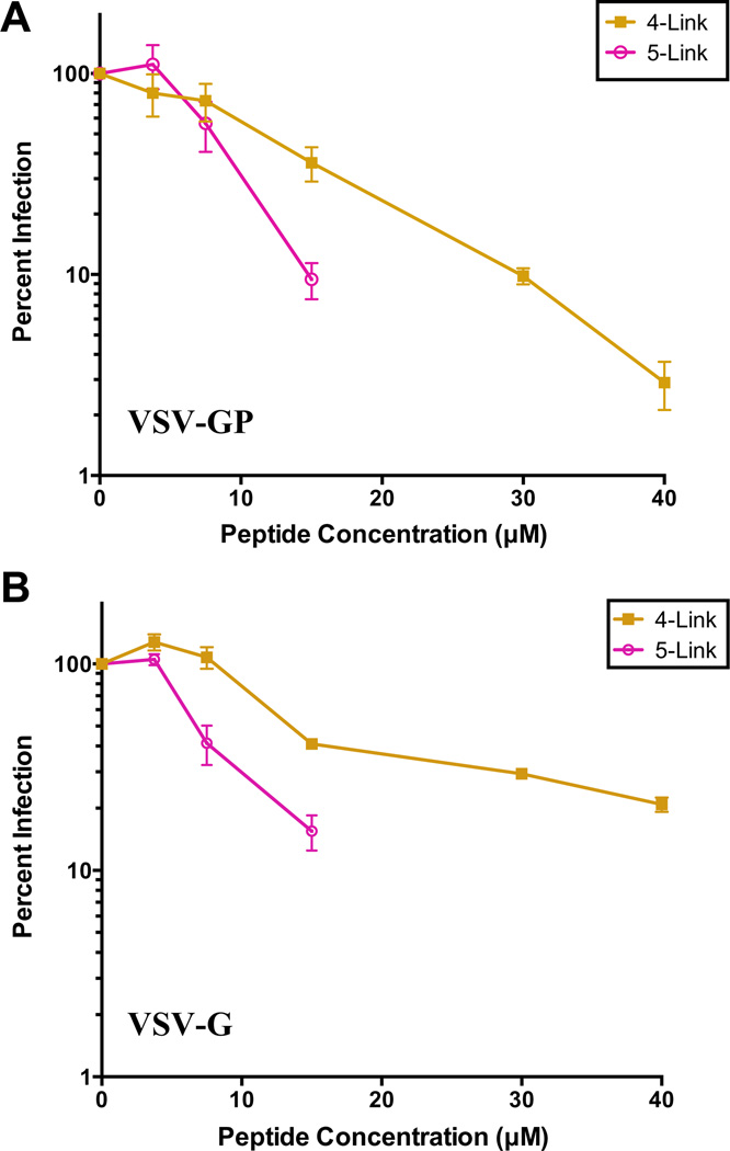
Peptide 4-Link provided potent neutralization of VSV-GP, with ~99% reduction (2 logs) in infection at 40 µM (Fig. 2). At this concentration, 4-Link was well tolerated by Vero cells as scored by visual inspection of the cells (data not shown) and a commercial cell viability assay (see Supplementary data). The potency of 4-Link is moderately higher than our previous studies on Tat-Ebo, which afforded 99% reduction only at concentrations of 75 µM.12 Interestingly, 4-Link was able to inhibit infection of VSV-G as well, though to a lesser extent than VSV-GP at high concentrations. At 40 µM, ~80% (<1 log) reduction in VSV-G infection was observed whereas 2 logs of inhibition were observed in VSV-GP. Therefore, there is some specificity for 4-Link activity toward the EBOV GP. Vero cells incubated with concentrations of 5-Link exceeding 15 µM showed signs of toxicity by visual inspection (not shown), which prevented assessment of antiviral activity at higher concentrations. At lower concentrations, 5-Link inhibited both VSV-GP and VSV-G with similar potency. It is interesting to note that that 4-Link and 5-Link had activity against VSV-G, since the parent compound Tat-Ebo was highly specific for VSV-GP over VSV-G (Ref. 12). It is possible that incorporation of side chain–side chain crosslinks results in general effects of cellular toxicity (as in the case of 5-Link) or disruption of endsomal uptake mechanisms (4-Link).
Figure 2.
Inhibition of VSV-GP (A) or VSV-G (B) entry by side chain–side chain crosslinked peptides.
We next sought to explore the structural properties of 1-Chol, 2-Chol, 4-Link, and 5-Link to determine if their propensities to adopt α-helical conformation were correlated with activity. Circular dichroism (CD) spectra indicated surprisingly that 1-Chol and 2-Chol adopt a strong α-helical conformation at pH 4.6 and pH 7.1 (Fig. 3). In contrast, Lys-Ebo, whose sequence is similar to that of 1-Chol but does not contain the cholesterol-conjugated cysteine, did not exhibit an α-helical signature. This result was unexpected; the cholesterol moiety in 1-Chol and 2-Chol is separated from the CHR segment by a flexible tripeptide linker (~GSG~) and a tetralysine segment in both peptides. Therefore, it is unlikely that the cholesterol induces α-helical conformation by direct contacts with any of the side chain residues in the CHR segment. Another possibility is that the cholesterol group induces limited aggregation of the peptide to form α-helical bundles. However, we found that the CD spectrum for 1-Chol was similar over a 4.1–23 µM range (see Supplementary data), suggesting that any aggregation-induced structural transitions do not take place in this concentration range.
Figure 3.
(A and B) CD spectra of peptides in 10 mM NaOAc, pH 4.6 (A), and 10 mM NaH2PO4, pH 7.1 (B). Peptide concentrations ranged from 36 µM to 75 µM. (C) α-Helical wheel depiction of 4-Link and 5-Link. Potential stabilizing (blue) or destabilizing (red) interactions are shown as well as side chain–side chain crosslinks. Primary sequence and numbering shown above; ‘X’ indicates positions involved in crosslinks.
Both 4-Link and 5-Link had partial α-helical characteristic at pH 7.1 as indicated by a negative band at ~208 nm and a shoulder at 222 nm (Fig. 3B). It is interesting to note that 5-Link had stronger α-character than did 4-Link, indicating that placement of the side chain–side chain crosslink has structural effects. This behavior contrasts with previous results on Tat-Ebo indicating this peptide is unstructured in aqueous solution.12 Therefore, we conclude that the thioetheramide side chain–side chain crosslink enhances α-helical conformation in 4-Link and 5-Link. However, we found that 4-Link and 5-Link had diminished α-helical character at pH 4.6 relative to pH 7.1. α-Helical wheel analysis suggests this pH-dependent behavior may be due to side chain–side chain interactions between Arg residues of the Tat segment and acidic or His residues of the CHR segment. For example, Arg9 (at a c position) has the capacity to form α-helix stabilizing salt bridges with Glu16 (another c position) and Asp19 (an f position) at neutral pH. However, such interactions would be diminished under conditions where the acidic residues are protonated. Arg11 (at an e position) may form unfavorable electrostatic interactions with His18 (an e position) under conditions where the imidazolium group is protonated.
Here we describe the synthesis and evaluation of modified C-peptides for the inhibition of EBOV GP-mediated cell entry. Although potent, the inhibitory effect of cholesterol-conjugated C-peptides 1-Chol and 2-Chol had little specificity for the glycoprotein of EBOV (VSV-GP vs VSV-G) or the native CHR sequence (1-Chol vs 3-Chol). It seems reasonable that some aspect of inhibition in these peptides arises from delivery of the CHR segment to the sites of membrane fusion. However, these data also suggest additional effects, related specifically to the combination of peptide and cholesterol, contribute to this activity. The fact that inhibition with both VSV-GP and VSV-G is observed indicates that this non-specific component may include mechanisms of cellular uptake or endosomal maturation. Curiously, cholesterol conjugation enhances α-helical structure in a concentration-independent manner. Whether this structural effect plays a role in the broad inhibitory activity remains to be determined. These results further suggest that caution must be exercised in considering cholesterolconjugated C-peptides as a therapeutic antiviral approach.13–16,19 Furthermore, we found modest enhancement in activity with the side chain–side chain crosslinked peptide 4-Link relative to its linear analog Tat-Ebo. CD indicates an enhancement in α-helical structure, which may play a role in this inhibitory activity. However, it is expected that 4-Link would function at pHs of the late endosome (pH 4.5–5.5), and under these conditions structure was not promoted. These results provide novel insights into C-peptide inhibition of the filoviruses.
Supplementary Material
Acknowledgments
We thank Wei Wang and the Albert Einstein College of Medicine Chemical Biology Core Facility for assistance with synthesis of the cholesterol-conjugated peptides, and Rohit Jangra for assistance with cellular toxicity and viral entry assays. This work was funded by the NIH (AI090249 to J.R.L. and AI088027 to K.C.). J.F.K. was supported in part by Medical Scientist Training Program T32-GM007288.
Footnotes
Supplementary data
Supplementary data associated with this article can be found, in the online version, at http://dx.doi.org/10.1016/j.bmcl.2013.07.056.
References and notes
- 1.Feldmann H, Geisbert TW. Lancet. 2011;377:849. doi: 10.1016/S0140-6736(10)60667-8. [DOI] [PMC free article] [PubMed] [Google Scholar]
- 2.Lee JE, Saphire EO. Future Virol. 2009;4:621. doi: 10.2217/fvl.09.56. [DOI] [PMC free article] [PubMed] [Google Scholar]
- 3. http://www.cdc.gov/ncidod/dvrd/spb/mnpages/dispages/ebola.htm.
- 4.Lee JE, Saphire EO. Curr. Opin. Struct. Biol. 2009;19:408. doi: 10.1016/j.sbi.2009.05.004. [DOI] [PMC free article] [PubMed] [Google Scholar]
- 5.Miller EH, Chandran K. Curr. Opin. Virol. 2012;2:206. doi: 10.1016/j.coviro.2012.02.015. [DOI] [PMC free article] [PubMed] [Google Scholar]
- 6.Weissenhorn W, Carfi A, Lee KH, Skehel JJ, Wiley DC. Mol. Cell. 1998;2:605. doi: 10.1016/s1097-2765(00)80159-8. [DOI] [PubMed] [Google Scholar]
- 7.Malashkevich VN, Schneider BJ, McNally ML, Milhollen MA, Pang JX, Kim PS. Proc. Natl. Acad. Sci. U.S.A. 1999;96:2662. doi: 10.1073/pnas.96.6.2662. [DOI] [PMC free article] [PubMed] [Google Scholar]
- 8.Carette JE, Raaben M, Wong AC, Herbert AS, Obernosterer G, Mulherkar N, Kuehne AI, Kranzusch PJ, Griffin AM, Ruthel G, Dal Cin P, Dye JM, Whelan SP, Chandran K, Brummelkamp TR. Nature. 2011;477:340. doi: 10.1038/nature10348. [DOI] [PMC free article] [PubMed] [Google Scholar]
- 9.Eckert DM, Kim PS. Annu. Rev. Biochem. 2001;70:777. doi: 10.1146/annurev.biochem.70.1.777. [DOI] [PubMed] [Google Scholar]
- 10.Harrison SC. Nat. Struct. Mol. Biol. 2008;15:690. doi: 10.1038/nsmb.1456. [DOI] [PMC free article] [PubMed] [Google Scholar]
- 11.Koellhoffer JF, Malashkevich VN, Harrison JS, Toro R, Bhosle RC, Chandran K, Almo SC, Lai JR. Biochemistry. 2012;51:7665. doi: 10.1021/bi300976m. [DOI] [PMC free article] [PubMed] [Google Scholar]
- 12.Miller EH, Harrison JS, Radoshitzky SR, Higgins CD, Chi X, Dong L, Kuhn JH, Bavari S, Lai JR, Chandran K. J. Biol. Chem. 2011;286:15854. doi: 10.1074/jbc.M110.207084. [DOI] [PMC free article] [PubMed] [Google Scholar]
- 13.Ingallinella P, Bianchi E, Ladwa NA, Wang YJ, Hrin R, Veneziano M, Bonelli F, Ketas TJ, Moore JP, Miller MD, Pessi A. Proc. Natl. Acad. Sci. U.S. A. 2009;106:5801. doi: 10.1073/pnas.0901007106. [DOI] [PMC free article] [PubMed] [Google Scholar]
- 14.Porotto M, Yokoyama CC, Palermo LM, Mungall B, Aljofan M, Cortese R, Pessi A, Moscona A. J. Virol. 2010;84:6760. doi: 10.1128/JVI.00135-10. [DOI] [PMC free article] [PubMed] [Google Scholar]
- 15.Porotto M, Rockx B, Yokoyama CC, Talekar A, Devito I, Palermo LM, Liu J, Cortese R, Lu M, Feldmann H, Pessi A, Moscona A. PLoS Pathog. 2010;6:e1001168. doi: 10.1371/journal.ppat.1001168. [DOI] [PMC free article] [PubMed] [Google Scholar]
- 16.Lee KK, Pessi A, Gui L, Santoprete A, Talekar A, Moscona A, Porotto M. J. Biol. Chem. 2011;286:42141. doi: 10.1074/jbc.M111.254243. [DOI] [PMC free article] [PubMed] [Google Scholar]
- 17.Cardoso RM, Brunel FM, Ferguson S, Zwick M, Burton DR, Dawson PE, Wilson IA. J. Mol. Biol. 2007;365:1533. doi: 10.1016/j.jmb.2006.10.088. [DOI] [PubMed] [Google Scholar]
- 18.Ingale S, Gach JS, Zwick MB, Dawson PE. J. Pept. Sci. 2010;16:716. doi: 10.1002/psc.1325. [DOI] [PubMed] [Google Scholar]
- 19.Pessi A, Langella A, Capitò E, Ghezzi S, Vicenzi E, Poli G, Ketas T, Mathieu C, Cortese R, Horvat B, Moscona A, Porotto M. PLoS One. 2012;7:e36833. doi: 10.1371/journal.pone.0036833. [DOI] [PMC free article] [PubMed] [Google Scholar]
Associated Data
This section collects any data citations, data availability statements, or supplementary materials included in this article.


