

Abstract
This study is a comparison between three methods that are frequently used for the surgical treatment of pilonidal disease all over the world: modified excision and repair, wide excision and secondary repair, and wide excision and flap. The first technique is done by our group for the first time, and has not been described previously in the literature. This is an interventional study performed at Omid, Sadr, and Rasoul Akram hospitals on patients who had undergone operation because of pilonidal sinus disease and met the inclusion criteria between 2004 and 2007. Exclusion criteria were (1) acute pilonidal sinus diseases, (2) history of pilonidal sinus surgery, (3) history of systemic diseases (DM, malignancy, etc.), and (4) pilonidal abscess. Essential information was extracted from complete medical archives. Any data not available in files or during follow-up visits (all patients supposed to be followed at least for 1 year) were gathered by a telephone interview. A total of 194 patients met the criteria and had complete archived files. Longer duration of hospital stay was found in the “wide excision and closing with flap” method comparing with two other methods (P < 0.05). Length of incapacity for work was not different between the “wide excision and modified repair” and “wide excision” (P > 0.5) methods, but longer for “wide excision and flap” in comparison with two others (P < 0.05). Healing time was significantly longer in the “wide excision” method in comparison with two other methods (P < 0.05). However, “wide excision and modified repair” method had the least healing time between all above techniques, except for length of leaving the office. All the three recurrences (1.5 %) occurred in the wide excision and flap method (P < 0.05). The frequency of postoperative complications was 2 (3.3 %) in wide excision and modified repair, 15 (18.5 %) in wide excision, and 17 (32.7 %) in wide excision and flap closure; these differences in complications were statistically significant (P < 0.05). Our results show that the wide excision and modified repair technique, which has been described for the first time, is an acceptable method due to a low recurrence rate and better wound outcomes comparing with wide excision alone and wide excision and flap techniques for the surgical treatment of pilonidal sinus disease.
Keywords: Pilonidal sinus, Complication, Excision and modified repair technique
Introduction
Sacrococcygeal pilonidal disease (SPD) is a common condition that usually occurs in younger patients. Sometimes it causes discomfort that may interfere with education or work due to poor hygiene and malodor or itching for a long period. It may cause severe pain and abscess formation as well [8, 13]. Even malignant transformation has been described in this condition in the literature [10]. The etiology is still uncertain, but it relates to the implantation of loose hairs into the depth of the natal cleft due to frequent microtraumas. Important factors are the nature of the hair itself, implantation force and the vulnerability of the skin [1, 9], and perhaps the vacuum effect of natal cleft during sitting.
More than half of the patients present with a sacrococcygeal abscess [7, 19]. Multiple surgical treatment options exist: simple incision and drainage, lying open, marsupialization, excision and primary closure, or rhomboid excision and the Limberg flap procedure [3, 4, 5, 15, 17, 18]. Simple excisional techniques are associated with high morbidity and recurrence due to the presence of the natal cleft [2, 6, 10, 14, 16] and its nature.
The present study is designed to compare the treatment outcomes between three methods being commonly used in the surgical treatment for pilonidal disease in our hospitals (in Rasoul, Omid, and Sadr hospitals): (1) modified excision and repair, (2) wide excision, and (3) wide excision and flap (Limberg).
Patients and Methods
This interventional study was performed at Omid, Sadr, and Rasoul Akram hospitals on patients who had undergone elective operation because of pilonidal sinus disease and met the inclusion criteria between 2004 and 2007. So each patient who had undergone operation because of pilonidal sinus by one of the techniques—(1) wide excision and modified repair, (2) wide excision, and (3) wide excision and closure with flap (Limberg)—enrolled in this study. However, the following criteria did not exist:
Acute pilonidal sinus diseases and highly complicated sinuses
History of pilonidal sinus surgery
History of systemic diseases (DM, malignancy, etc.)
The patients were selected randomly: Saturday and Sunday, the first technique; Monday and Tuesday, the second technique; Wednesday and Thursday, the third technique.
Essential information was extracted from complete medical archives and entered in prepared checklists. Any data missing in files or follow-ups (all patients supposed to be followed at least for 1 year) needed to be were searched by calling patients, and similarly, from surgical results gathered by a telephone interview [11, 12].
The physician visiting the patients postoperatively was not aware about the technique (unless he guesses himself) and was not a member of our team, in order to reduce the bias.
Surgical Techniques
Under general or spinal anesthesia in prone position, two strips of adhesive tape are anchored snugly and symmetrically about 10 cm from the midline at the level of the sinus and pulled down and fastened beneath the table. This spreads the intergluteal fold for better visualization of the operative area. A routine skin preparation follows after the skin is carefully shaved. A unilateral ovoid incision is made around the opening of the sinus tract about 1 cm away from either side (Fig. 1). Firm pressure and outward pull makes the skin taut and controls the bleeding. An Allis forceps is placed at the lateral angle of the skin then a skin flap is made, and the sinus is cut out en bloc (Fig. 2).
Fig. 1.
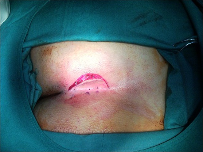
Lateral incision
Fig. 2.
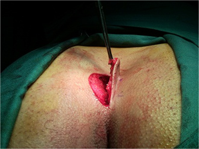
Flap released the cyst is still remaining
The subcutaneous tissue is excised downward and laterally to the fascia underneath. Great care is taken to protect the fascia from the incision, as it offers the only defense against deep spread of infection. Small, pointed hemostate clamps should be used to clamp the bleeding vessels in order to make sure that the smallest amount of tissue destruction is made. Electrocoagulation may be used to control bleeding and to keep the amount of buried suture material minimum. Some prefer to avoid any suture material by using compression or electrocoagulation to control all the bleeding points. Extreme care should be taken in the dissection of the lower end of the incision, as many small, troublesome vessels are encountered frequently that tend to retract when divided. After careful inspection of the wound to make sure that all sinus tracts have been removed, the subcutaneous fat is undercut at its junction with the underlying fascia. This undercutting should extend only far enough to allow approximation of the edges without tension. After all bleeding points are controlled, the wound should be thoroughly washed with saline. The chances of primary healing are greatly enhanced if the field is absolutely dry with several stitches, particularly the one that fixes the flap to the underlying fascia, which is the main stitch that distinguishes this method from others (Fig. 3).
Fig. 3.

Stitches that fix the flap to the fascia
If unexpected infection has been encountered, the wound should be kept open. In uncomplicated sinuses, the wound is closed after all bleedings are controlled, with a skin flap. Four stitches fix the flap to the fascia and subcutaneous tissue (Fig. 4).
Fig. 4.

Stitches that fix the flap to the fascia
Then sutures are introduced 1 cm or a little more, from margins of the wound, to include the full thickness of contralateral skin and subcutaneous tissue to close the flap (Fig. 5). A dressing is applied with great care, and the sutures are allowed to remain in place for 10–14 days (Fig. 6).
Fig. 5.

Fixing the flap to the skin of buttock
Fig. 6.

One week after surgery
Postoperative Care
Complete protection against contamination is essential. Early ambulation is advisable, but sitting upon the incision on a hard chair is not. The patient should be encouraged always to sit on a cushion or to sit to the side on one buttock or the other during the early postoperative period. The diet is not restricted, but a low-residue diet to decrease the chances of contamination from a bowel movement and preventing constipation may be beneficial. Oral analgesics are prescribed in the presence of pain. No specific antibiotic is routinely administered unless with a clear indication. Regardless of the method used, frequent and repeated dressings are indicated to avoid possible early recurrence and prolonged discomfort and disability. The importance of keeping all hair removed from the intergluteal fold until healing is complete cannot be overemphasized. Depilatory agents may be used several times per month provided that pretesting for sensitivity to the agent has been negative.
In our case, all patients were scheduled for regular postoperative visits by a person out of our team. The results were gathered in particular forms and each patient got a number for further analysis.
Statistical Analysis
Results were presented as mean ± SD or median ± SD and frequency (%). Differences in sex, complications, and recurrence rate were analyzed with the chi-square test. Healing time, length of hospital stay, duration of incapacity to work, and number of punctures were analyzed with the Mann–Whitney U test in two groups and Kruskal–Wallis H test among three groups (normal distribution was not detected) . Finally the ANOVA test was used to compare mean among more than two groups for continuous data. A P value < 0.05 was considered statistically significant. SPSS-16 was used to analyze gathered data.
Results
A total of 282 patients with pilonidal sinus disease were reviewed in which only 194 (68.7 %) patients met the criteria and had complete archived files. From now onward, we will talk about this group of 194 patients. The mean age in all patients was 24 ± 6.5 years. Most of them were males (n = 152; 78.4 %) and male-to-female ratio was 3.6. The median of disease duration was 1 ± 1.9 (14 days to 17 years). The median length of hospital stay, length of incapacity for work, and healing time were 2 ± 1.7 (1–11 days), 7 ± 3.9 (7–21 days), 28 ± 12.8 (7–60 days), respectively.
The recurrence of disease was found only in 3 (1.5 %) of all. The most frequent number of detected skin orifices was 1 in 91 (46.9 %). No complications were found in 160 (77.6 %), and among the postoperative complications, bleeding was the most frequent (n = 12; 7.2 %); the list of other complications is shown in Table 1. The frequency of surgery methods was as follow:
Wide excision and modified repair (n = 61; 31.4 %)
Wide excision (n = 81; 41.8 %)
Wide excision and closed with flap (n = 52; 26.8 %).
Table 1.
The frequency of postoperative complications in all patients are shown
| Complications | Frequency | Percent |
|---|---|---|
| No complications | 160 | 77.6 |
| Bleeding | 15 | 7.2 |
| Infection | g | 4.3 |
| chronic pain | 10 | 4.8 |
| Seroma | 7 | 3.4 |
| Hematoma | 3 | 1.5 |
| Necrosis | 2 | 0.9 |
| Total | 206 | 100.0 |
Finally, most of the patients had overall satisfaction (n = 173; 89.2 %) regardless of any surgery method. The mean age was not statistically different in three surgical methods and neither the distribution of sex (P > 0.05), but the mean “duration of pilonidal sinus disease” was higher in the wide excision and modified repair method in comparing with two other methods (P < 0.05) (Table 2). The results of the following variables were compared according to surgical methods (Tables 3 and 4): postoperative complication, recurrence of disease, length of hospital stay (day), length of incapacity for work (day), healing time (days), and number of puncture. As shown in the table, “longer duration of hospital stay” was found in the wide excision and flap method in comparing with two other methods (P < 0.05), but “length of incapacity for work” was not different between the “wide excision and modified repair” and “wide excision” methods (P > 0.5). Healing time was significantly longer in the wide excision method in comparison with two other methods (P < 0.05). However, the wide excision and modified repair method had the lowest value in all the above three elapsed times. The median number of skin orifices found in the wide excision and modified repair method was more than in the wide excision method (P < 0.05), while other comparisons did not differ in the median number of orifices (P > 0.05). All the recurrences (n = 3; 1.5 %) were reported in the wide excision and flap method (P < 0.05). Types of postoperative complications according to surgical methods were as follows: 2 (3.3 %) in wide excision and modified repair, 15 (18.5 %) in wide excision, and 17 (32.7 %) in wide excision and flap.
Table 2.
The comparison of age, gender and duration of pilonidal sinus disease are shown according to surgical methods
| Variables | Wide excision & rnodified repair | Wide excision | Wide excision & closed with flap | p-value | |
|---|---|---|---|---|---|
| N = 61 | N = 81 | N = 52 | |||
| Age | mean ± sd | 24 ± 6.6 | 24 ± 7.3 | 24 ± 5 | 0.91a |
| Gender | Male | 48(78.7 %) | 60(74.1 %) | 44(84.6 %) | 0.35b |
| Female | 13(21.3 %) | 21(25.9 %) | 8(15.4 %) | ||
| Duration of Pilonidal sinus disease year | mean ± sd | 1.71 ± 1.91 | 1.57 ± 2.22 | 1.10 ± 0.93 | 0.006c |
aANOVA test, b Chi2 test, cKruskal-Wallis Test
Table 3.
The comparison of duration of hospital stay , incapacity for work and healing time , number of punctures ,recurrence ,postoperative complications and patients satisfaction are shown according to surgical methods
| Variables | Wide excision & rnodified repair | Wide excision | Wide excision & closed with flap | p-value | |
|---|---|---|---|---|---|
| N = 61 | N = 81 | N = 52 | |||
| Length of hospital stay (day) | mean ± sd | 1.21 ± 0.48 | 2.40 ± 0.77 | 4.78 ± 1.58 | 0.001a |
| Length of incapacity for work (day) | mean ± sd | 7.34 ± 1.27 | 7.33 ± 0.94 | 15.13 ± 2.76 | 0.001a |
| Healing time (days) | mean ± sd | 11.11 ± 2.27 | 35.03 ± 9.53 | 28.84 ± 8.22 | 0.001a |
| Number of punctures | mean + sd | 2 ± 0.9 | 1 ± 0.9 | 1.5 ± 1 | 0.09a |
| Recurrence | Yes | 0 % | 0 % | 3(5.8 %) | 0.017b |
| No | 59(100.0 %) | 81(100.0 %) | 49(94.2 %) | ||
| Postoperative complication | Yes | 2(3.3 %) | 15(18.5 %) | 17(32.7 %) | 0.001b |
| No | 59(96.7 %) | 66(81.5 %) | 35(67.3 %) | ||
| Patient satisfaction | Yes | 59(96.7 %) | 73(90.1 %) | 41(78.8 %) | 0.009b |
| No | 2(3.3 %) | 8(9.9 %) | 11(21.2 %) | ||
aKruskal-Wallis Test, bChi2 test
Table 4.
The Bivariate statistical significance of duration of hospital stay, incapacity for work and healing time, number of punctures, recurrence, postoperative complications and patients satisfaction are shown according to surgical methods
| Variables | Surgical methods | ||
|---|---|---|---|
| I versus 2 | I versus 3 | 2 versus 3 | |
| Length of hospital stay | 0.0001a | 0.0001a | 0.0001a |
| Length of incapacity for work | 0.63a | 0.0001a | 0.0001a |
| Healing time | 0.0001a | 0.0001a | 0.0001a |
| Number of punctures | 0.02a | 0.61a | 0.25a |
| Recurrence | 1b | 0.043b | 0.029b |
| Postoperative complication | 0.006b | 0.0001a | 0.06b |
1: Wide excision & modified repair
2: ‘Wide Excision’
3: ‘Wide excision & closed with flap’
aMann-Whitney test, bChi2 test
Conclusion
Based on the data presented, we conclude that the wide excision and modified repair technique is superior to the wide excision and wide excision and flap techniques for the surgical treatment of pilonidal sinus disease with respect to no recurrence and the least frequency of postoperative complications and shorter healing time and length of hospital stay in comparison with two other techniques. Also most frequent satisfaction was declared by patients from this surgical method. This is an introduction to this method, and for suggestion of this method as a superior method more surveys with more samples and comparison with other famous techniques seem to be essential.
References
- 1.Karydakis GE. Easy and successful treatment of pilonidal sinus after explanation of its causative process. Aust N Z J Surg. 1992;62:385–389. doi: 10.1111/j.1445-2197.1992.tb07208.x. [DOI] [PubMed] [Google Scholar]
- 2.Lee HC, Ho YH, Seow CF, Eu KW, Nyam D. Pilonidal disease in Singapore: clinical features and management. Aust N Z J Surg. 2000;70:196–198. doi: 10.1046/j.1440-1622.2000.01785.x. [DOI] [PubMed] [Google Scholar]
- 3.Manterola C, Barroso M, Araya JC, Fonseca L. Pilonidal disease: 25 cases treated by the Dufourmentel technique. Dis Colon Rectum. 1991;34:649–652. doi: 10.1007/BF02050344. [DOI] [PubMed] [Google Scholar]
- 4.Morell V, Charlton BL, Deshmukh N. Surgical treatment of pilonidal disease: comparison of three different methods in fifty-nine cases. Mil Med. 1991;156:144–146. [PubMed] [Google Scholar]
- 5.Hassan HK, Francis IM, Neglen P. Primary closure or secondary granulation after excision of pilonidal sinus? Acta Chir Scand. 1990;156:695–699. [PubMed] [Google Scholar]
- 6.Sondenaa K, Nesvik I, Anderson E, Natas O, Soreide JA. Patient characteristics and symptoms in chronic pilonidal sinus disease. Int J Colorectal Dis. 1995;10(1):39–42. doi: 10.1007/BF00337585. [DOI] [PubMed] [Google Scholar]
- 7.Berry DP. Pilonidal sinus disease. J Wound Care. 1992;1(3):29–32. doi: 10.12968/jowc.1992.1.3.29. [DOI] [PubMed] [Google Scholar]
- 8.Price ML, Griffiths WAD. Normal body hair: a review. Clin Exp Dermatol. 1985;10:87–97. doi: 10.1111/j.1365-2230.1985.tb00534.x. [DOI] [PubMed] [Google Scholar]
- 9.Trent JT, Kirsner RS. Wounds and malignancy. Adv Skin Wound Care. 2003;16(1):31–34. doi: 10.1097/00129334-200301000-00014. [DOI] [PubMed] [Google Scholar]
- 10.Sondenaa K, Nesvik I, Andersen E, et al. Bacteriology and complications of chronic pilonidal sinus treatment with excision and primary suture. Int J Colorectal Dis. 1995;10(3):161–166. doi: 10.1007/BF00298540. [DOI] [PubMed] [Google Scholar]
- 11.Solla JA, Rothenberger DA. Chronic pilonidal disease. An assessment of 150 cases. Dis Col Rec. 1990;33(9):758–761. doi: 10.1007/BF02052321. [DOI] [PubMed] [Google Scholar]
- 12.Hodgkin W. Pilonidal sinus disease. J Wound Care. 1998;7(9):481–483. doi: 10.12968/jowc.1998.7.9.481. [DOI] [PubMed] [Google Scholar]
- 13.Jones DJ. ABC of colorectal diseases. Pilonidal sinus. BMJ. 1992;305:410–412. doi: 10.1136/bmj.305.6850.410. [DOI] [PMC free article] [PubMed] [Google Scholar]
- 14.Sondenaa K, Diab R, Nesvik I. Influence of failure of primary wound healing on subsequent recurrence of pilonidal sinus. Eur J Surg. 2002;168(11):614–618. doi: 10.1080/11024150201680007. [DOI] [PubMed] [Google Scholar]
- 15.Senapati A, Cripps NP, Thompson MR, Franzini DA. Bascom’s operation in the day-surgical management of symptomatic pilonidal sinus. Br J Surg. 2000;87:1067–1070. doi: 10.1046/j.1365-2168.2000.01472.x. [DOI] [PubMed] [Google Scholar]
- 16.Allen-Marsh TG. Pilonidal sinus: finding the right track for treatment. Br J Surg. 1990;77:123–132. doi: 10.1002/bjs.1800770203. [DOI] [PubMed] [Google Scholar]
- 17.Keighley MR (1999) Pilonidal sinus. In: Keighley MRB, Williams N (eds) Surgery of the anus, rectum and colon, p 539–563
- 18.Guyuron B, Dinner MI, Dowden RV. Excision and grafting in the treatment of recurrent pilonidal sinus disease. Surg Gynecol Obstet. 1983;156:201–204. [PubMed] [Google Scholar]
- 19.Sondenaa K, Anderson E, Nesvik I, Soreide A. Patient characteristics and symptoms in chronic pilonidal sinus disease. Int J Colorect Dis. 1995;10:39–42. doi: 10.1007/BF00337585. [DOI] [PubMed] [Google Scholar]