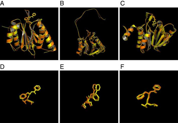
Figure 4.

Schematic representations of the structural overlay corresponding to the crystal structure of the yeast 20S proteasome bortezomib inhibitor complex [PDB code:2F16] (orange) and the model of the Pf 20S proteasome with docked bortezomib inhibitor (yellow) in the catalytic sites of (A) β1, (B) β2 and (C) β5 subunits. Magnified images of the bortezomib inhibitors (in stick representation) are shown for the above subunits in (D), (E) and (F), respectively.
