Abstract
Background:
Estimation of age is important in forensic sciences as a way to establish the identity of human remains. Of the various parts of the body used in age estimation, teeth are the least affected by the taphonomic process. Their durability means that they are sometimes the only body part available for study. Several methods of age estimation have been studied using bone and teeth, and among them, tooth wear and apposition of secondary dentine are the currently available non-destructive methods.
Objectives:
The purpose of the study was to determine the age of adults by using Kvaal's method as well as to establish the relationship of chronological age and dental age with its reliability and limitations on digital panoramic radiographs.
Materials and Methods:
The present study was based on panoramic radiographs that consisted of two groups. One hundred orthopantomographs with Kvaal's criteria (Group A) and 50 orthopantomographs without Kvaal's criteria (Group B) were included. Various parameters were measured and the result was analyzed by means of SPSS-12.0 program statistical data.
Result and Conclusion:
On the basis of Kvaal's criteria, the difference between chronological age and real age was 8.3 years. This suggests that the accuracy of this method depends on the precision of measurements and quality and number of the orthopantomographs.
Keywords: Age estimation, Kvaal's method, orthopantomographs, panoramic radiographs, pulp/tooth area ratio, secondary dentine
Introduction
Age is one of the essential factors in establishing the identity of a person. Age can be estimated in different ways by using chronological age, skeletal age and dental age.[1] Estimation of the age of sub-adult individuals at death is currently based on the synostosis of secondary ossification centers and the development and eruption status of the teeth. However, determination of the age of adults is more complex.[2] Therefore, in that manner, the speciality of forensic odontology plays a small but significant role.[3] The identification of dental remains is of primary importance as teeth are the most durable and resilient parts of the skeleton[4] and, with their physiologic variations, pathoses and effects of therapy,[5] they resist the influence of many factors and taphonomic process and disintegrate very slowly. They are sometimes the only body part available for study and this makes teeth very suitable for dental age estimation.[6,7]
As far as dental age estimation is concerned, tooth development is a complex process that takes place from early fetal life to approximately 20 years of age. Both developmental and regressive changes to the tooth can be related to chronological age.[7] Age estimation up to puberty can be performed by development process, dental radiographs (intraoral periapical radiographs, bitewing radiographs, orthopantomographs) or by a combined radiographic technique of the third molar tooth staging development and hand wrist and cervical vertebrae radiographs.[8] But, after third molar development, it becomes increasingly difficult to assess age accurately. Only aging process and regressive changes of teeth are helpful at adult age.
Several methods have been developed to estimate age based on dental tissue and tooth morphology, like morphologic, radiographic, histological and biochemical methods. Some of these methods are especially developed to estimate the age at death as they require sectioning, while others may also be used in clinical situations. Majority of the cases concerning age estimation are performed on living people. Morphological and radiographic methods (Schour and Massler's method, Demirjian's method and Kvaal's method) are useful in living individuals at adolescent and adult age, whereas histological and biochemical methods (Gustafson's and Johanson's method, Bang and Ramm method, aspartic acid racemization and cemental annulation technique) are useful in dead victims.[7,9] The use of radiographs for age estimation is characteristic of techniques that involve observation of the morphologically distinct stages of mineralization. Such determinations are also based on the degree of formation of root and crown structures, the stage of eruption and the intermixture of primary and adult dentitions.[5]
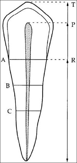
Kvall et al. reported a method in 1995[10] that allows estimation based on morphological measurements of two-dimensional radiographic features of individual teeth. The measurements include comparisons of pulp and root length, pulp and tooth length, tooth and root length and pulp and root widths at three defined levels [Figure 1]. This method is less discriminatory than other methods, but has the important advantage of being non-invasive, not requiring extraction of teeth, being useful for examination and regression analysis of all data performed, with age as the dependent variable.[11]
Figure 1.

Diagram showing tooth measurements according to Kvaal et al.: T, tooth length; R, root length on the mesial surface; P, maximum pulp length; A, root and pulp width at enamel–cementum junction (ECJ); B, root and pulp width midway between measurement levels A and C; C, root and pulp width midway between apex and ECJ
The purpose of the study is to determine the age of adults by measuring the pulp/tooth ratio from digital panaromic radiographs using Kvaal's method as well as to establish the relationship of chronological age and dental age with its reliability and limitations of Kvaal's method on digital panoramic radiographs.
Materials and Methods
The present prospective study was based on 150 digital panoramic radiographs. The patients were selected from the Oral Medicine and Radiology Department whose orthopantomographs were taken as a part of routine diagnosis and treatment purpose only. Patients’ age group was between 20 and 55 years, irrespective of their sex, gender and religion. Patients’ birth dates were noted after analyzing their specific identity proofs. The radiographs were taken using a Kodak 8000 C panoramic and cephalometric machine at 71-73 voltage and 10 mA. Exposure time was 13-14 s with 4096 bits grey scale level.
Patients’ radiographs selection criteria were as follows:
Only high-quality panoramic radiographs, with respect to angulations, contrast and correct positioning, were included in this study.
Radiographs should be free from any artifacts.
Radiographs should not show any developmental anomalies of teeth related to size, shape and structure of teeth.
The analytic study was carried out on six teeth irrespective of the right or left side. They are maxillary central incisor (11/21), maxillary lateral incisor (12/22), maxillary second premolar (15/25), mandibular lateral incisor (32/42), mandibular canine (33/43) and mandibular first premolar (34/44). The resultant radiographs were divided into two groups.
Group A contained 100 patients’ radiographs (based on Kvaal's criteria) without malaligned teeth, severe overlapping of adjacent teeth, badly rotated teeth, carious lesions, restorative fillings, root canal treatment, periapical pathoses and root resorption in selected study.
Group B contained 50 patients’ radiographs (not based on Kvaal's criteria) that did not match the above-mentioned criteria.
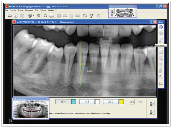
All digital orthopantomograms, originally obtained in DICOM format, were analyzed using the “Kodak Dental Imaging Software.” This software program permits not only to view and manipulate digital X–rays but also to obtain the quantification of relative distance in number of pixels between two different reference points after defining their relative position in the X and Y axes on the digital image. Contrast, brightness and full screen views were adjusted for study teeth without any distortion and magnification and even in individual teeth dimensions [Figure 2].
Figure 2.

Measurement of mandibular teeth by dragging cursor from one point to other, with three measurements at a time with different color indications
After getting all 10 parameters (T, P, R, A, B, C, M, W, L, W–L), mean value of ratios for each tooth, three maxillary teeth, three mandibular teeth and all six teeth were calculated for both groups. Statistical analysis was performed by means of SPSS–12.0 program. Pearson correlation coefficients between chronological age and the ratios were also calculated. From the calculated mean values, mean difference, standard deviation, standard error of mean between chronological and calculated age and regression equations were determined. After establishing the regression equation, it was applied in individual radiographs and age was calculated.
Results
Pearson correlation coefficient ratio between the chronological age and the calculated ratios based on length and width measurements for all teeth and each tooth of both groups was calculated. For Group A, all coefficient ratios of all parameters (T, P, R, A, B, C, M, W, L, W–L) significantly decreased in each tooth, in combined maxillary teeth, in combined mandibular teeth and all six teeth as the age increased, except R value (pulp/tooth ratio), that gave a positive correlation with age in maxillary individual teeth. For Group B, the correlation coefficient ratio showed some variations. All values were significantly decreased as per age, except ratios R, A, B, C, W and L; those were increased for all six teeth. Ratio T, P and M significantly decreased in all six teeth [Table 1].
Table 1.
Pearson correlation ratio between real age and calculated ratios for all teeth

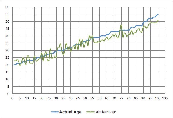
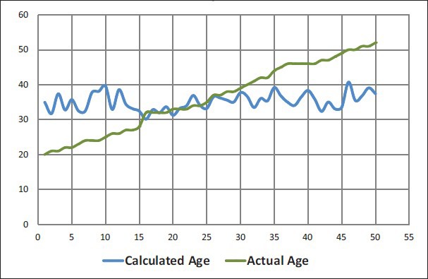
Regression equations were made of all six teeth, all maxillary teeth, for all mandibular teeth and of individual teeth for both groups. A regression equation for all six teeth was 109.26–196.804(M)–63.39(W–L). Coefficient of determination (r2) for all six teeth was 0.29 with standard error of estimate of 8.3 years. For Group B, the regression equation for all six teeth was −76.017 + 128.366(M)−57.817(W–L). Coefficient of determination (r2) for all six teeth was 0.12 with standard error of estimate of 9.45 years. Standard deviation and other statistical measures between chronological age and estimated age for both groups were also made [Table 2, Graphs 1 and 2].
Table 2.
Regression formula for age in years and statistical measures of difference between real age and estimated age based on regression equation for both groups

Graph 1.

Comparison of calculated age with actual age – Group A
Graph 2.

Comparison of calculated age with actual age – Group B
Discussion
Estimation of age is important in forensic sciences as a way to establish the identity of human remains. Chronological age assessment based solely on dental factors can be a reliable indicator of an individual's age, and it is often feasible because teeth may persist long after other parts of the skeleton have disintegrated.[12] Examination of dental radiographs of fully developed teeth is rarely advocated for use in age estimation. It is, however, a simple, non-destructive method that can be employed both on living individuals and on the unknown dead, either in identification cases or in archaeological investigations.[10]
Based on these age-related changes, a variety of methods for dental age estimation were proposed. Quantification of these morphological changes nearly always requires extraction (indirect measurement) with or without preparation of microscopic sections. These methods are time-consuming and expensive, and the destructive approach may not be acceptable for ethical, religious, cultural or scientific reasons.[13,14] Thus, a radiologic technique like the one developed by Kvaal et al. is one of the few that can be used. It is based only on the size of the pulp in relation to the whole tooth and gives a measure of the secondary dentin formation. Therefore, techniques that have been or are being developed for age estimation in living individuals mostly rely on dentin by using radiological imaging of teeth.[14,15]
The teeth were selected by the criterion that teeth from both jaws were included. They would preferably have included molars as well, but the preliminary study clearly demonstrated that accurate measurements of multi-rooted teeth were difficult to perform, and for the same reason maxillary first premolars (which frequently have two roots) were likewise excluded. In the preliminary study, measurements from the maxillary canine demonstrated the lowest correlation coefficients with age, which is consistent with the results found when measurements were made on radiographs of extracted teeth. The mandibular second premolars are frequently found to have been lost early in life, possibly as a result of orthodontic treatment. In the small preliminary sample, significant differences between the ratios from the left and right mandibular central incisors were observed. For these reasons, all these three types of teeth were not included in the present study. An earlier investigation based on manual measurements indicated that maxillary second premolars, laterals and centrals, and mandibular first premolars, canines and laterals were most often present in older patients and were significantly correlated with age.[10] For the same reason, these six teeth were selected.
The present study, based on the non-destructive method using Kvaal's method, on 150 panoramic radiographs represents an important contribution to the already existing age determination methods in adults. The advantage of this method is that it can be applied to living persons. Furthermore, orthopantomographs also provide information of the individuals’ identity and other age-related features such as third molar development, number of teeth and periodontal recession. However, the number of cases in this study was too low to investigate other age-related parameters. In present study, total length of tooth (T), pulp (P) and root (R) was measured. The length of the pulp chamber seems to be influenced by several individual factors (e.g. chewing habits), inducing tertiary dentine production at the roof of the chamber and therefore cannot be used for age determination.[16] To compensate for differences in magnification and angulations on radiographs, the ratios of all measured parameters were taken (T/R, P/R, P/T, etc.).
In the present study, apposition of secondary dentine was estimated by measuring pulp/tooth length and width ratios (T, P, R, A, B, C, M, W, L, W–L). However, it is partly dependent on the anatomy of the tooth and pulp. To reduce the effect of unusual anatomy of one tooth, the results become more accurate if three teeth in the maxilla and the mandible, respectively, are used, or, even better, when all six teeth are used in one formula. In these cases, the individual rather than the tooth is the unit.[14] In the present study, the correlation ratios are significantly decreased in case of combined all six and individual three maxillary and mandibular teeth for Group A, but, in Group B, the ratios were positive due to anatomical variation in teeth length as well as in width.
In the radiographs of the present study, the bone overshadowed the apical third of the tooth so that the width from this area of the tooth could not be measured with sufficient accuracy. The ratios between the pulp and the root have also been used in a previous study of age estimation from tooth measurements. As the size of the pulp is reduced with age, the correlation coefficient between age and the ratios is negative, whereas the inverse ratio would give a positive correlation coefficient. The present study showed that the correlation between age and the ratio of tooth to root length (T) was significant for all types of teeth, indicating that attrition on the occlusal surface was the factor that could be related to age, which is in accordance to the literature. For that reason, T factor was excluded while measuring the mean value of all ratios (M). Briefly, in Group A, all ratios regarding T, P, R, A, B, C, M, W, L and W–L significantly reduced in individual as well as all combined three teeth and six teeth due to deposition of secondary dentine, but, in Group B, all coefficient ratios P, R, A, B, C, M, W, L except T ratio were increasing with age, which was similar to the suggested data.[10,17]
In the present study, Group A radiographs were taken without any pathology according to Kvaal's method. Non-ideal radiographs with caries, dental fillings, crowns and periapical pathology were taken in Group B to examine their effects on standard Kvaal's method as well as to determine whether these radiographs can be used to estimate age by Kvaal's method or not. The present study showed 8.3 years difference between chronological age and real age as per the established regression formula in Group A, which showed a 0.3-year difference to the suggested data as compared with the technique that was applied on intraoral radiographs (Kvaal and T. Solheim).[10] On panaromic radiographs, it showed a variation of 1-1.2 years (Bosmans and Willems).[17] In Group B, a 9.45-year difference was noted, which showed only 1-1.2 year difference in non-ideal radiographs when compared with Group A. This suggests that the accuracy of Kvaal's method depends on the precision of the measurements and the quality and number of the orthopantomographs without any artifacts, restorations or malalignments.[16,17]
In our study, we have used digital orthopantomographs for assessment of age parallel to Bosmans et al.'s study,[17] whereas the original study by Kvaal et al.[10] used the conventional intraoral periapical radiographs for obtaining the regression formulae. It is true that anterior teeth images are inherently distorted in a panoramic radiograph due to the projection geometry while comparing conventional intraoral periapical radiographs. Thus, it can be a limitation for digital orthopantomograph. However, simultaneously, it offers the possibility and advantage of evaluation of all the teeth along with alveolar bone in both the jaws and their required measurements to be made on a single radiograph. Furthermore, accuracy, reproducibility and precision of such technique is most important. Any difference found can lead to many variables, including precision of methods, age distribution, sample size and statistical approach, while the acceptability of intraoral radiographs is dependent on the techniques used and the practical training of the personnel.
Footnotes
Source of Support: Nil
Conflict of Interest: None declared
References
- 1.Willems G, Moulin-Romsee C, Solheim T. Non-destructive dental-age calculation methods in adults: intra- and inter-observer effects. Forensic Sci Int. 2002;126:221–6. doi: 10.1016/s0379-0738(02)00081-6. [DOI] [PubMed] [Google Scholar]
- 2.González-Colmenares G, Botella-López MC, Moreno-Rueda G, Fernández-Cardenete JR. Age estimation by a dental method: A comparison of lamendin's and prince and ubelaker's technique. J Forensic Dent Sci. 2007;52:1156–60. doi: 10.1111/j.1556-4029.2007.00508.x. [DOI] [PubMed] [Google Scholar]
- 3.Pretty IA, Sweet D. Look at forensic dentistry, Part 1: The role of teeth in the determination of human identity. Br Dent J. 2001;190:359–66. doi: 10.1038/sj.bdj.4800972. [DOI] [PubMed] [Google Scholar]
- 4.Gorea RK, Singh A, Singla U. Age estimation from the physiological changes of teeth. J Indian Forensic Sci. 2004;26:94–6. [Google Scholar]
- 5.Avon SL. Forensic odontology: The roles and responsibilities of the dentist. J Can Dent Assoc. 2004;70:453–8. [PubMed] [Google Scholar]
- 6.Ogino T, Ogino H. Application to forensic odontology of aspartic acid racemization in unerupted and supernumerary teeth. J Dent Res. 1988;67:1319–22. doi: 10.1177/00220345880670101501. [DOI] [PubMed] [Google Scholar]
- 7.Reppien K, Sejrsen B, Lynnerup N. Evaluation of post-mortem estimated dental age versus real age: A retrospective 21-year survey. Forensic Sci Int. 2006;159S:S84–8. doi: 10.1016/j.forsciint.2006.02.021. [DOI] [PubMed] [Google Scholar]
- 8.Raut DL, Mody RN. Radiographic evaluation of cervical vertebrae, carpal metacarpal bones and mandibular third molar during adolescence and in young adults. J Indian Acad Oral Med Radiol. 2006;18:24–9. [Google Scholar]
- 9.Shafer's textbook of oral pathology. 5th ed. Philadelphia: Elsevier Publishers; 2008. Acharya and Sivapathasundharam; pp. 1213–5. [Google Scholar]
- 10.Kvaal SI, Kolltveit KM, Thomsen IO, Solheim T. Age estimation of adults from dental radiographs. Forensic Sci Int. 1995;74:175–85. doi: 10.1016/0379-0738(95)01760-g. [DOI] [PubMed] [Google Scholar]
- 11.Forensic Dentistry. 2nd ed. USA: CRC Press Taylor and Francis Group; 2010. Senn and Stimson; p. 282. [Google Scholar]
- 12.Valenzuela A, Martin-De Las Heras S, Mandojana JM, De Dios Luna J, Valenzuela M, Villanueva E. Multiple regression models for age estimation by assessment of morphologic dental changes according to teeth source. Am J Forensic Med Pathol. 2002;23:386–9. doi: 10.1097/00000433-200212000-00018. [DOI] [PubMed] [Google Scholar]
- 13.Vandevoort F, Bergmans L, Cleynenbreugel J, Bielen D, Lambrechts P, Wevers M, et al. Age calculation using x-ray microfocus computed tomographical scanning of teeth: A pilot study. J Forensic Dent Sci. 2004;49:1–4. [PubMed] [Google Scholar]
- 14.Solheim T, Vonen A. Dental age estimation, quality assurance and age estimation of asylum seekers in Norway. Forensic Sci Int. 2006;159S:S56–60. doi: 10.1016/j.forsciint.2006.02.016. [DOI] [PubMed] [Google Scholar]
- 15.Yang F, Jacobs R, Willems G. Dental age estimation through volume matching of teeth imaged by cone-beam CT. Forensic Sci Int. 2006;159S:S78–83. doi: 10.1016/j.forsciint.2006.02.031. [DOI] [PubMed] [Google Scholar]
- 16.Paewinsky E, Pfeiffer H, Brinkmann B. Quantification of secondary dentine formation from orthopantomograms-a contribution to forensic age estimation methods in adults. Int J Legal Med. 2005;119:27–30. doi: 10.1007/s00414-004-0492-x. [DOI] [PubMed] [Google Scholar]
- 17.Bosmans N, Ann P, Aly M, Williems G. The application of Kvaal's dental age calculation technique on panoramic dental radiographs. Forensic Sci Int. 2005;153:208–12. doi: 10.1016/j.forsciint.2004.08.017. [DOI] [PubMed] [Google Scholar]


