Abstract
Rationale: The pathophysiologic causes of obstructive sleep apnea (OSA) likely vary among patients but have not been well characterized.
Objectives: To define carefully the proportion of key anatomic and nonanatomic contributions in a relatively large cohort of patients with OSA and control subjects to identify pathophysiologic targets for future novel therapies for OSA.
Methods: Seventy-five men and women with and without OSA aged 20–65 years were studied on three separate nights. Initially, the apnea-hypopnea index was determined by polysomnography followed by determination of anatomic (passive critical closing pressure of the upper airway [Pcrit]) and nonanatomic (genioglossus muscle responsiveness, arousal threshold, and respiratory control stability; loop gain) contributions to OSA.
Measurements and Main Results: Pathophysiologic traits varied substantially among participants. A total of 36% of patients with OSA had minimal genioglossus muscle responsiveness during sleep, 37% had a low arousal threshold, and 36% had high loop gain. A total of 28% had multiple nonanatomic features. Although overall the upper airway was more collapsible in patients with OSA (Pcrit, 0.3 [−1.5 to 1.9] vs. −6.2 [−12.4 to −3.6] cm H2O; P <0.01), 19% had a relatively noncollapsible upper airway similar to many of the control subjects (Pcrit, −2 to −5 cm H2O). In these patients, loop gain was almost twice as high as patients with a Pcrit greater than −2 cm H2O (−5.9 [−8.8 to −4.5] vs. −3.2 [−4.8 to −2.4] dimensionless; P = 0.01). A three-point scale for weighting the relative contribution of the traits is proposed. It suggests that nonanatomic features play an important role in 56% of patients with OSA.
Conclusions: This study confirms that OSA is a heterogeneous disorder. Although Pcrit-anatomy is an important determinant, abnormalities in nonanatomic traits are also present in most patients with OSA.
Keywords: respiratory physiology, arousal, muscle, upper airway pathophysiology, sleep-disordered breathing
At a Glance Commentary
Scientific Knowledge on the Subject
Previous studies have established that there are likely multiple causes of obstructive sleep apnea (OSA). However, most of these studies were limited to investigating one mechanism in isolation, involved relatively small numbers of participants, and were not performed using detailed physiologic measurements. Accordingly, before the current study, the proportion of patients with OSA in whom nonanatomic pathophysiologic features are present was unknown.
What This Study Adds to the Field
This is the largest comprehensive physiologic study to date showing that the causes of OSA are multifactorial and not just anatomically driven. One or more nonanatomic pathophysiologic traits are present in 69% of patients with OSA. We propose a three-point (Passive critical closing pressure of the upper airway, Arousal threshold, Loop gain, and Muscle responsiveness [PALM]) scale to help guide future investigation aimed at developing novel targeted therapies for OSA according to pathophysiologic characterization.
Obstructive sleep apnea (OSA) is a common breathing disorder characterized by repetitive narrowing and closure of the upper airway during sleep (1). OSA is associated with major adverse health outcomes including increased cardiovascular risk (2). The first-line treatment for OSA, continuous positive airway pressure (CPAP), is highly efficacious in reducing sleep-disordered breathing events. However, about half of all patients with OSA who try CPAP therapy are either completely intolerant or only partially adherent (3). Accordingly, new treatments for OSA are clearly required.
If new treatments for OSA are to be effective, understanding the various underlying causes is essential (4, 5). Key pathophysiologic causes likely include (1) an anatomically compromised or collapsible upper airway (high passive critical closing pressure of the upper airway [Pcrit]) (6); (2) inadequate responsiveness of the upper-airway dilator muscles during sleep (minimal increase in EMG activity to negative pharyngeal pressure) (7, 8); (3) waking up prematurely to airway narrowing (a low respiratory arousal threshold) (9–13); and (4) having an oversensitive ventilatory control system (high loop gain) (13–15). Indeed, small physiologic studies have demonstrated interventions that lower Pcrit (16–18), increase the electrical activity to genioglossus (19, 20), increase the arousal threshold (9), or lower loop gain (21, 22) can reduce OSA severity. However, these important pathophysiologic traits have not been measured collectively in afflicted individuals. This study aimed to quantify carefully each of these phenotypic traits in a relatively large cohort of patients with OSA and a group of healthy control subjects. We hypothesized that the relative contribution of each trait would vary among patients, with the emergence of different phenotypic subgroups. In identifying and characterizing these subgroups, the ultimate goal is to provide insight for the development of novel therapeutic approaches that target underlying mechanisms in individual patients with OSA. Some of the results of these studies have been previously reported in abstract form (23–25).
Methods
Subjects
Informed written consent, as approved by the Partners’ Healthcare Institutional Review Board, was obtained in 90 men and women (69 patients with OSA who had been treated with CPAP for ≥3 months and 21 healthy control subjects). Other than OSA, defined as an apnea-hypopnea index (AHI) greater than 10 events per hour of sleep, participants were healthy and were not taking any medications known to affect sleep or the other parameters measured in the study. Data addressing separate aims involving a small number of participants from the current study have been previously reported (26, 27).
Measurements and Equipment
Polysomnography.
Electroencephalograms, electrooculograms, and surface submentalis EMGs were applied to enable sleep staging and score arousals (28, 29). Chest and abdominal motion bands, a position monitor, finger pulse oximetry, and airflow monitoring (thermistor plus nasal pressure) were also applied to enable respiratory event detection according to standard criteria (30).
Key physiologic measurements.
Two Teflon-coated stainless steel fine-wire intramuscular electrodes (Cooner Wire Company, Chatsworth, CA) with 2 mm of Teflon removed from the tip were inserted into the largest upper-airway dilator muscle, the genioglossus, by a 25-gauge needle 3 to 4 mm on either side of the frenulum to a depth of approximately 1.5 cm after surface anesthesia (4% lidocaine HCl) to create a bipolar EMG recording. Both nostrils were decongested (0.05% oxymetazoline HCl). The clearer nostril was anesthetized (4% lidocaine HCl) and an epiglottic pressure (Pepi) catheter (model MCP-500; Millar, Houston, TX) was advanced 1 to 2 cm below the base of the tongue under direct visualization. The catheter was taped to the nostril and passed through a port in a CPAP mask (Gel Mask; Philips Respironics, Murrysville, PA). The mask was attached to a pneumotachograph (model 3700A; Hans Rudolf Inc., Kansas City, MO) and differential pressure transducers (Validyne Corporation, Northbridge, CA) for measurement of airflow and mask pressure.
Protocol
Participants were studied on three separate occasions approximately 1 week apart. Initially, patients withheld their CPAP use for a single night and a standard overnight sleep study was performed to quantify the AHI. Participants were encouraged to sleep on their back for at least 4 hours on the diagnostic night. On the two physiology nights (random order), subjects were studied between approximately 10:30 pm and 5:00 am while lying in the supine posture on therapeutic CPAP for the patients with OSA or at least 4 cm H2O in the control subjects. This holding pressure was increased during sleep to eliminate any sign of inspiratory airflow limitation, according to the epiglottic-airflow relationship, as required in both groups.
Swallows and tongue protrusions against the top teeth were performed before sleep to determine maximal genioglossus EMG activity for each participant (31). After stable non-REM sleep was established, progressive CPAP drops for up to 3 minutes were applied to induce varying degrees of upper-airway collapse using a modified CPAP device capable of delivering ± 20 cm H2O (Philips Respironics) for measurement of (1) Pcrit (6), (2) genioglossus muscle responsiveness (7), and (3) the respiratory arousal threshold (32). Steady-state loop gain was measured on a separate night also using CPAP drops (21, 27) as described below.
Data Analysis and Statistical Procedures
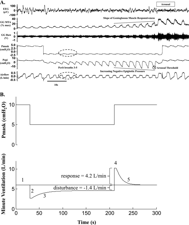
CPAP compliance history was measured objectively by inbuilt compliance meter. To determine the Pcrit in each participant, linear regression was performed between peak inspiratory airflow and mask pressure for breaths three to five after each CPAP drop if the breaths were flow-limited (33). Raw genioglossus EMG was rectified, moving-time-averaged (100 milliseconds), and expressed as a percentage of maximum activity (31). Linear regression was performed between peak genioglsssus EMG versus nadir epiglottic pressure for each artifact-free breath before arousal or sudden restoration of airflow during CPAP drops. Genioglossus muscle responsiveness was defined as the slope of the relationship between these two variables (7). The respiratory arousal threshold was defined as the average nadir epiglottic pressure immediately before cortical arousal (>3 seconds of high frequency activity on the EEG) for all CPAP drops lasting greater than or equal to 10 seconds that terminated in an arousal and had greater than or equal to 2 cm H2O decrement in epiglottic pressure in the preceding 30 seconds (9, 32). Figure 1A shows an example of a CPAP drop highlighting how each of these three pathophysiologic traits was defined. Loop gain was quantified as the ventilatory response to a disturbance in ventilation (21, 27). The disturbance was produced by the CPAP drop, as shown in the schematic diagram in Figure 1B.
Figure 1.
(A) Example of a continuous positive airway pressure (CPAP) drop highlighting the variables of interest for calculation of Pcrit, genioglossus muscle responsiveness, and the respiratory arousal threshold. (B) Determination of loop gain from a CPAP drop. (1) Before the drop, the patient’s airway is open and ventilation is at eupnea. (2) When CPAP is dropped, the upper airway narrows and limits ventilation. (3) As a result, CO2 increases and, in many individuals, activates and stiffens the pharyngeal muscles and increases ventilation slightly (although it typically remains below eupnea). In this schematic example, the disturbance is a change in ventilation of −1.4 L/min. (4) The response to this disturbance is determined by reopening the airway with CPAP and measuring the ventilatory overshoot, which is 4.2 L/min. Therefore, the loop gain is 4.2 ÷ −1.4 = −3 (i.e., for every liter per minute reduction in ventilation, there is a threefold increase in ventilatory drive). (5) After the airway is reopened and the excess CO2 is blown off, ventilation returns back to eupnea. Refer to the text and reference (27) for further detail. GG = genioglossus; MTA = 100 millisecond moving-time-average of the rectified raw electromyographic activity; Pepi = epiglottic pressure; Pcrit = critical closing pressure of the upper airway; Pmask = mask pressure.
Similar to previous studies that characterized the various sleep-disordered breathing severities according to Pcrit (6, 34), to provide pathophysiologic insight Pcrit was separated into four categories: (1) control subjects with a highly negative Pcrit (<−5 cm H2O); (2) control subjects and patients with OSA with overlapping Pcrit values (<−5 to −2 cm H2O); and patients with OSA with (3) moderately (−2 to +2 cm H2O) to (4) highly collapsible upper airways (>+2 cm H2O). Poor genioglossus muscle responsiveness was defined as a less than 0.1% of maximum increase in EMG activity per centimeter of water of negative epiglottic pressure. Awakening to greater than −15 cm H2O of negative epiglottic pressure constituted a low respiratory arousal threshold as previously defined (9). High loop gain was defined as less than −5, based on physiologic insight from a previous intervention study (21) and model simulations (27).
For each outcome variable where the distribution of the delta between conditions was normally distributed (according to a Shapiro-Wilk test), statistical comparisons were performed using Student paired t tests. Nonnormally distributed data were compared using a nonparametric Mann-Whitney test. Analysis of variance was used to compare phenotypic traits between Pcrit groups (SigmaPlot, San Jose, CA). Group data are reported as mean ± SD or median with interquartile range for nonnormally distributed variables. Statistical significance was defined as P less than 0.05.
Results
Subject Characteristics
Four of the control subjects and 11 of the patients with OSA did not complete all three nights of the protocol and were excluded from data analysis. Reasons for incomplete data included the diagnosis of OSA in one of the control subjects, and insufficient sleep in three of the control subjects and six of the patients with OSA. The remaining five patients with OSA were excluded because of the presence of central respiratory events, failure to attend, uncontrolled hypertension, use of an antidepressant, and a technical problem. Anthropometric, baseline sleep, and CPAP compliance characteristics for the 75 participants that completed all three nights are displayed in Table 1.
TABLE 1.
ANTHROPOMETRIC, SLEEP, AND CPAP COMPLIANCE CHARACTERISTICS
| Patients with OSA (n = 58) | Control Subjects (n = 17) | |
|---|---|---|
| Sex |
15 ♀ |
10 ♀ |
| Age, yr |
47 ± 11 |
38 ± 12 |
| Body mass index, kg/m2 |
35 ± 6 |
26 ± 4 |
| Median AHI, number of events per hour sleep |
39 (19–65) |
3 (2–4) |
| AHI range, number of events per hour sleep |
11–112 |
0–10 |
| Sao2 nadir, % |
82 (77–86) |
91 (89–93) |
| CPAP compliance, h/night | 6.4 ± 1.6 | — |
Definition of abbreviations: AHI = apnea-hypopnea index; CPAP = continuous positive airway pressure; OSA = obstructive sleep apnea.
Values are mean ± SD or median and interquartile range in parentheses.
Phenotypic Traits
A total of 27 ± 12 CPAP drops per subject were delivered during sleep. Of these, 6 ± 5 caused an immediate arousal (within <10 seconds) and 2 ± 4 were excluded from the analysis because of poor signal quality. The average reduction in the CPAP holding level required to derive the phenotypic traits was 6 ± 2 cm H2O. All four traits could be quantified in 52 subjects, three in 22 subjects, and two in one subject. Group and individual data for each phenotypic trait are displayed in Figures 2 and 3. There was substantial between-subject variability in both patients and control subjects.
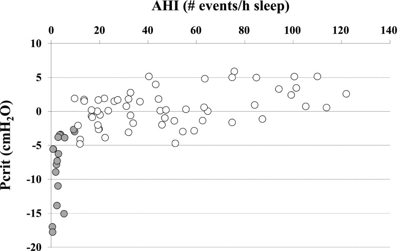
Figure 2.
Critical closing pressure of the upper airway (Pcrit) versus the apnea-hypopnea index (AHI). Grey circles represent individuals with an AHI less than 10 events per hour of sleep.
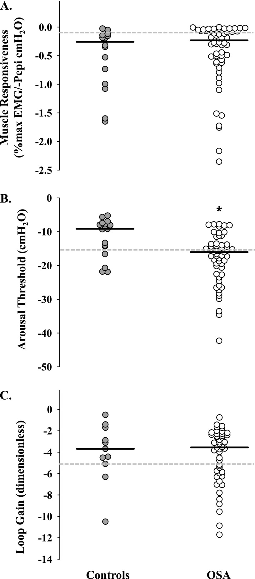
Figure 3.
Phenotypic trait scatter plots for (A) slope of genioglossus muscle responsiveness versus negative epiglottic pressure, (B) the respiratory arousal threshold, and (C) loop gain. Note the large degree of between-subject variability within patients with OSA and control subjects and considerable between-group overlap for each of the pathophysiologic parameters. Horizontal black lines represent median values. Values above the dashed grey lines in A (more positive) and B (more positive) indicate poor muscle responsiveness and a low arousal threshold, respectively. Values below the dashed grey line in C (more negative) indicate a high loop gain. Refer to text and Tables 2 and 3 for further detail. *Significant difference between groups (P < 0.05). OSA = obstructive sleep apnea; Pepi = epiglottic pressure.
Patients with OSA versus Control Subjects
Pcrit was more positive in the patients with OSA compared with the control subjects (0.3 [−1.5 to 1.9] vs. −6.2 [−12.4 to −3.6]; P < 0.001) (Figure 2). Patients with a Pcrit less than +2 cm H2O all had severe OSA (AHI >30 events per hour of sleep) (Figure 2; Table 2). Conversely, those with Pcrit values less than −5 cm H2O did not have OSA (AHI <10 events per hour of sleep) (Figure 2). However, there was substantial overlap in Pcrit between patients with OSA and control subjects in the subatmospheric range between −2 and −5 cm H2O. AHI varied considerably within this subgroup (Figure 2) as did the nonanatomic phenotypic traits (Table 2).
TABLE 2.
PHENOTYPIC TRAITS IN INDIVIDUALS WITH UPPER AIRWAY COLLAPSIBILITY BETWEEN −5 AND −2 cm H2O
| AHI (Number of Events per Hour of Sleep) | Pcrit (cm H2O) | GG Response (% Maximum EMG/−cm H2O Pepi) | Arousal Threshold (cm H2O) | Loop Gain (Dimensionless) |
|---|---|---|---|---|
| Control subjects | ||||
| 2.8 |
−3.8 |
−1.597 |
−7.4 |
−4.5 |
| 3.5 |
−3.5 |
−0.194 |
−5.2 |
— |
| 3.9 |
−3.4 |
−1.650 |
−13.3 |
— |
| 5.6 |
−3.9 |
−0.187 |
−21.9 |
−3.1 |
| 9.3 |
−2.7 |
−0.052 |
−8.4 |
−5.1 |
| 9.7 |
−3.0 |
−0.321 |
−20.7 |
−1.7 |
| Patients with OSA | ||||
| 11.1 |
−2.1 |
−2.166 |
−22.5 |
−10.9 |
| 11.9 |
−4.8 |
−1.726 |
−15.7 |
−5.9 |
| 12 |
−4.1 |
−0.008 |
−14.8 |
−4.5 |
| 17.9 |
−3.9 |
−0.711 |
−22.7 |
−6.0 |
| 19.3 |
−2.0 |
−1.513 |
−13.8 |
−8.8 |
| 19.4 |
−2.6 |
−0.019 |
−7.9 |
−2.5 |
| 19.6 |
−2.6 |
−0.061 |
−7.8 |
−6.3 |
| 31.9 |
−3.1 |
−0.272 |
−23.5 |
−11.7 |
| 39 |
−4.7 |
−0.026 |
−18.5 |
−3.1 |
| 39.3 |
−3.0 |
−0.042 |
−26.5 |
−5.7 |
| 59.1 | −2.9 | −0.085 | −10.4 | — |
Definition of abbreviations: AHI = apnea-hypopnea index; GG = genioglossus; OSA = obstructive sleep apnea; Pcrit = pharyngeal critical closing pressure; Pepi = epiglottic pressure.
Values in bold indicate poor muscle responsiveness, a low respiratory arousal threshold, or a high loop gain.
On average, the respiratory arousal threshold was significantly higher (more negative) in patients with OSA versus control subjects (P < 0.01) (Figure 3B). Nonetheless, approximately one-third of patients with OSA had a low arousal threshold according to previously defined thresholds (9) (Figure 3B). The proportion of CPAP drops that caused immediate arousals (within <10 seconds) was higher in those with a low compared with a high arousal threshold (23 [13–33] vs. 15 [8–22] %; P = 0.02) despite lower magnitude CPAP drops (5 [4–6] vs. 7 [6–7] cm H2O; P < 0.01). Conversely, there was no systematic difference in the slope of genioglossus muscle responsiveness (P = 0.41) or loop gain (P = 0.68) between patients with OSA and control subjects (Figures 3A and 3C). However, approximately one-third of patients with OSA had poor muscle responsiveness (Figure 3A). Similarly, 36% of patients with OSA had a high loop gain (Figure 3C).
Anatomic Subgroups
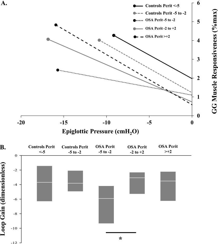
Group data according to Pcrit category for each of the phenotypic traits are displayed in Figure 4. When grouped according to upper-airway collapsibility (Pcrit) characterization, loop gain was almost twice as high (more negative) in patients with OSA with a Pcrit less than −2 cm H2O versus those with a Pcrit greater than +2 cm H2O (P = 0.01) (Figure 4B). Similarly, loop gain tended to be higher in the patients with OSA with a Pcrit less than −2 cm H2O compared with control subjects (P = 0.06) (Figure 4B) and patients with OSA with a Pcrit greater than +2 cm H2O (P = 0.07) (Figure 4B).
Figure 4.
Phenotypic traits according to upper airway collapsibility (critical closing pressure of the upper airway [Pcrit]) category. Control subjects with a Pcrit less than −5 cm H2O (n = 11; 65%) had a median apnea-hypopnea index (AHI) of 2 (1–3) events per hour of sleep. Control subjects with a Pcrit between −5 and −2 cm H2O (n = 6; 35%) had a median AHI of 5 (3–9) events per hour of sleep. Patients with obstructive sleep apnea (OSA) with a Pcrit between −5 and −2 cm H2O (n = 11; 19%) had a median AHI of 19 (12–39) events per hour of sleep. Patients with OSA with a Pcrit between −2 and +2 cm H2O (n = 33; 58%) had a median AHI of 32 (19–56) events per hour of sleep. Patients with OSA with a Pcrit greater than +2 cm H2O (n = 13; 23%) had a median AHI of 76 (48–100) events per hour of sleep. (A) The dots indicate the median arousal threshold, which is higher in the OSA groups. The two control groups have a relatively low arousal threshold, but note how they can achieve a high level of genioglossus (GG) muscle activity because of their steep slope and high starting point. Thus, combined with a relatively noncollapsible upper airway, these individuals probably do not need a higher arousal threshold to prevent OSA. The patients with OSA with a Pcrit greater than +2 have a steep muscle responsiveness slope and a high arousal threshold and thus can achieve the greatest amount of muscle activity. However, they are limited by their highly collapsible upper airway. The OSA group with a relatively uncollapsible upper airway (Pcrit between −5 and −2 cm H2O) has the poorest muscle activation response, which is likely to be an issue for this group. (B) Loop gain is substantially higher in the patients with OSA with a Pcrit between −5 and −2 cm H2O, in which ventilatory motor output increases sixfold for every liter per minute reduction in ventilation. The other groups exhibit only a threefold to fourfold increase and thus are much more stable. Horizontal white lines indicate median values and the grey portions of the box plot represent the 25th and 75th centiles. *Significant difference between groups (P < 0.05).
Discussion
The findings of this study highlight the multifactorial pathophysiology of OSA. Upper airway collapsibility and anatomy is an important determinant of the presence or absence of OSA and its severity. In our cohort, those with a Pcrit greater than +2 cm H2O invariably have severe OSA, whereas those with a Pcrit below −5 cm H2O do not have OSA. However, approximately one in five of the patients with OSA required less than −2 cm H2O to collapse their upper airway during sleep. Within the −2 to −5 cm H2O Pcrit range, the AHI varies considerably (some individuals do not have OSA). This finding suggests that other nonanatomic pathophysiologic traits are particularly important in contributing to the presence or absence of OSA and its severity within this group.
Phenotypic Traits: Between-Group Differences, Relationship with AHI, and Interactions with Pcrit
There is considerable intersubject variability and overlap between patients with OSA and control subjects for each of the four pathophysiologic traits measured in this study. As previously demonstrated (6, 16, 34–36), and consistent with the importance of upper-airway collapsibility in OSA pathogenesis, Pcrit is considerably more positive in patients with OSA compared with control subjects. Similarly, the broad distribution of Pcrit values, the widespread relationship between Pcrit and AHI, and the sizable between-group overlap observed in the current study are consistent with previous reports (6, 34).
Neuromuscular Compensation
An inability to mount an adequate increase in neural drive to the upper airway dilator muscles during sleep in response to negative collapsing pressure contributes to OSA pathogenesis (4, 8, 11). More than one-third of the patients with OSA studied generated less than a 0.1% increase in their maximal genioglossus activity in response to a 1 cm H2O decrement in negative pharyngeal pressure. Poor genioglossus responsiveness during sleep is particularly common in patients with OSA with a mild to moderately collapsible upper airway. Accordingly, this trait is likely to be important in mediating the presence or absence of apnea and its severity in these individuals. However, muscle responsiveness is not systematically different in patients with OSA compared with control subjects. This finding is consistent with previous studies demonstrating comparable or increased neural responsiveness to negative collapsing pressures in patients with OSA (7, 37, 38). However, the ability to translate upper airway neural drive to mechanical airway opening may be compromised in some patients with OSA (7, 39–41).
Arousal Threshold
Direct comparisons of the respiratory arousal threshold between patients with OSA and control subjects have not been previously performed. However, an increased arousal threshold in patients with OSA is in accordance with prior between-study comparisons in patients with severe OSA and healthy control subjects (42). CPAP therapy decreases the arousal threshold in patients with severe OSA (42–45). However, the current findings indicate that despite objectively documented regular CPAP use, the arousal threshold remains higher in patients with OSA compared with control subjects. Nonetheless, more than one-third of CPAP-treated patients with OSA have a low arousal threshold. In these individuals, waking up prematurely is predicted to contribute to their OSA by disrupting sleep continuity and limiting the opportunity for adequate upper airway muscle recruitment to restore airflow during sleep (4, 9, 10, 12).
Loop Gain
Previous studies have not directly compared loop gain in patients with OSA and nonsnoring control subjects (14, 15). However, both of these studies concluded that loop gain is likely to be important in OSA pathogenesis for some patients but not others according to their degree of upper-airway collapsibility (15) or apnea severity (14). Consistent with this notion, loop gain was not systematically different in patients with OSA compared with control subjects in the current study but was almost 50% higher (more negative) in patients with a Pcrit less than −2 cm H2O compared with those with more positive Pcrit values. Loop gain also tended to be higher in patients with OSA than control subjects with a similar Pcrit. This finding suggests that loop gain is likely to be particularly important in OSA pathogenesis in those with mild vulnerability to upper-airway collapse but less so in those with a highly collapsible upper airway, in whom biomechanical predisposition may prevail.
Taken together, these findings indicate that the way in which these traits interact to cause or prevent OSA is highly complex and likely varies considerably between individuals. The degree of upper airway collapsibility is particularly important in mediating the relative importance of each of the nonanatomic traits. Indeed, many healthy individuals without OSA also display vulnerability in the nonanatomic phenotypic traits, particularly arousal threshold. However, in the absence of an inherently collapsible upper airway, these traits in and of themselves are unlikely to cause clinically important sleep-disordered breathing.
The PALM Scale, Simplified Approaches, and Potential Novel Targets for Therapy
In carefully defining each of these four pathophysiologic traits, several key themes have emerged. In this paper we present the PALM (Pcrit, arousal threshold, loop gain, and muscle responsiveness) scale to assist in categorizing patients with OSA according to anatomic and nonanatomic phenotypic traits (Table 3). The scale provides insight into the proportion and characteristics of patients with OSA in each category. To stimulate future research, it also highlights the potential to target new and existing therapies based on specific mechanisms in individual patients with OSA.
TABLE 3.
Pcrit, AROUSAL THRESHOLD, LOOP GAIN, AND MUSCLE RESPONSIVENESS SCALE (THE PALM SCALE)
| PALM Category | Proportion of Patients | Category Cut-Offs | Patient Features | Possible Treatment Targets |
|---|---|---|---|---|
| 1 |
23% |
Pcrit greater than +2 cm H2O |
Highly collapsible upper airway |
Major anatomic or mechanical intervention likely required (e.g., CPAP) |
| 62% have one or more nonanatomic traits in the vulnerable* range | ||||
| 23% have poor muscle responsiveness | ||||
| 38% have a low arousal threshold | ||||
| 29% have high loop gain | ||||
| 23% have two or more potentially contributing factors | ||||
| All have severe OSA: AHI = 76 (53–100); range, 31–122 events per hour; REM/non-REM = 0.8† | ||||
| BMI = 37 ± 6; range, 28–45 kg/m2; age = 46 ± 11; range, 24–60 y; 8%♀ | ||||
| 2 |
58% |
Pcrit −2 to +2 cm H2O |
Moderately collapsible upper airway |
Candidate for one or a combination of targeted therapies |
| Overall severe OSA: AHI = 32 (19–55); wide range, 10–112 events per hour; REM/non-NREM = 1.2† | ||||
| BMI = 35 ± 6; range, 23–46 kg/m2; age = 47 ± 11; range, 20–65 y; 33%♀ | ||||
| 2a |
36% |
Pcrit −2 to +2 cm H2O without nonanatomic vulnerability |
Moderately collapsible upper airway; primarily anatomically driven |
Anatomic intervention (e.g., CPAP, mandibular advancement splint, upper airway surgery, positional therapy, or weight loss) |
| None have nonanatomic traits in the vulnerable* range | ||||
| 2b |
64% |
Pcrit −2 to +2 cm H2O with nonanatomic vulnerability |
Moderately collapsible upper airway and |
A combination of anatomic and nonanatomic interventions is likely required (e.g., mandibular advancement splint or weight loss plus oxygen or a sleep consolidation aid) |
| 100% have one or more nonanatomic traits in the vulnerable* range | ||||
| 52% have poor muscle responsiveness | ||||
| 48% have a low arousal threshold | ||||
| 50% have high loop gain | ||||
| 33% have two or more potentially contributing factors | ||||
| 3 | 19% | Pcrit less than −2 cm H2O | Some vulnerability to upper airway collapse |
Candidate for one or a combination of targeted therapies with an increased likelihood that nonanatomic interventions (e.g., oxygen or a sleep consolidation aid) would be beneficial in these patients |
| 100% have one or more nonanatomic traits in the vulnerable* range | ||||
| 55% have poor muscle responsiveness | ||||
| 45% have a low arousal threshold | ||||
| 70% have high loop gain | ||||
| 55% have two or more potentially contributing factors | ||||
| Overall mild-moderate OSA: AHI = 19 (13–37); range, 11–59 events per hour; REM/non-REM = 3.3† | ||||
| BMI = 34 ± 5; range, 26–42 kg/m2; age = 47 ± 12; range, 26–64 y; 27%♀ |
Definition of abbreviations: AHI = apnea-hypopnea index; BMI = body mass index; CPAP = continuous positive airway pressure; OSA = obstructive sleep apnea; Pcrit = pharyngeal critical closing pressure.
Definitions of “vulnerable” for nonanatomic traits: poor muscle responsiveness; greater than −0.1% maximum genioglossus electromyographic activity/−cm H2O epiglottic pressure, low arousal threshold; greater than −15 cm H2O, and high loop gain; less than −5 dimensionless. Refer to the text for further detail.
REM/non-REM AHI ratio was only calculated in patients who had greater than 30 minutes of REM sleep during their diagnostic study (64% of PALM category 1, 58% of PALM category 2, and 46% of PALM category 3 patients).
Approximately one in four patients with OSA falls into PALM scale 1. These patients have a high Pcrit, invariably have severe OSA, and approximately two-thirds have vulnerability in one or more of the other three phenotypic traits. Although targeting one or more nonanatomic traits (muscle responsiveness, arousal threshold, or loop gain) with novel therapies in these patients may reduce OSA severity, it is highly unlikely that this approach is sufficient to overcome the extent of upper airway collapsibility present in these patients. Accordingly, CPAP or a major anatomic intervention is likely to be required for this group of patients.
PALM scale category 2 constitutes almost three out of five of the patients with OSA studied. These patients have a moderately collapsible upper airway with a wide range of OSA severities, although on average OSA is moderately severe. Within category 2, there are two types of patients: those in whom the nonanatomic phenotypic traits are unlikely to predispose to OSA, and those in whom one or more of the nonanatomic traits is likely to be important in the pathogenesis.
In category 2a, which constitutes approximately one-third of category 2 patients, an anatomic intervention is likely to be required. In addition to CPAP, other interventions, such as a mandibular advancement splint, upper airway surgery, positional therapy, or weight loss, may be beneficial. The ability of each of these non-CPAP approaches to reduce OSA severity effectively to normal limits depends on the baseline upper airway collapsibility and the ability of the intervention to reduce collapsibility in a particular individual. For example, on average, mandibular advancement therapy reduces Pcrit by 2.3 cm H2O (18), uvulopalatopharyngoplasty surgery by 3.3 cm H2O (17), supine to lateral position by 2.2 cm H2O (46), and a 17% reduction in body mass index reduces Pcrit by 7.5 cm H2O (16).
In the category 2b patients, a primarily nonanatomic intervention may be beneficial. For example, oxygen therapy reduces loop gain and OSA severity by approximately 50% in patients with OSA with high loop gain (22), acetazolamide reduces loop gain and OSA severity by about 50% (21), and stabilizing or increasing CO2 reduces OSA severity by more than 30% in patients with high loop gain (by controller gain) (47). A roughly doubling of genioglossus EMG activity reduces Pcrit by 1.6 cm H2O (48), whereas a standard dose of eszopiclone increases the arousal threshold and reduces OSA severity by approximately 45% in patients with OSA with a low arousal threshold (9). Reductions in AHI by these approaches may be more pronounced than in category 2a patients, although this assumption clearly requires investigation. The extent to which reductions in AHI may occur with nonanatomic interventions is also likely to be influenced importantly by the individual’s baseline upper airway collapsibility. Ultimately, a combination of an anatomic intervention (e.g., mandibular advancement) and a nonanatomic intervention (e.g., oxygen therapy) may be required to achieve complete therapeutic use for many category 2b patients with non-CPAP approaches.
Approximately one in five patients with OSA falls into PALM scale category 3. These patients have some vulnerability to upper airway collapse. However, negative pharyngeal pressure is required and many non-OSA individuals have comparable Pcrit values. OSA severity tends to be mild to moderate and more severe in REM in these patients. All of the category 3 patients have one or more of the nonanatomic traits that are likely to be contributing importantly to their apnea pathogenesis. Therapeutic approaches that target these specific traits are anticipated to be highly effective in this subgroup of patients. Ultimately, the use of the PALM scale categorization to assist in optimizing patient selection and treatment effectiveness needs to be assessed formally by adequately powered targeted intervention studies.
Methodologic and Future Study Considerations
The generalizability of the current findings is limited by several features of the patient population and study design. First, the patients with OSA were highly compliant with CPAP therapy. Our goal was to minimize the confounding effects of OSA (sleep fragmentation, hypoxemia) to determine “inherent” characteristics of each of the pathophysiologic traits (43) rather than disease consequences. Accordingly, the magnitude of the pathophysiologic traits defined in this cohort, and the proportion of patients in each category, may vary in untreated patients. In addition, the traits may vary over time; with aging; between sexes; and with other events including sleep deprivation, changes in autonomic activation, and with exposure to intermittent hypoxia. However, the effect of CPAP treatment on each of these traits across the spectrum of disease remains uncertain. For example, sleep deprivation and CPAP treatment can reduce chemosensitivity (43, 49). Accordingly, the proportion of patients with OSA with loop gain abnormalities in untreated patients with OSA may be higher than reported in the current study. Interestingly, the proportion of patients with OSA with a low arousal threshold in the current cohort is comparable with that of untreated patients with OSA (9). This finding suggests that CPAP therapy may do little to alter the arousal threshold in these individuals. Nonetheless, further investigation is required to characterize each of the phenotypic traits in untreated patients, including those who are noncompliant with CPAP therapy.
Second, data were acquired in non-REM sleep. Thus, the role of these pathophysiologic traits in REM sleep remains unknown. The patients with OSA studied were also predominantly obese. Comparing pathophysiologic traits between obese and nonobese individuals in future studies is likely to be insightful. It is also important to note that the passive Pcrit is a composite measure of overall upper airway collapsibility during sleep. Although anatomy is clearly an important determinant of Pcrit, other imaging techniques are required to assess upper airway anatomy per se.
Finally, despite the complexity of the measurements performed and our careful approach to data collection, there remain other mechanisms that are also likely to be important in OSA pathophysiology. These include but are not limited to surface tension forces (50), lung volume effects (51), fluid shifts (52, 53), body-head position (46, 54), hormonal effects (55, 56), and ventilatory responses to transient stimuli (57). Although many of these mechanisms may be mediated through one or more of the four traits defined in this study, these and other currently unknown factors must be important in either protecting against or perpetuating OSA for certain individuals. For example, in examining the individual data in Table 2 of patients with OSA and control subjects with similar upper airway collapsibility, overall the patients with OSA seem to have greater vulnerability across the three measured nonanatomic phenotypic traits. However, despite vulnerability in all three traits, one of the control subjects (AHI of 9.3 events per hour) does not have OSA and must be protected by other currently unknown mechanisms. Determining these factors is clearly an important objective for future research.
Summary
The causes of OSA vary among patients. Upper airway collapsibility and anatomy is fundamentally important in OSA pathogenesis. However, other traits, such as upper airway muscle responsiveness during sleep, the respiratory arousal threshold, and loop gain, may also play an important role in OSA pathogenesis for certain patients. How these and other pathophysiologic factors interact with upper airway collapsibility and anatomy ultimately determines the presence or absence of OSA and its severity. The three-point PALM scale defined in this study constitutes an important initial step in categorizing individual patients with OSA based on several key pathophysiologic traits. The objective of the PALM scale is to provide a conceptual framework in which to stimulate future hypothesis-driven testing of therapies that target specific underlying mechanisms in individuals. Ultimately, novel approaches that can easily and reliably define the varying phenotypic traits in clinical practice are required. A simplified method in which these traits can be acquired in a single night study is one such approach (21, 27, 58).
Acknowledgments
Acknowledgement
The authors are grateful for contributions and technical support on this project provided by Erik Smales, Geoffrey Kehlman, Karen Stevenson, Lauren Hess, Louise Dover, Pamela DeYoung, Salonee Parikh, and Scott Smith. Barbara Toson also provided invaluable statistical advice.
Footnotes
Supported by NIH 5R01HL048531. The authors have also received funding support from the AHA (0840159N, 10SDG3510018); the ARC (60702); the NHMRC of Australia (510392, 1049814); and the NIH (RO1 102231, R01 HL090897, NIH K24 HL 093218, NIH P01 HL 095491, NIH R01HL110350, and NIH R01 HL085188). The Harvard Catalyst is funded by UL1 RR 025758-01. A modified continuous positive airway pressure machine was provided by Philips Respironics and was used to obtain the physiologic measurements performed in this study.
Author Contributions: D.J.E. wrote the manuscript and all of the authors contributed to the final version. D.J.E., A.S.J., and A.W. contributed to experimental design and collected and analyzed the data. A.M. contributed to experimental design and data interpretation. D.P.W. was responsible for the study conception, design, and data interpretation.
Originally Published in Press as DOI: 10.1164/rccm.201303-0448OC on May 30, 2013
Author disclosures are available with the text of this article at www.atsjournals.org.
References
- 1.Young T, Palta M, Dempsey J, Skatrud J, Weber S, Badr S. The occurrence of sleep-disordered breathing among middle-aged adults. N Engl J Med. 1993;328:1230–1235. doi: 10.1056/NEJM199304293281704. [DOI] [PubMed] [Google Scholar]
- 2.Yaggi HK, Concato J, Kernan WN, Lichtman JH, Brass LM, Mohsenin V. Obstructive sleep apnea as a risk factor for stroke and death. N Engl J Med. 2005;353:2034–2041. doi: 10.1056/NEJMoa043104. [DOI] [PubMed] [Google Scholar]
- 3.Kribbs NB, Pack AI, Kline LR, Smith PL, Schwartz AR, Schubert NM, Redline S, Henry JN, Getsy JE, Dinges DF. Objective measurement of patterns of nasal CPAP use by patients with obstructive sleep apnea. Am Rev Respir Dis. 1993;147:887–895. doi: 10.1164/ajrccm/147.4.887. [DOI] [PubMed] [Google Scholar]
- 4.White DP. Pathogenesis of obstructive and central sleep apnea. Am J Respir Crit Care Med. 2005;172:1363–1370. doi: 10.1164/rccm.200412-1631SO. [DOI] [PubMed] [Google Scholar]
- 5.Eckert DJ, Malhotra A. Pathophysiology of adult obstructive sleep apnea. Proc Am Thorac Soc. 2008;5:144–153. doi: 10.1513/pats.200707-114MG. [DOI] [PMC free article] [PubMed] [Google Scholar]
- 6.Gleadhill IC, Schwartz AR, Schubert N, Wise RA, Permutt S, Smith PL. Upper airway collapsibility in snorers and in patients with obstructive hypopnea and apnea. Am Rev Respir Dis. 1991;143:1300–1303. doi: 10.1164/ajrccm/143.6.1300. [DOI] [PubMed] [Google Scholar]
- 7.Jordan AS, Wellman A, Heinzer RC, Lo YL, Schory K, Dover L, Gautam S, Malhotra A, White DP. Mechanisms used to restore ventilation after partial upper airway collapse during sleep in humans. Thorax. 2007;62:861–867. doi: 10.1136/thx.2006.070300. [DOI] [PMC free article] [PubMed] [Google Scholar]
- 8.Loewen AH, Ostrowski M, Laprairie J, Maturino F, Hanly PJ, Younes M. Response of genioglossus muscle to increasing chemical drive in sleeping obstructive apnea patients. Sleep. 2011;34:1061–1073. doi: 10.5665/SLEEP.1162. [DOI] [PMC free article] [PubMed] [Google Scholar]
- 9.Eckert DJ, Owens RL, Kehlmann GB, Wellman A, Rahangdale S, Yim-Yeh S, White DP, Malhotra A. Eszopiclone increases the respiratory arousal threshold and lowers the apnoea/hypopnoea index in obstructive sleep apnoea patients with a low arousal threshold. Clin Sci (Lond) 2011;120:505–514. doi: 10.1042/CS20100588. (accelerated publication) [DOI] [PMC free article] [PubMed] [Google Scholar]
- 10.Younes M. Role of arousals in the pathogenesis of obstructive sleep apnea. Am J Respir Crit Care Med. 2004;169:623–633. doi: 10.1164/rccm.200307-1023OC. [DOI] [PubMed] [Google Scholar]
- 11.Younes M, Ostrowski M, Atkar R, Laprairie J, Siemens A, Hanly P. Mechanisms of breathing instability in patients with obstructive sleep apnea. J Appl Physiol. 2007;103:1929–1941. doi: 10.1152/japplphysiol.00561.2007. [DOI] [PubMed] [Google Scholar]
- 12.Longobardo GS, Evangelisti CJ, Cherniack NS. Analysis of the interplay between neurochemical control of respiration and upper airway mechanics producing upper airway obstruction during sleep in humans. Exp Physiol. 2008;93:271–287. doi: 10.1113/expphysiol.2007.039917. [DOI] [PubMed] [Google Scholar]
- 13.Younes M. Role of respiratory control mechanisms in the pathogenesis of obstructive sleep disorders. J Appl Physiol. 2008;105:1389–1405. doi: 10.1152/japplphysiol.90408.2008. [DOI] [PubMed] [Google Scholar]
- 14.Younes M, Ostrowski M, Thompson W, Leslie C, Shewchuk W. Chemical control stability in patients with obstructive sleep apnea. Am J Respir Crit Care Med. 2001;163:1181–1190. doi: 10.1164/ajrccm.163.5.2007013. [DOI] [PubMed] [Google Scholar]
- 15.Wellman A, Jordan AS, Malhotra A, Fogel RB, Katz ES, Schory K, Edwards JK, White DP. Ventilatory control and airway anatomy in obstructive sleep apnea. Am J Respir Crit Care Med. 2004;170:1225–1232. doi: 10.1164/rccm.200404-510OC. [DOI] [PMC free article] [PubMed] [Google Scholar]
- 16.Schwartz AR, Gold AR, Schubert N, Stryzak A, Wise RA, Permutt S, Smith PL. Effect of weight loss on upper airway collapsibility in obstructive sleep apnea. Am Rev Respir Dis. 1991;144:494–498. doi: 10.1164/ajrccm/144.3_Pt_1.494. [DOI] [PubMed] [Google Scholar]
- 17.Schwartz AR, Schubert N, Rothman W, Godley F, Marsh B, Eisele D, Nadeau J, Permutt L, Gleadhill I, Smith PL. Effect of uvulopalatopharyngoplasty on upper airway collapsibility in obstructive sleep apnea. Am Rev Respir Dis. 1992;145:527–532. doi: 10.1164/ajrccm/145.3.527. [DOI] [PubMed] [Google Scholar]
- 18.Ng AT, Gotsopoulos H, Qian J, Cistulli PA. Effect of oral appliance therapy on upper airway collapsibility in obstructive sleep apnea. Am J Respir Crit Care Med. 2003;168:238–241. doi: 10.1164/rccm.200211-1275OC. [DOI] [PubMed] [Google Scholar]
- 19.Eastwood PR, Barnes M, Walsh JH, Maddison KJ, Hee G, Schwartz AR, Smith PL, Malhotra A, McEvoy RD, Wheatley JR, et al. Treating obstructive sleep apnea with hypoglossal nerve stimulation. Sleep. 2011;34:1479–1486. doi: 10.5665/sleep.1380. [DOI] [PMC free article] [PubMed] [Google Scholar]
- 20.Schwartz AR, Barnes M, Hillman D, Malhotra A, Kezirian E, Smith PL, Hoegh T, Parrish D, Eastwood PR. Acute upper airway responses to hypoglossal nerve stimulation during sleep in obstructive sleep apnea. Am J Respir Crit Care Med. 2012;185:420–426. doi: 10.1164/rccm.201109-1614OC. [DOI] [PMC free article] [PubMed] [Google Scholar]
- 21.Edwards BA, Sands SA, Eckert DJ, White DP, Butler JP, Owens RL, Malhotra A, Wellman A. Acetazolamide improves loop gain but not the other physiological traits causing obstructive sleep apnoea. J Physiol. 2012;590:1199–1211. doi: 10.1113/jphysiol.2011.223925. [DOI] [PMC free article] [PubMed] [Google Scholar]
- 22.Wellman A, Malhotra A, Jordan AS, Stevenson KE, Gautam S, White DP. Effect of oxygen in obstructive sleep apnea: role of loop gain. Respir Physiol Neurobiol. 2008;162:144–151. doi: 10.1016/j.resp.2008.05.019. [DOI] [PMC free article] [PubMed] [Google Scholar]
- 23.Eckert DJ, Jordan AS, Malhotra A, White DP, Wellman A. Variability in the pathophysiological phenotypic causes of obstructive sleep apnea: targets for novel therapeutic approaches. Sleep. 2011;34:A159. (abstract) [Google Scholar]
- 24.Eckert DJ, Jordan AS, Malhotra A, White DP, Wellman A. Characterization of pathophysiological phenotypic traits in obstructive sleep apnea: avenues for novel treatment strategies. Am J Respir Crit Care Med. 2011;183:A3690. (abstract) [Google Scholar]
- 25.Eckert DJ, Jordan AS, Malhotra A, White DP, Wellman A. Defining phenotypic causes of obstructive sleep apnea: identification of novel therapeutic targets. Am J Respir Crit Care Med. 2013;187:A6120. doi: 10.1164/rccm.201303-0448OC. (abstract) [DOI] [PMC free article] [PubMed] [Google Scholar]
- 26.Jordan AS, Eckert DJ, Wellman A, Trinder JA, Malhotra A, White DP. Termination of respiratory events with and without cortical arousal in obstructive sleep apnea. Am J Respir Crit Care Med. 2011;184:1183–1191. doi: 10.1164/rccm.201106-0975OC. [DOI] [PMC free article] [PubMed] [Google Scholar]
- 27.Wellman A, Eckert DJ, Jordan AS, Edwards BA, Passaglia CL, Jackson AC, Gautam S, Owens RL, Malhotra A, White DP. A method for measuring and modeling the physiological traits causing obstructive sleep apnea. J Appl Physiol. 2011;110:1627–1637. doi: 10.1152/japplphysiol.00972.2010. [DOI] [PMC free article] [PubMed] [Google Scholar]
- 28.Rechtschaffen A, Kales A.A manual of standardized terminology, techniques, and scoring system for sleep stages of human subjects. Los Angeles: Brain Information Service/ Brain Research Institute, UCLA; 1968 [Google Scholar]
- 29.American Sleep Disorders Association. EEG arousals: Scoring rules and examples: a preliminary report from the sleep disorders atlas task force of the American Sleep Disorders Association. Sleep. 1992;15:173–184. [PubMed] [Google Scholar]
- 30.AASM. Sleep-related breathing disorders in adults: recommendations for syndrome definition and measurement techniques in clinical research. The report of an American Academy of Sleep Medicine task force. Sleep. 1999;22:667–689. [PubMed] [Google Scholar]
- 31.Mezzanotte WS, Tangel DJ, White DP. Waking genioglossal electromyogram in sleep apnea patients versus normal controls (a neuromuscular compensatory mechanism) J Clin Invest. 1992;89:1571–1579. doi: 10.1172/JCI115751. [DOI] [PMC free article] [PubMed] [Google Scholar]
- 32.Heinzer RC, White DP, Jordan AS, Lo YL, Dover L, Stevenson K, Malhotra A. Trazodone increases arousal threshold in obstructive sleep apnoea. Eur Respir J. 2008;31:1308–1312. doi: 10.1183/09031936.00067607. [DOI] [PMC free article] [PubMed] [Google Scholar]
- 33.Jordan AS, Wellman A, Edwards JK, Schory K, Dover L, MacDonald M, Patel SR, Fogel RB, Malhotra A, White DP. Respiratory control stability and upper airway collapsibility in men and women with obstructive sleep apnea. J Appl Physiol. 2005;99:2020–2027. doi: 10.1152/japplphysiol.00410.2004. [DOI] [PMC free article] [PubMed] [Google Scholar]
- 34.Kirkness JP, Schwartz AR, Schneider H, Punjabi NM, Maly JJ, Laffan AM, McGinley BM, Magnuson T, Schweitzer M, Smith PL, et al. Contribution of male sex, age, and obesity to mechanical instability of the upper airway during sleep. J Appl Physiol. 2008;104:1618–1624. doi: 10.1152/japplphysiol.00045.2008. [DOI] [PMC free article] [PubMed] [Google Scholar]
- 35.Isono S, Remmers JE, Tanaka A, Sho Y, Sato J, Nishino T. Anatomy of pharynx in patients with obstructive sleep apnea and in normal subjects. J Appl Physiol. 1997;82:1319–1326. doi: 10.1152/jappl.1997.82.4.1319. [DOI] [PubMed] [Google Scholar]
- 36.Eastwood PR, Szollosi I, Platt PR, Hillman DR. Comparison of upper airway collapse during general anaesthesia and sleep. Lancet. 2002;359:1207–1209. doi: 10.1016/S0140-6736(02)08224-7. [DOI] [PubMed] [Google Scholar]
- 37.Eckert DJ, Lo YL, Saboisky JP, Jordan AS, White DP, Malhotra A. Sensorimotor function of the upper-airway muscles and respiratory sensory processing in untreated obstructive sleep apnea. J Appl Physiol. 2011;111:1644–1653. doi: 10.1152/japplphysiol.00653.2011. [DOI] [PMC free article] [PubMed] [Google Scholar]
- 38.Berry RB, White DP, Roper J, Pillar G, Fogel RB, Stanchina M, Malhotra A. Awake negative pressure reflex response of the genioglossus in OSA patients and normal subjects. J Appl Physiol. 2003;94:1875–1882. doi: 10.1152/japplphysiol.00324.2002. [DOI] [PubMed] [Google Scholar]
- 39.Patil SP, Schneider H, Marx JJ, Gladmon E, Schwartz AR, Smith PL. Neuromechanical control of upper airway patency during sleep. J Appl Physiol. 2007;102:547–556. doi: 10.1152/japplphysiol.00282.2006. [DOI] [PubMed] [Google Scholar]
- 40.Dantas DA, Mauad T, Silva LF, Lorenzi-Filho G, Formigoni GG, Cahali MB. The extracellular matrix of the lateral pharyngeal wall in obstructive sleep apnea. Sleep. 2012;35:483–490. doi: 10.5665/sleep.1730. [DOI] [PMC free article] [PubMed] [Google Scholar]
- 41.Brown EC, Cheng S, McKenzie DK, Butler JE, Gandevia SC, Bilston LE. Respiratory movement of upper airway tissue in obstructive sleep apnea. Sleep. 2013;36:1069–1076. doi: 10.5665/sleep.2812. [DOI] [PMC free article] [PubMed] [Google Scholar]
- 42.Berry RB, Kouchi KG, Der DE, Dickel MJ, Light RW. Sleep apnea impairs the arousal response to airway occlusion. Chest. 1996;109:1490–1496. doi: 10.1378/chest.109.6.1490. [DOI] [PubMed] [Google Scholar]
- 43.Loewen A, Ostrowski M, Laprairie J, Atkar R, Gnitecki J, Hanly P, Younes M. Determinants of ventilatory instability in obstructive sleep apnea: inherent or acquired? Sleep. 2009;32:1355–1365. doi: 10.1093/sleep/32.10.1355. [DOI] [PMC free article] [PubMed] [Google Scholar]
- 44.Boudewyns A, Sforza E, Zamagni M, Krieger J. Respiratory effort during sleep apneas after interruption of long-term CPAP treatment in patients with obstructive sleep apnea. Chest. 1996;110:120–127. doi: 10.1378/chest.110.1.120. [DOI] [PubMed] [Google Scholar]
- 45.Haba-Rubio J, Sforza E, Weiss T, Schroder C, Krieger J. Effect of CPAP treatment on inspiratory arousal threshold during NREM sleep in OSAS. Sleep Breath. 2005;9:12–19. doi: 10.1007/s11325-005-0002-5. [DOI] [PubMed] [Google Scholar]
- 46.Ong JS, Touyz G, Tanner S, Hillman DR, Eastwood PR, Walsh JH. Variability of human upper airway collapsibility during sleep and the influence of body posture and sleep stage. J Sleep Res. 2011;20:533–537. doi: 10.1111/j.1365-2869.2011.00925.x. [DOI] [PubMed] [Google Scholar]
- 47.Xie A, Teodorescu M, Pegelow DF, Teodorescu MC, Gong Y, Fedie JE, Dempsey JA. Effects of stabilizing or increasing respiratory motor outputs on obstructive sleep apnea. J Appl Physiol. 2013;115:22–33. doi: 10.1152/japplphysiol.00064.2013. [DOI] [PMC free article] [PubMed] [Google Scholar]
- 48.Jordan AS, White DP, Owens RL, Eckert DJ, Rahangdale S, Yim-Yeh S, Malhotra A. The effect of increased genioglossus activity and end-expiratory lung volume on pharyngeal collapse. J Appl Physiol. 2010;109:469–475. doi: 10.1152/japplphysiol.00373.2010. [DOI] [PMC free article] [PubMed] [Google Scholar]
- 49.White DP, Douglas NJ, Pickett CK, Zwillich CW, Weil JV. Sleep deprivation and the control of ventilation. Am Rev Respir Dis. 1983;128:984–986. doi: 10.1164/arrd.1983.128.6.984. [DOI] [PubMed] [Google Scholar]
- 50.Kirkness JP, Madronio M, Stavrinou R, Wheatley JR, Amis TC. Relationship between surface tension of upper airway lining liquid and upper airway collapsibility during sleep in obstructive sleep apnea hypopnea syndrome. J Appl Physiol. 2003;95:1761–1766. doi: 10.1152/japplphysiol.00488.2003. [DOI] [PubMed] [Google Scholar]
- 51.Stadler DL, McEvoy RD, Sprecher KE, Thomson KJ, Ryan MK, Thompson CC, Catcheside PG. Abdominal compression increases upper airway collapsibility during sleep in obese male obstructive sleep apnea patients. Sleep. 2009;32:1579–1587. doi: 10.1093/sleep/32.12.1579. [DOI] [PMC free article] [PubMed] [Google Scholar]
- 52.Redolfi S, Arnulf I, Pottier M, Lajou J, Koskas I, Bradley TD, Similowski T. Attenuation of obstructive sleep apnea by compression stockings in subjects with venous insufficiency. Am J Respir Crit Care Med. 2011;184:1062–1066. doi: 10.1164/rccm.201102-0350OC. [DOI] [PubMed] [Google Scholar]
- 53.Redolfi S, Yumino D, Ruttanaumpawan P, Yau B, Su MC, Lam J, Bradley TD. Relationship between overnight rostral fluid shift and obstructive sleep apnea in nonobese men. Am J Respir Crit Care Med. 2009;179:241–246. doi: 10.1164/rccm.200807-1076OC. [DOI] [PubMed] [Google Scholar]
- 54.Walsh JH, Maddison KJ, Platt PR, Hillman DR, Eastwood PR. Influence of head extension, flexion, and rotation on collapsibility of the passive upper airway. Sleep. 2008;31:1440–1447. [PMC free article] [PubMed] [Google Scholar]
- 55.Rowley JA, Zhou XS, Diamond MP, Badr MS. The determinants of the apnea threshold during NREM sleep in normal subjects. Sleep. 2006;29:95–103. doi: 10.1093/sleep/29.1.95. [DOI] [PubMed] [Google Scholar]
- 56.Killick R, Wang D, Hoyos CM, Yee BJ, Grunstein RR, Liu PY. The effects of testosterone on ventilatory responses in men with obstructive sleep apnea: a randomised, placebo-controlled trial. J Sleep Res. 2013;22:331–336. doi: 10.1111/jsr.12027. [DOI] [PubMed] [Google Scholar]
- 57.Jordan AS, Eckert DJ, Catcheside PG, McEvoy RD. Ventilatory response to brief arousal from non-rapid eye movement sleep is greater in men than in women. Am J Respir Crit Care Med. 2003;168:1512–1519. doi: 10.1164/rccm.200302-150OC. [DOI] [PubMed] [Google Scholar]
- 58.Wellman A, Edwards BA, Sands SA, Owens RL, Nemati S, Butler J, Passaglia CL, Jackson AC, Malhotra A, White DP. A simplified method for determining phenotypic traits in patients with obstructive sleep apnea. J Appl Physiol. 2013;114:911–922. doi: 10.1152/japplphysiol.00747.2012. [DOI] [PMC free article] [PubMed] [Google Scholar]


