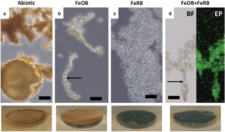
Figure 5.

Light microscope images of biofilms/corrosion products after 14 d exposures in amended ASW with and without additions of FeOB Mariprofundus sp. DIS-1 and FeRB S. frigidimarina. Associated macroscopic images of surfaces of CS (1.59 cm diameter) in situ after 14 d are shown at the bottom. Abundant orange iron oxides were present in the abiotic controls (a), but cells were not observed in this treatment. Cells were observed in association with stalk structures of iron oxide (indicated by arrows) in treatments containing FeOB (b and d). Abiotic treatments (a) and those containing only FeRB (c) did not contain stalk structures of iron oxide. Note abundant cels of planktonic FeRB in images (c) and (d). The image (d) shows the same sample examined using brightfield (BF) and epifluorescence (EP) microscopy stained with the nucleic acid stain Syto13. Scale bars represent 10 μm.
