Abstract
Objectives:
To evaluate the shielding effect of thyroid collar for digital panoramic radiography.
Methods:
4 machines [Orthopantomograph® OP200 (Instrumentarium Dental, Tuusula, Finland), Orthophos CD (Sirona Dental Systems GmbH, Bensheim, Germany), Orthophos XG Plus (Sirona Dental Systems GmbH) and ProMax® (Planmeca Oy, Helsinki, Finland)] were used in this study. Average tissue-absorbed doses were measured using thermoluminescent dosemeter chips in an anthropomorphic phantom. Effective organ and total effective doses were derived according to the International Commission of Radiological Protection 2007 recommendations. The shielding effect of one collar in front and two collars both in front and at the back of the neck was measured.
Results:
The effective organ doses of the thyroid gland obtained from the 4 panoramic machines were 1.12 μSv for OP200, 2.71 μSv for Orthophos CD, 2.18 μSv for Orthophos XG plus and 2.20 μSv for ProMax, when no thyroid collar was used. When 1 collar was used in front of the neck, the effective organ doses of the thyroid gland were 1.01 μSv (9.8% reduction), 2.45 μSv (9.6% reduction), 1.76 μSv (19.3% reduction) and 1.70 μSv (22.7% reduction), respectively. Significant differences in dose reduction were found for Orthophos XG Plus and ProMax. When two collars were used, the effective organ doses of the thyroid gland were also significantly reduced for the two machines Orthophos XG Plus and ProMax. The same trend was observed in the total effective doses for the four machines.
Conclusions:
Wearing a thyroid collar was helpful when the direct digital panoramic imaging systems were in use, whereas for the indirect digital panoramic imaging systems, the thyroid collar did not have an extra protective effect on the thyroid gland and whole body.
Keywords: panoramic radiography, digital panoramic radiography, radiation dosimetry, effective dose, thyroid gland
Introduction
Although radiation dose has been reduced to a certain degree by using digital techniques, it is still a main concern in daily dental care, especially with the potential risk of cancer from diagnostic X-ray, which was revealed in recent studies.1,2 Digital panoramic radiography has been widely used for the past 30 years. To take a panoramic radiograph, the tube head of a panoramic machine rotates one cycle around the head of a patient. During this procedure, the front and back areas of the upper and lower jaws and the upper part of the neck are irradiated. In the head and neck regions, the thyroid gland is the organ where the adverse effects from radiation exposure are likely to occur owing to its location and the larger dose it may receive during a dental radiation exposure. It is well known that the thyroid collar is an effective shielding device for the protection of the thyroid gland from exposures to intraoral radiographs.3,4 However, with respect to panoramic radiography, the results from previous studies on the shielding effects of a thyroid collar are controversial. Block et al5 found that a thyroid collar could not reduce thyroid dose, but Sikorski and Taylor6 reported that the thyroid collar was protective during exposures to panoramic radiography. Since these two studies, no similar study has been performed, and panoramic radiography has moved from analogue to digital. The aim of the present study was to measure and evaluate the shielding effect of a thyroid collar during digital panoramic examination.
Materials and methods
Panoramic machines used
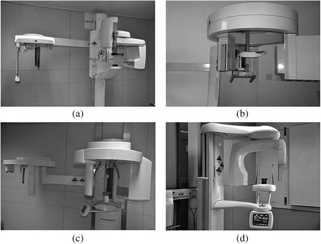
Four panoramic machines, Orthopantomograph® OP200 (Instrumentarium Dental, Tuusula, Finland), Orthophos CD (Sirona Dental Systems GmbH, Bensheim, Germany), Orthophos XG Plus (Sirona Dental Systems GmbH) and ProMax® (Planmeca Oy, Helsinki, Finland), were used in this study. OP200 and Orthophos CD are indirect digital image capturing systems that use phosphor storage plate (PSP) as image receptors. By using these machines, digital images can be obtained only after an exposed receptor is scanned by a specially designed scanner. Orthophos XG Plus and ProMax, however, use charge-coupled devices (CCDs) as image detectors, which can be used to get a digital image immediately after an exposure is made. The photographs of the four machines are shown in Figure 1. The exposure time, tube voltage and tube current used for panoramic examination are shown in Table 1.
Figure 1.
Images of the four panoramic machines (a) Orthopantomograph® OP200 (Instrumentarium Dental, Tuusula, Finland), (b) Orthophos CD (Sirona Dental Systems GmbH, Bensheim, Germany), (c) Orthophos XG Plus (Sirona Dental Systems GmbH) and (d) ProMax® (Planmeca Oy, Helsinki, Finland)
Table 1.
Exposure parameters of the four panoramic machines
| Machine | Image receptor | Exposure time (s) | Tube voltage (kVp) | Tube current (mA) |
| Orthopantomograph® OP200 | PSP | 17.6 | 66 | 10 |
| Orthophos CD | PSP | 13.9 | 71 | 15 |
| Orthophos XG plus | CCD | 14.1 | 69 | 15 |
| ProMax® | CCD | 16.0 | 66 | 12 |
CCD, charge-coupled device; PSP, phosphor storage plate.
Orthopantomograph OP200 is manufactured by Instrumentarium Dental, Tuusula, Finland; Orthophos CD by Sirona Dental Systems GmbH, Bensheim, Germany; Orthophos XG plus by Sirona Dental Systems GmbH; and ProMax by Planmeca Oy, Helsinki, Finland.
The phantom
An adult human male anthropomorphic phantom (ART-210; Radiology Support Device, Inc, Long Beach, CA) was used in this study. The phantom had tissue-equivalent X-ray attenuating characteristics and closely conformed to the specifications of the International Commission on Radiation Units and Measurements.
Thyroid collar shielding technique
The panoramic radiograph was performed after having a 0.35 mm Pb thyroid collar (model HRNG-I; Beijing Huaren Health Science & Technology Developing Co, Ltd, Beijing, China) in front of the neck of the phantom. To obtain maximum shielding effect, exposures with two collars placed both in front and at the back of the neck were also carried out. Thus, for each panoramic machine, three exposures were completed as follows:
without collar around the neck
with one collar around tightly in front of the neck
with two collars around tightly both in front and at the back of the neck.
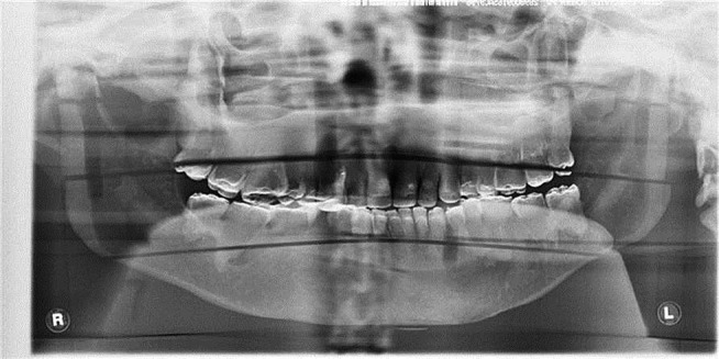
The placement of thyroid collars around the neck of the phantom is shown in Figure 2. A sample radiograph of the phantom with one thyroid collar around the neck is shown in Figure 3.
Figure 2.
Photo of the thyroid collar and the placement of thyroid collars around the neck of phantom
Figure 3.

A sample image of the phantom
The measurement of absorbed dose
Thermoluminescent dosemeter (TLD) chips (LiF:Mg, Cu, P) were used to measure the absorbed doses. Before the study, all dosemeters were calibrated using a 60Co source. Three chips were positioned at each of the 21 locations within the head and neck regions of the phantom. The method presented by Qu et al7 was used to position the TLD chips (Table 2). Before loading, the TLDs were annealed at 240 °C for 10 min and then cooled immediately to ambient temperature. All TLDs were read within 90 min after each exposure using a BR2000D reader (Beijing Bochuangte Science & Technology Development Co, Ltd, Beijing, China). The consistency of the dose measurement by the TLD system was proved by Qu et al.8
Table 2.
Locations of TLD dosemeter chips
| Phantom level | Phantom location | TLD ID |
| 2 | Calvarium anterior | 1 |
| 2 | Calvarium right | 2 |
| 2 | Calvarium posterior | 3 |
| 2 | Mid-brain | 4 |
| 3 | Pituitary | 5 |
| 4 | Right orbit | 6 |
| 4 | Left orbit | 7 |
| 3 | Right lens of the eye | 8 |
| 3 | Left lens of the eye | 9 |
| 5 | Left cheek | 10 |
| 6 | Right parotid | 11 |
| 6 | Left parotid | 12 |
| 6 | Right ramus | 13 |
| 6 | Center cervical spine | 14 |
| 7 | Left back of the neck | 15 |
| 7 | Right mandible body | 16 |
| 7 | Left mandible body | 17 |
| 7 | Right submandibular gland | 18 |
| 7 | Left submandibular gland | 19 |
| 9 | Thyroid | 20 |
| 9 | Oesophagus | 21 |
TLD, thermoluminescent dosemeter.
During each scanning, six non-irradiated TLDs were kept outside the scanning room to measure the background radiation dose, which was subtracted from the measured dose values later. To ensure that even small radiation doses could be measured, the phantom was exposed five times during each examination protocol without changing the position of the phantom. It was assumed that the radiation dose delivered for each exposure was the same when the panoramic machine was well maintained. Measured values from TLDs at different positions within a tissue or organ were divided by five to express the average tissue-absorbed dose per examination in microgray (μGy).
Effective dose calculation
The average absorbed dose and the percentage of a tissue or organ irradiated during an examination (Table 3) were used to calculate the radiation-weighted dose (HT) in microsievert. Using the tissue weighting factors (WT, Table 4) recommended by the International Commission on Radiological Protection (ICRP) in 2007, the effective organ dose (microsievert) could be calculated as the product of the equivalent dose and the relevant ICRP tissue weighting factors. The total effective dose (E) was the sum of all the effective organ doses (i.e. E = ∑WT × HT). The effective dose can give a broad indication of the level of detriment to health from radiation exposure.
Table 3.
Estimated percentage of tissue irradiated and TLDs used to calculate mean absorbed dose to a tissue or organ
| Tissue or organ | Fraction irradiated (%) | TLD ID |
| Bone marrow | 16.5 | |
| Mandible | 1.3 | 13, 16, 17 |
| Calvaria | 11.8 | 1, 2, 3 |
| Cervical spine | 3.4 | 14 |
| Thyroid | 100.0 | 20 |
| Oesophagus | 10.0 | 21 |
| Skin | 5.0 | 8, 9, 10, 15 |
| Bone surfacea | 16.5 | |
| Mandible | 1.3 | 13, 16, 17 |
| Calvaria | 11.8 | 1, 2, 3 |
| Cervical spine | 3.4 | 14 |
| Salivary glands | 100.0 | |
| Parotid | 100.0 | 11, 12 |
| Submandibular | 100.0 | 18, 19 |
| Brain | 100.0 | 4, 5 |
| Remainder | ||
| Lymphatic nodes | 5.0 | 11–14, 16–19, 21 |
| Muscle | 5.0 | 11–14, 16–19, 21 |
| Extrathoracic airway | 100.0 | 6, 7, 11–14, 16–19, 21 |
| Oral mucosa | 100.0 | 11–13, 16–19 |
MEACR, mass energy absorption coefficient ratio; TLD, thermoluminescent dosemeter.
MEACR = −0.0618 × 2/3 kV peak + 6.9406 using data taken from National Bureau of Standards handbook no. 85.9
Bone surface dose = bone marrow dose × bone/muscle MEACR.
Table 4.
Tissue weighting factors for the calculation of effective radiation dose, according to the International Commission of Radiological Protection (ICRP) 2007 recommendations
| Tissue | WT | ∑WT |
| Bone marrow, colon, lung, stomach, breast and remainder tissuesa | 0.12 | 0.72 |
| Gonads | 0.08 | 0.08 |
| Bladder, oesophagus, liver, thyroid | 0.04 | 0.16 |
| Bone surface, brain, salivary glands, skin | 0.01 | 0.04 |
| Total | 1.00 |
ICRP 2007 commendations.11
Remainder tissue: adrenals, extrathoracic region, heart, gall bladder, kidneys, lymphatic nodes, muscle, oral mucosa, pancreas, prostrate, small intestine, spleen, thymus and uterus/cervix.
Statistical analysis
One-way analysis of variance was used to assess the effective organ doses and the total effective doses resulting from each protocol. A difference of p < 0.05 was considered significant.
Results
Table 5 shows the equivalent doses to tissue/organs of the four machines. Tables 6 and 7 show the effective doses. For the panoramic machine OP200, the effective organ dose of thyroid was 1.12 μSv when the thyroid collar was not used, and the total effective dose was 10.73 μSv. When one collar was used, the effective organ dose of the thyroid and the total effective dose were 1.01 μSv and 10.26 μSv, respectively. No significant differences were found for the effective doses measured with and without the use of one thyroid collar (p = 0.07 for the thyroid dose and p = 0.423 for the total effective dose). When two thyroid collars were used, the effective doses were not further reduced (p = 0.29 for the thyroid dose, p = 0.482 for the total effective dose). Similar results were obtained for the panoramic machine Orthophos CD.
Table 5.
Mean equivalent dose to tissue or organs and total equivalent dose during panoramic exposure of the four machines (μSv)
| Machine | Bone marrow | Thyroid | Oesophagus | Skin | Bone surface | Salivary glands | Brain | Remainder tissues or organs | Total dose | |||
| Lymphatic nodes | Extrathoracic region | Muscles | Oral mucosa | |||||||||
| OP200 | ||||||||||||
| a | 9.94 | 27.89 | 1.97 | 4.16 | 40.34 | 311.78 | 10.03 | 12.16 | 200.46 | 12.16 | 282.51 | 913.3924 |
| b | 9.05 | 25.20 | 1.83 | 4.33 | 36.71 | 308.48 | 11.29 | 11.60 | 191.37 | 11.60 | 270.99 | 882.4716 |
| c | 8.83 | 26.56 | 1.87 | 4.15 | 35.80 | 297.89 | 10.06 | 11.36 | 187.40 | 11.36 | 265.69 | 860.9635 |
| Orthophos CD | ||||||||||||
| a | 10.74 | 67.87 | 4.25 | 10.14 | 43.57 | 419.17 | 9.41 | 13.93 | 230.78 | 13.93 | 318.72 | 1142.5140 |
| b | 11.43 | 58.87 | 4.29 | 6.77 | 46.35 | 449.58 | 12.89 | 14.81 | 245.43 | 14.81 | 338.83 | 1204.0510 |
| c | 11.95 | 55.53 | 3.47 | 7.44 | 48.47 | 442.33 | 15.00 | 14.95 | 247.81 | 14.95 | 342.74 | 1204.6570 |
| Orthophos XG plus | ||||||||||||
| a | 15.99 | 54.60 | 3.89 | 8.16 | 64.85 | 604.05 | 18.41 | 20.45 | 337.49 | 20.45 | 471.52 | 1619.8630 |
| b | 10.78 | 43.95 | 2.81 | 6.36 | 43.74 | 384.17 | 12.68 | 13.42 | 222.39 | 13.42 | 309.34 | 1063.0580 |
| c | 10.94 | 41.56 | 2.91 | 6.56 | 44.37 | 425.72 | 11.23 | 14.44 | 238.62 | 14.44 | 334.59 | 1145.3610 |
| ProMax® | ||||||||||||
| a | 14.53 | 54.95 | 4.05 | 5.10 | 58.95 | 939.55 | 15.12 | 30.25 | 496.98 | 30.25 | 739.14 | 2388.8740 |
| b | 13.95 | 42.60 | 3.11 | 4.93 | 56.59 | 796.95 | 16.21 | 26.49 | 435.40 | 26.49 | 644.62 | 2067.3220 |
| c | 13.59 | 45.45 | 3.37 | 4.80 | 55.11 | 839.01 | 13.84 | 27.27 | 448.01 | 27.27 | 665.21 | 2142.9170 |
a, Without collar around the neck; b, with one collar tightly around the front of the neck; c, with two collars tightly around both the front and the back of the neck.
Orthopantomograph OP200 is manufactured by Instrumentarium Dental, Tuusula, Finland; Orthophos CD by Sirona Dental Systems GmbH, Bensheim, Germany; Orthophos XG plus by Sirona Dental Systems GmbH; and ProMax by Planmeca Oy, Helsinki, Finland.
Table 6.
Effective doses of thyroid with and without application of thyroid collar (μSv)
| Machine | No collar | One collar | Two collars |
| OP200 | 1.12 | 1.01 | 1.06 |
| Orthophos CD | 2.71 | 2.45 | 2.17 |
| Orthophos XG plus | 2.18 | 1.76a | 1.66a |
| ProMax® | 2.20 | 1.70a | 1.82a |
Significant differences between doses obtained with and without the use of the thyroid collar(s).
Orthopantomograph OP200 is manufactured by Instrumentarium Dental, Tuusula, Finland; Orthophos CD by Sirona Dental Systems GmbH, Bensheim, Germany; Orthophos XG plus by Sirona Dental Systems GmbH; and ProMax by Planmeca Oy, Helsinki, Finland.
Table 7.
Total effective doses with and without application of thyroid collar (μSv)
| Machine | No collar | One collar | Two collars |
| OP200 | 10.73 | 10.26 | 9.89 |
| Orthophos CD | 14.33 | 14.93 | 14.52 |
| Orthophos XG plus | 19.06 | 12.79a | 13.53a |
| ProMax® | 26.26 | 22.71a | 23.49a |
Significant differences between doses obtained with and without the use of the thyroid collar(s).
Orthopantomograph OP200 is manufactured by Instrumentarium Dental, Tuusula, Finland; Orthophos CD by Sirona Dental Systems GmbH, Bensheim, Germany; Orthophos XG plus by Sirona Dental Systems GmbH; and ProMax by Planmeca Oy, Helsinki, Finland.
With respect to the panoramic machine Orthophos XG Plus, when the thyroid collars were not used, the thyroid and the total effective doses were 2.18 μSv and 19.06 μSv, respectively. When one or two thyroid collars were in use, the effective organ dose of the thyroid was reduced to 1.76 μSv (19.3% reduction) or 1.66 μSv (23.9% reduction), and the total effective dose was reduced to 12.79 μSv (33% reduction) or 13.53 μSv (29% reduction), respectively. The dose reductions were significant. When the dose reduction effect of one or two thyroid collars was analysed, no significant differences were found for both the total effective dose (p = 0.128) and the effective organ dose of the thyroid (p = 0.354).
The dose reduction was similar when the thyroid collars were used for the panoramic machine ProMax. The effective organ dose of thyroid and the total effective dose were reduced significantly when one thyroid collar (p = 0.004 for effective organ dose of thyroid, p = 0.002 for total effective dose) or two thyroid collars (p = 0.015 for effective organ dose of thyroid, p = 0.008 for total effective dose) were used. No further dose reduction was observed when the use of one and two thyroid collars was compared (p = 0.399 for effective organ dose of thyroid, p = 0.361 for the total effective dose).
Discussion
For the past 30 years, digital panoramic radiography has been used worldwide. However, the shielding effect of the thyroid collar during digital panoramic examination has not been reported. The present study revealed that the shielding effect of the thyroid collars differs with the use of different digital panoramic modalities.
When the panoramic machines OP200 and Orthosphos CD were used, the shielding effect of the thyroid collar was not significant, irrespective of whether one or two thyroid collars were used. This may be explained by the use of a vertical beam collimation set-up by the manufacturer. By using a collimation, the thyroid may be effectively protected from a primary beam.10
When the direct digital panoramic machines Orthophos XG Plus and ProMax were used, a significant shielding effect of the thyroid collar was observed. When one collar was used for taking radiographs with Orthophos XG Plus, 19% of the thyroid dose and 33% of the total effective dose could be reduced. Putting on two thyroid collars, both in front and at the back of the neck, however, could not further reduce the total effective dose but slightly reduced the thyroid dose. The use of a thyroid collar, irrespective of whether one or two, was also helpful in reducing the dose of other organs. A similar dose reduction trend was observed with the use of the panoramic machine ProMax.
The radiation dose of panoramic radiography has been a concern previously. Some studies were carried out to assess the risk of diagnostic radiation10,12–14. However, the methods and the tissue weighting factors used in these studies are not the same and could not be directly compared. ICRP periodically reassesses the risk of ionizing radiation by looking at new data from exposures of the human population. For calculating the effective dose, tissue weighting factors used in the ICRP 1990 formula were based largely on cancer mortality data. The 2007 tissue weighting factors incorporate additional incidence and mortality data that have been available.11 Salivary glands and brain were judged to be sufficient to warrant weighting as individually named tissues. Three new tissues (from the extrathoracic region, lymphatic nodes and oral mucosa) have been added to the remainder tissues. Therefore, the ICRP 2007 publications should be used to calculate the effective dose to estimate the exposure risk in the maxillofacial region because it shows additional evidence on the risk of cancer in soft tissues. Only Ludlow et al12 used the tissue weighting factors from ICRP 2007 recommendation to calculate the effective dose of panoramic radiography. In that study, the measured effective dose was 14.2 μSv for Orthophos XG and 24.3 μSv for ProMax, respectively. These data are similar to the data obtained from the present study and validate both studies. In the studies performed by Danforth and Clark,10 Gijbels et al13 and Gavala et al,14 the tissue weighting factors from ICRP 1990 recommendations were used to calculate the effective dose. These studies showed that the effective radiation dose was between 3.85 μSv and 38 μSv for panoramic examinations.
In the present study, the digital panoramic machines OP200, Orthophos CD, Orthophos XG Plus and ProMax were used. The OP200 and Orthophos CD are a type of panoramic machine that uses a phosphor plate as an image detector, whereas Orthophos XG Plus and ProMax have CCD detectors to capture images. Since the advent of immediate display of captured images, most machines today have CCD detectors. The OP200 and Orthophos CD were the only machines available with PSP detectors when the study was performed. Both the total effective dose and the effective organ dose were reduced significantly for Orthophos XG Plus and ProMax, when a thyroid collar was used, in contrast to the other two machines with PSP detectors. From the results, it can be inferred that the panoramic machine using PSP detector is superior to the one with a CCD detector in terms of radiation protection.
Although the present study shows that a thyroid collar should be used when direct digital panoramic imaging systems are used, the thyroid collar is not widely adopted in clinics. The main reason is two-fold: one owing to the fact that the image of the mandible is often disturbed by the thyroid collar and the other because many oral radiologists believe that the radiation to the thyroid is already shielded by the use of a collimator installed in machines. The present study, however, shows that this is only partly true. For the CCD-based direct digital panoramic imaging systems, the use of a thyroid collar is still recommended. The results also suggest that it may be possible for manufacturers to modify the geometry or collimation of CCD-based direct digital panoramic imaging systems such that the radiation dose could be reduced to a maximum degree.
In conclusion, wearing thyroid collar is helpful when the direct digital panoramic imaging systems, such as Orthophos XG Plus and ProMax, are used, whereas for the indirect digital panoramic imaging systems, e.g. OP200 and Orthophos CD, thyroid collar does not add an extra protective effect on the thyroid gland and the whole body.
Acknowledgments
The authors would like to express their sincere thanks to Dr Hao Wang for allowing them to use the panoramic machine Orthophos CD for the study.
References
- 1.Brenner DJ, Doll R, Goodhead DT, Hall EJ, Land CE, Little JB, et al. Cancer risks attributable to low doses of ionizing radiation: assessing what we really know. Proc Natl Acad Sci USA 2003; 100: 13761–13766 10.1073/pnas.2235592100 [DOI] [PMC free article] [PubMed] [Google Scholar]
- 2.Sadetzki S, Mandelzweig L. Childhood exposure to external ionising radiation and solid cancer risk. Br J Cancer 2009; 100: 1021–1025 10.1038/sj.bjc.6604994 [DOI] [PMC free article] [PubMed] [Google Scholar]
- 3.Rush ER, Thompson NA. Dental radiography technique and equipment: how they influence the radiation dose received at the level of the thyroid gland. Radiography 2007; 13: 214–220 [Google Scholar]
- 4.Marshall NW, Faulkner K, Clarke P. An investigation into the effect of protective devices on the dose to radiosensitive organs in the head and neck. Br J Radiol 1992; 65: 799–802 [DOI] [PubMed] [Google Scholar]
- 5.Block AJ, Goepp RA, Mason EW. Thyroid radiation dose during panoramic and cephalometric dental X-ray examinations. Angle Orthod 1977; 47: 17–24 [DOI] [PubMed] [Google Scholar]
- 6.Sikorski PA, Taylor KW. The effectiveness of the thyroid shield in dental radiology. Oral Surg Oral Med Oral Pathol 1984; 58: 225–236 [DOI] [PubMed] [Google Scholar]
- 7.Qu X, Li G, Zhang Z, Ma X. Thyroid shields for radiation dose reduction during cone beam computed tomography scanning for different oral and maxillofacial regions. Eur J Radiol 2012; 81: 376–380 10.1016/j.ejrad.2011.11.048 [DOI] [PubMed] [Google Scholar]
- 8.Qu XM, Li G, Ludlow JB, Zhang ZY, Ma XC. Effective radiation dose of ProMax 3D cone-beam computerized tomography scanner with different dental protocols. Oral Surg Oral Med Oral Pathol Oral Radiol Endod 2010; 110: 770–776 10.1016/j.tripleo.2010.06.013 [DOI] [PubMed] [Google Scholar]
- 9.Ludlow JB, Ivanovic M. Comparative dosimetry of dental CBCT devices and 64-slice CT for oral and maxillofacial radiology. Oral Surg Oral Med Oral Pathol Oral Radiol Endod 2008; 106: 106–114 10.1016/j.tripleo.2008.03.018 [DOI] [PubMed] [Google Scholar]
- 10.Danforth RA, Clark DE. Effective dose from radiation absorbed during a panoramic examination with a new generation machine. Oral Surg Oral Med Oral Path Oral Radiol Endod 2000; 89: 236–243 10.1067/moe.2000.103526 [DOI] [PubMed] [Google Scholar]
- 11.Valentin J. The 2007 recommendations of the International Commission on Radiological Protection. ICRP publication 103. Ann ICRP 2007; 37: 1–332 10.1016/j.icrp.2007.10.003 [DOI] [PubMed] [Google Scholar]
- 12.Ludlow JB, Davies-Ludlow LE, White SC. Patient risk related to common dental radiographic examinations: the impact of 2007 International Commission on Radiological Protection recommendations regarding dose calculation. J Am Dent Assoc 2008; 139: 1237–1243 [DOI] [PubMed] [Google Scholar]
- 13.Gijbels F, Jacobs R, Bogaerts R, Debaveye D, Verlinden S, Sanderink G. Dosimetry of digital panoramic imaging. Part I: patient exposure. Dentomaxillofac Radiol 2005; 34: 145–149 [DOI] [PubMed] [Google Scholar]
- 14.Gavala S, Donta C, Tsiklakis K, Boziari A, Kamenopoulou V, Stamatakis HC. Radiation dose reduction in direct digital panoramic radiography. Eur J Radiol 2009; 71: 42–48 10.1016/j.ejrad.2008.03.018 [DOI] [PubMed] [Google Scholar]


