

Abstract
Myxococcus xanthus is a Gram-negative bacterium that has a complex life cycle including a temporal sequence of cellular aggregation, mound formation, and myxosporulation. During development, protein S (molecuar weight 23,000) is induced and accumulates in very large amounts. Protein S was found in the soluble fraction of early developmental extracts and in the insoluble fraction in later extracts. This insoluble form of protein S can be solubilized by the addition of 1 M NaCl at 0°C to extracts from aggregated cells (mound stage) or by the addition of 1 M NaCl at 30°C to mature spores. Salt extraction (1 M NaCl) of protein S from mature spores was partially inhibited by the addition of Mg2+ and almost completely inhibited by the addition of Ca2+. The viability of spores was not changed by a salt extraction that removed their protein S. Examination of thin sections of mature spores and extracted spores by electron microscopy suggested that the protein S-deficient spores lacked a spore surface coat about 300 A thick. Purified protein S will spontaneously self-assemble onto protein S-deficient spores after removal of the NaCl by dialysis or by addition of 10 mM Ca2+ to undialyzed samples. Glycerol-induced spores did not contain protein S and did not serve as primers for assembly of protein S. Quantitation of the self-assembly process showed almost stoichiometric binding of protein S to the protein S-deficient spores until saturation at 3.3 × 10⁶ molecules per spore, a value 1.35 times higher than the normal level of proteins S found in mature spores. Protein S in the “reconstituted” spores was as protease resistant and sonication resistant as the protein S of native spores. Electron microscopy of the reconstituted spores revealed the assembly of new material on the spore surface. Adjacent spores were sometimes observed to be fused to each other through a common protein S layer. These results suggest that protein S serves a function in spore—spore interaction in the fruiting body.
Keywords: spores, fruiting body, sodium dodecyl sulfate gel electrophoresis, calcium ion
Full text
PDF

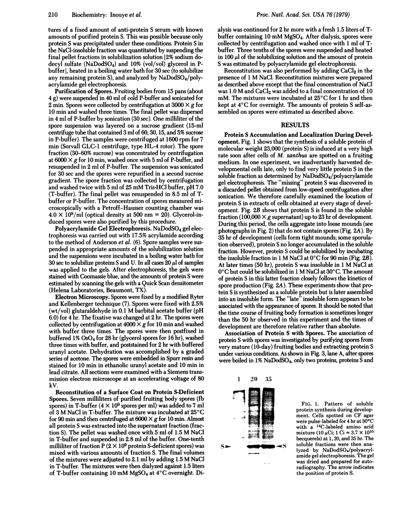


Images in this article


Selected References
These references are in PubMed. This may not be the complete list of references from this article.
- Anderson C. W., Baum P. R., Gesteland R. F. Processing of adenovirus 2-induced proteins. J Virol. 1973 Aug;12(2):241–252. doi: 10.1128/jvi.12.2.241-252.1973. [DOI] [PMC free article] [PubMed] [Google Scholar]
- Aronson A. I., Fitz-James P. Structure and morphogenesis of the bacterial spore coat. Bacteriol Rev. 1976 Jun;40(2):360–402. doi: 10.1128/br.40.2.360-402.1976. [DOI] [PMC free article] [PubMed] [Google Scholar]
- Bacon K., Eiserling F. A. A unique structure in microcysts of Myxococcus xanthus. J Ultrastruct Res. 1967 Dec;21(5):378–382. doi: 10.1016/s0022-5320(67)80147-3. [DOI] [PubMed] [Google Scholar]
- Campos J. M., Geisselsoder J., Zusman D. R. Isolation of bacteriophage MX4, a generalized transducing phage for Myxococcus xanthus. J Mol Biol. 1978 Feb 25;119(2):167–178. doi: 10.1016/0022-2836(78)90431-x. [DOI] [PubMed] [Google Scholar]
- DWORKIN M., GIBSON S. M. A SYSTEM FOR STUDYING MICROBIAL MORPHOGENESIS: RAPID FORMATION OF MICROCYSTS IN MYXOCOCCUS XANTHUS. Science. 1964 Oct 9;146(3641):243–244. doi: 10.1126/science.146.3641.243. [DOI] [PubMed] [Google Scholar]
- DiRienzo J. M., Nakamura K., Inouye M. The outer membrane proteins of Gram-negative bacteria: biosynthesis, assembly, and functions. Annu Rev Biochem. 1978;47:481–532. doi: 10.1146/annurev.bi.47.070178.002405. [DOI] [PubMed] [Google Scholar]
- Hagen D. C., Bretscher A. P., Kaiser D. Synergism between morphogenetic mutants of Myxococcus xanthus. Dev Biol. 1978 Jun;64(2):284–296. doi: 10.1016/0012-1606(78)90079-9. [DOI] [PubMed] [Google Scholar]
- Inouye S., Wang S., Sekizawa J., Halegoua S., Inouye M. Amino acid sequence for the peptide extension on the prolipoprotein of the Escherichia coli outer membrane. Proc Natl Acad Sci U S A. 1977 Mar;74(3):1004–1008. doi: 10.1073/pnas.74.3.1004. [DOI] [PMC free article] [PubMed] [Google Scholar]
- LOWRY O. H., ROSEBROUGH N. J., FARR A. L., RANDALL R. J. Protein measurement with the Folin phenol reagent. J Biol Chem. 1951 Nov;193(1):265–275. [PubMed] [Google Scholar]
- RYTER A., KELLENBERGER E., BIRCHANDERSEN A., MAALOE O. Etude au microscope électronique de plasmas contenant de l'acide désoxyribonucliéique. I. Les nucléoides des bactéries en croissance active. Z Naturforsch B. 1958 Sep;13B(9):597–605. [PubMed] [Google Scholar]
- Sudo S. Z., Dworkin M. Comparative biology of prokaryotic resting cells. Adv Microb Physiol. 1973;9:153–224. doi: 10.1016/s0065-2911(08)60378-1. [DOI] [PubMed] [Google Scholar]