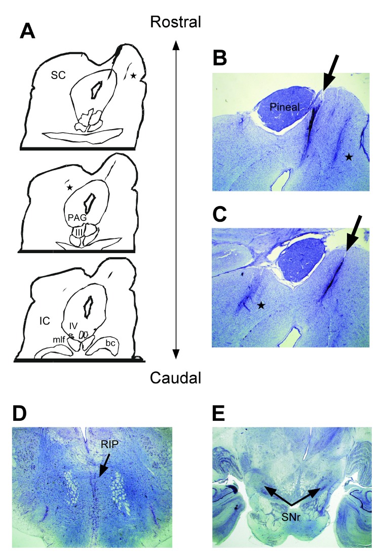
Figure 2. Anatomy of the rostral superior colliculus.
Panel A shows tracings of the superior colliculus (SC) and other structures in the brainstem relative to the lesion we found bisecting the commissure and the right rostral medial SC. Panels B & C show photomicrographs taken of the lesion as indicated by arrows. Two other electrode tracts are indicated by asterisks. Panels D & E show that the RIP and the SNr, respectively, of this monkey remained intact and undisturbed. Abbrevations: SC) superior colliculus, IC) inferior colliculus, PAG) periaqueductal gray, III) oculomotor nerve, IV) trochlear nerve, bc) brachium conjunctivum, RIP) raphe interpositus, SNr) substantia nigra reticulata.

