Abstract
An elderly woman presented with abdominal pain and vomiting, was known to have gallstones. A CT scan was arranged identifying gallstone ileus and cholecystitis. Ensuing sepsis precipitated fast atrial fibrillation delaying the planned laparotomy. Her symptoms subsequently resolved with conservative management. Ten days following admission her abdomen became distended. A repeat CT scan showed large bowel dilation with intramural air suggestive of obstruction and bowel ischaemia. Emergency laparotomy was performed identifying a large 23 mm gallstone impacted at the rectosigmoid junction (gallstone coleus). The stone was milked back to the transverse colon where it was retrieved and a transverse loop colostomy was formed. Gallstone ileus is rare; gallstone coleus is even rarer. On review of the published literature both entities have not been seen in the same patient during the same admission or indeed caused by the same gallstone.
Background
Gallstone ileus is rare, current consensus on management is with laparotomy and enterolithotomy. Controversy remains on whether to perform any biliary surgery at the same time (ie, closing the cholecysto-enteric fistula or cholecystectomy).1 This case is of particular interest as what started out as a rare gallstone ileus spontaneously resolved and developed 10 days later into a gallstone coleus. The obstruction was so pronounced that massive caecal dilation led to intramural air requiring decompression through a transverse loop colostomy. To the authors knowledge a case with all of these features has not been published previously. Knowledge of this entity is essential to prevent misdiagnosis and the subsequent morbidity and mortality associated with its sequlae.
Case presentation
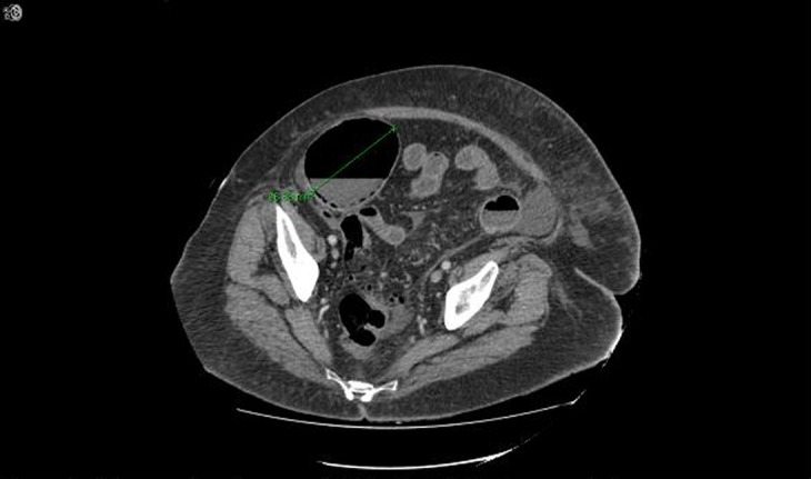
An 80-year-old Caucasian woman presented with severe abdominal pain and vomiting for 5 days. Her medical history included atrial fibrillation and asymptomatic gallstones. Her admission blood tests revealed raised inflammatory markers and a mildly raised bilirubin. Her abdomen was distended and tenderness was elicited in the epigastric region. A CT scan was arranged for a probable diagnosis of cholecystitis. The CT scan identified cholecystitis and a gallstone ileus. Intravenous antibiotics were started and a plan was made for a laparotomy to treat the gallstone ileus. Prior to theatre she developed fast atrial fibrillation due to sepsis, medical advice was sought and digoxin was started. While waiting for haemodynamic stability to return the abdominal pain settled, her inflammatory markers normalised and bowel movements started. It was decided to observe for resolution. Four days after the initial CT scan (5 days after initial admission), a repeat CT was performed confirming resolution of the small bowel obstruction with continued cholecystitis. Over the proceeding 4 days her abdomen became distended again, an abdominal X-ray was performed which identified large bowel obstruction. A third CT scan was arranged showing grossly distended large bowel with intramural air in the caecum suggestive of large bowel ischaemia (figure 1).
Figure 1.

CT scan showing caecal dilation with intramural air.
Investigations
Admission blood tests: C reactive protein 382 mg/L, white cell count 11.0×10⁹/L, bilirubin 26 μmol/L (rest of the liver function tests normal).
CT scan day 1 of admission: contracted thick-walled gallbladder, filling defects in keeping with gallstones, pneumobilia, duodenum in proximity to gallbladder—possible fistulation, mild small bowel dilation with transition at terminal ileum and a collapsed colon.
CT scan day 5 of admission: resolution of the small bowel obstruction with continued cholecystitis.
CT scan day 10 of admission: grossly distended large bowel up to the mid-descending colon with intramural air in the caecum suggestive of large bowel ischaemia.
Differential diagnosis
Cholecystitis
Gallstone ileus
Ischaemic bowel
Gallstone coleus
Treatment
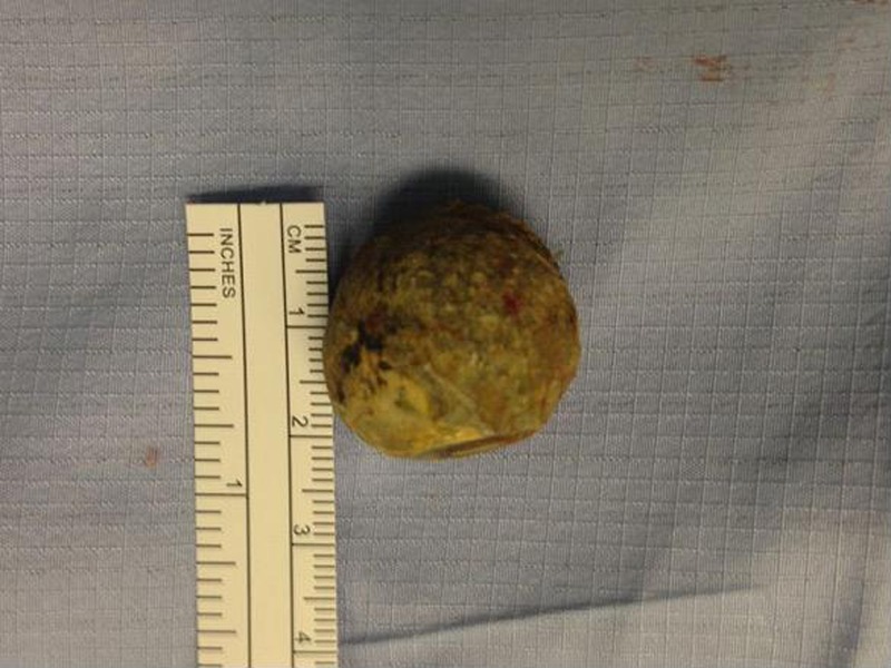
Intravenous antibiotics were started for cholecystitis. An emergency laparotomy found grossly dilated large bowel and a large 23 mm gallstone (figure 2) lodged at the rectosigmoid junction which was narrowed due to diverticular stricturing. The gallstone was milked back to the transverse colon and a mid-transverse loop colostomy was formed to decompress the massively distended caecum—following which the abdomen was closed. Postoperatively the patient was admitted to the intensive care unit for 10 days where she remained intubated for 48 h and required ionotropic support.
Figure 2.

Photograph of the gall stone found during operation.
Outcome and follow-up
Full recovery and discharge home.
Discussion
Gallstone ileus is rare, yet multiple case reports have been published on this subject. It is caused by erosion of a gallstone into the small bowel forming a cholecystoenteric fistula. The gallstone typically lodges at the narrowed ileocaecal junction causing small bowel obstruction. This causes a characteristic triad of signs that can be seen on a plain film; pnuemobilia, an ectopic gallstone and small bowel obstruction (Riglers triad). These signs should be actively sought if the patient has a history of biliary colic and develops obstructive symtoms. It has a high mortality (12–27%) due to delayed or misdiagnosis.2 Recurrent gallstone ileus has been reported in 5% of patients.3 Treatment is with enterolithotomy and either immediate or delayed cholecystectomy. Gallstone coleus is rarer still with only a handful of published cases on this disease.4–8 Cholecystocolonic fistula is usually the cause of this entity. There is no consensus in the literature on how to treat this condition with some performing enterolithotomy,6 some performing colectomy/hartmanns procedure4 7 and one case retrieving the stone through colonoscopy.5 The case reported here highlights the possibility of a single gallstone fistulating into the small bowel causing gallstone ileus overcoming the narrowing at the ileocaecal junction only to impact and obstruct at the narrowed rectosigmoid junction causing a gallstone coleus. This was due to diverticular stricturing. We also presented an alternative operative management with enterotomy and loop colostomy due to massive large bowel dilation. A case such as this has not been presented previously.
Learning points.
Gallstone ileus is rare and but gallstone coleus is rarer still, knowledge of these entities is essential to prevent misdiagnosis and the subsequent morbidity and mortality associated with their sequlae.
Management of gallstone ileus/coleus is with early removal of the impacted stone most frequently through laparotomy and enterotomy.
There is no consensus currently on treatment of gallstone coleus. If the site of impaction is of questionable viability a bowel resection or a defunctioning procedure may be necessary (such as the loop colostomy performed in this case).
Footnotes
Contributors: WB wrote the case report, ME and MJH were involved in proof reading and advised on the content.
Competing interests: None.
Patient consent: Obtained.
Provenance and peer review: Not commissioned; externally peer reviewed.
References
- 1.Williams G, Ravikumar R. The operative management of gallstone ileus. Ann R Coll Surg Engl 2010;2013:279–81 [DOI] [PMC free article] [PubMed] [Google Scholar]
- 2.Rojas-Rojas DJ, Martínez-Ordaz JL, Romero-Hernández T. Biliary ileus: 10-year experience. Case series. Cir Cir 2012;2013:228–32 [PubMed] [Google Scholar]
- 3.Hayes N, Saha S. Recurrent gallstone ileus. Clin Med Res 2012;2013:236–9 [DOI] [PMC free article] [PubMed] [Google Scholar]
- 4.Van Kerschaver O, Van Maele V, Vereecken L, et al. Gallstone impacted in the rectosigmoid junction causing a biliary ileus and a sigmoid perforation. Int Surg 2009;2013:63–6 [PubMed] [Google Scholar]
- 5.Doddi S, Basu NN, Kamal T, et al. Large bowel obstruction due to gallstone: ‘gallstone coleus’. Grand Rounds 2007;2013:36–8 http://www.grandrounds-e-med.com/articles/gr070009/gr070009.pdf (accessed 3 Jul 2013) [Google Scholar]
- 6.Vaughan-Shaw PG, Talwar A. Gallstone ileus and fatal gallstone coleus: the importance of the second stone. BMJ Case Reports 2013;2013: pii: bcr2012008008. [DOI] [PMC free article] [PubMed] [Google Scholar]
- 7.Athwal TS, Howard N, Belfield J, et al. Large bowel obstruction due to impaction of a gallstone. BMJ Case Rep. Published online: 10 Feb 2012. doi:10.1136/bcr.11.2011.5100 [DOI] [PMC free article] [PubMed] [Google Scholar]
- 8.Ranga N. Large bowel and small bowel obstruction due to gallstones in the same patient. BMJ Case Rep 2011; 2013 pii: bcr0920103372 [DOI] [PMC free article] [PubMed] [Google Scholar]


