

Abstract
Steroid hormones act in specific regions of the brain to alter behavior and physiology. While it has been well established that the bioavailability of the steroid and the expression of its receptor is critical to understanding steroid action in brain, the importance of nuclear receptor coactivators in brain is becoming more apparent. This review will focus on the function of the p160 family of coactivators, which includes steroid receptor coactivator-1 (SRC-1), SRC-2 and SRC-3, in steroid receptor action in brain. The expression, regulation and function of these coactivators in steroid-dependent gene expression in brain and behavior will be discussed.
Keywords: steroid receptor coactivator-1 (SRC-1/NcoA-1), SRC-2 (GRIP1/TIF2/NCoA-2), androgen receptor, estrogen receptor, progestin receptor, sex behavior, hypothalamus, steroid hormones
Introduction
Steroid hormones act in the brain to profoundly influence behavior and physiology. These hormones elicit many of their biological effects by binding to their respective receptors, which are members of the steroid/nuclear receptor superfamily of transcriptional activators. Receptors for estrogens (ER), progestins (PR) and androgens (AR) can act in a classic genomic mechanism of action by binding directly to target DNA to alter transcription (1). In addition, these receptors can act in a rapid, non-classical manner that involve receptors located at the membrane that activate intracellular signaling pathways in brain (2-7) In the classic genomic mechanism of action, nuclear receptor coregulators enhance (coactivators) or repress (corepressors) the transcriptional activity of steroid receptors. Over 350 coregulators have been identified to function with the large superfamily of nuclear receptors (8). The knowledge of the function of these coregulators in behavior, physiology and disease is growing rapidly. This review will focus on the role of the p160 steroid receptor coactivator family in the classic genomic mechanism of ER and PR action in brain and the regulation of behavior.
Genomic mechanisms of ER and PR action
ER and PR, as well as other steroid receptors, have a modular domain structure consisting of a highly variable amino-terminal region (N-domain), a conserved central DNA binding domain (DBD) and a carboxy-terminal ligand binding domain (LBD) (1, 9). In general, steroid receptors have two transcriptional activation domains in the amino (AF-1) and carboxyl (AF-2) termini (10). Intracellular ER are expressed as two subtypes, α and β, which are transcribed from different genes (11, 12). These subtypes differ in their abilities to bind different ligands and regulate transcription (13, 14), distribution in brain (15-18), and regulation of behavior (19-23). In addition, there are ER splice variants (24-26) that bind differentially with coactivators (27) and may provide another level of regulation. In most species, PR are expressed in two forms; the full-length PR-B and the N-terminally truncated PR-A, which are encoded by the same gene but are under the regulation of alternate promoters and internal translation start sites (28, 29). Under certain cell and promoter contexts, human PR-B is a stronger transcriptional activator than PR-A (30-32) and PR-A can repress the transcriptional activity of PR-B. These differences are most likely due to an additional AF domain in the N-terminus of PR-B (33) and a transcriptional inhibitory region that has been identified in PR-A (32, 34), respectively. In further support of these differences in PR-A and PR-B, these two PR isoforms appear to have distinct functions in reproductive behavior and physiology (35, 36).
In the classic, ligand-dependent, genomic mechanism of action of ER, PR and other steroid receptors are complexed with several chaperone molecules, including heat shock proteins (hsp), in the absence of hormone. Upon binding hormone, receptors undergo a conformational change that causes dissociation of hsp and allow receptors to dimerize (37). Activated receptors bind directly to specific steroid response elements (SRE) and SRE-like sequences in the promoter regions of target genes (1, 9). Binding of receptors to DNA increases or decreases gene transcription by altering the rate of recruitment of general transcription factors and influencing the recruitment of RNA polymerase II to the initiation site (38, 39). It is generally thought that estrogens and progestins can act in brain via their respective receptors to alter neuronal gene transcription in a fashion similar to that described above, resulting in profound changes in behavior and physiology (4, 40, 41).
Nuclear receptor coregulators
Coregulators consist of coactivators and corepressors that are required for efficient transcriptional regulation by nuclear receptors (8, 42). Corepressors and their complexes associate with nuclear receptors when unliganded or bound to antagonists and serve to repress nuclear receptor transcription by recruiting corepressor complexes to the cis-active elements in the promoter and enhancers of target genes (42). Nuclear receptor coactivators, which are the focus of this review, dramatically enhance the transcriptional activity of ER and PR, as well as other nuclear receptors, by acting as bridging molecules between the receptor and the general transcription machinery and modifying chromatin within the promoter and enhancer regions by histone acetylation, methylation and phosphorylation (42, 43). Under most conditions, steroid receptors interact with coactivators in the presence of an agonist, but not in the absence of ligand or in the presence of an antagonist or a selective receptor modulator (44-47) but c.f. (48-50). In vitro studies indicate that recruitment of nuclear receptor coactivators is rate-limiting in steroid receptor-mediated gene transcription (42, 51). In further support of the importance of nuclear receptor coactivators in steroid-dependent transcription in vitro, squelching, or the repression of the transcriptional activity of one steroid receptor by another, is reversed by the addition of coactivators (44). Thus, a critical component of efficient steroid dependent transcription is the recruitment by receptors of nuclear receptor coactivators to the complex (8, 42). Finally, the significance of both coactivators and corepressors in a variety of diseases, including hormone-dependent cancer and some neurological disorders, is becoming more apparent (8).
The p160 steroid receptor coactivator family
The steroid receptor coactivator (SRC) family of p160 proteins consists of SRC-1 (NcoA-1), SRC-2 (GRIP1/TIF2/NCoA-2) and SRC-3 (AIB1/TRAM-1/ ACTR/RAC3/pCIP). Nuclear receptor coactivators, including the SRC coactivator family, share a general set of characteristics. The SRC family of coactivators physically interacts with steroid receptors, including ER, PR, AR and receptors for glucocorticoids (GR), in a ligand-dependent manner (43). The SRCs physically interact with agonist-bound receptors through centrally-located multiple LXXLL motifs (L, leucine; X, any amino acid) that make up nuclear receptor boxes. The SRCs and other coactivators do not bind DNA and thus distinguish them from traditional transcription factors. The C-terminus of the SRCs contains two activation domains: AD-1 and AD-2. The N-terminus contains a third activation domain (AD-3) and a basic helix loop helix-Per Arnt Sims (bHLH-PAS) motif, which is the most conserved domain within this family of proteins. The activation domains interact with secondary coactivators known as co-coactivators (43). These co-coactivators modify chromatin to facilitate binding of regulatory proteins and general transcription factors.
The p160 steroid receptor coactivator family in reproductive physiology and behavior
Expression and regulation in brain
Sex steroids, including estrogens, progestins and androgens, are required for brain development and reproductive behavior in rodents and birds. Therefore, both rodents and birds have been excellent models for studying coactivator function in brain. In male and female rodents, SRC-1 mRNA and protein are expressed at high levels in the cortex, hippocampus, cerebellum and hypothalamus (52-58, 59: Bian, 2011 #5215). In addition, the SRC-1 isoform, full length SRC-1a, is found in high levels in the rodent hypothalamus, whereas levels of the C-terminally truncated SRC-1e are higher in the nucleus accumbens, thalamus, and amygdala (54). Recently, this SRC-1a:SRC-1e ratio has been shifted in the central nucleus of the amygdala using antisense targeting the SRC-1e isoform, which may prove valuable in studying the functions of these SRC isoforms in brain (60). Expression of SRC-1 in the female rat brain appears to decline as the animal ages, suggesting a loss of steroid sensitivity (61). SRC-2 is highly expressed throughout the hippocampus, amygdala and hypothalamus, including the medial preoptic area (MPOA), ventral medial nucleus (VMN), arcuate nucleus (ARC), bed nucleus of the stria terminalis, supraoptic nucleus and suprachiasmatic nucleus (58, 62-64). While it is not known if a sex difference exists, SRC-3 is expressed predominantly in the hippocampus and very sparsely in the hypothalamus in both male and female rodents (58, 64).
The avian brain provides an excellent model for studying steroid action since singing and non-singing birds respond to steroids. Songbirds have a specific group of interconnected nuclei called the song control system that are required for singing and are sexually dimorphic and steroid-sensitive (65). . In the songbird zebra finch, AR and ER are expressed in the song control nuclei from early post-hatching ages (66-68). Injection of 17β-estradiol in early post-hatching females masculinizes the song system and makes the females capable of singing as adults (69-72). While the organizational effects of estrogens are limited to early development in zebra finches, in other songbirds, such as canaries, manipulation of these hormones during adulthood affects the size of song nuclei and song (73). In quail, a non-singing bird, steroids regulate both appetitive and consummatory (copulatory) male sexual behavior (74, 75). (76, 77)In males, AR and ER are expressed in the medial preoptic nucleus (POM) of quail and are required for both aspects of sexual behavior (74, 78). Members of the p160 family of steroid receptor coactivators, SRC-1 and SRC-2, are expressed in both songbirds and non-singing birds. SRC-1 mRNA is expressed as early as post-hatching day one (P1) in the telencephalon of zebra finches, and in the song control nuclei and hypothalami of adult canaries and zebra finches (79, 80). Interestingly, SRC-1 mRNA and protein show a male-biased expression in the song nucleus HVC of adult canaries and zebra finches, respectively (79, 80). In the quail brain, SRC-1 is expressed in the steroid sensitive areas, including the POM and bed nucleus of stria terminalis (79). Similar to SRC-1, quail POM expresses SRC-2 protein in a level that is similar in males and females (81).
In order for coactivators to function with steroid receptors, they must be expressed in the same cells. Estradiol-priming dramatically increases the expression of PR in a variety of rodent brain regions, including the MPOA, VMN, ARC and the midbrain central gray (82-87). We found that SRC-1 and SRC-2 are expressed in the majority of estradiol-induced PR cells in regions involved in female reproduction, including the VMN, MPOA and ARC in rats and mice (62, 88, 89). Given that virtually all estradiol-induced PR cells in the hypothalamus contain ERα (84, 85), these findings suggest that these coexpressing cells represent functional sites of interaction between steroid receptors and coactivators in brain (62, 88, 89). In further support, SRC-1 was found to be expressed in estrogen-sensitive proopiomelanocortin and steroidogenic factor-1 neurons in the arcuate nucleus and VMH, respectively (90).
It is thought that coactivators are modulators of cellular responsiveness to steroids. In support, SRC-1 knockout mice, while fertile, have decreased responsiveness in progestin target tissues (91) and partial resistance to thyroid hormone (92). It is important to note that in these mice SRC-2 is up-regulated in steroid sensitive tissues, including brain and testes, suggesting that increased expression of SRC-2 compensates for the loss of SRC-1 (91). Therefore, studying the regulation of coactivator expression is essential to understanding hormone action in brain. A number of studies indicate that hormones can regulate coactivator expression in rodent and bird brain. In rodents, SRC-1 is expressed in a sexually dimorphic manner in the pituitary gland, with males having higher mRNA (52) and protein (93) levels than females. In further support, male rodents have higher levels of SRC-1 than females in a number of brain regions, including the dorsomedial hypothalamus, ventromedial hypothalamus (VMH) and paraventricular nucleus (94). Ovariectomy decreases SRC-1 expression in the VMH, while estradiol reverses this effect (95). In the hypothalamus of cycling female rats, SRC-1 levels were lowest during diestrus, and highest at proestrus and estrus, suggesting that ovarian hormones up-regulate SRC-1 (96). In contrast, ovariectomy did not alter SRC-1 levels in the hippocampus, suggesting that ovarian hormones do not regulate SRC-1 expression in this brain region (97). Interestingly, the endocrine disruptor 4-methylbenzylidene camphor (4-MBC), which has estrogenic activity and impairs the thyroid axis, increases SRC-1 mRNA in the VMH and MPOA of female rats (98). Exposure of another endocrine disruptor, 3-benzylidene camphor (3-BC), during early development through adulthood increases SRC-1 mRNA levels in the MPO of both males and females (99). These effects of 4-MBC and 3-BC on SRC-1 could enhance their estrogenic effects and alter other nuclear receptor signaling pathways. Testosterone treatment does not alter SRC-1 expression in the MPOA, BNST, ARC and amygdala of castrated hamsters (100). However, testosterone decreases SRC-2 expression in hypothalamus of male rats (63). Finally, thyroid hormone decreases SRC-1 expression in rat cortex and dentate gyrus (101) and neonatal mouse cerebellum (102). In adult birds, testosterone increases SRC-1 expression in the quail hypothalamus (103), while administration of estradiol, testosterone or aromatase inhibitor has no effect on SRC-1 expression in zebra finches (80).
In addition to gonadal steroids, it appears that glucocorticoids and stress can influence SRC-1 expression. Treatment of male rats with the synthetic glucocorticoid, dexamethasone, reduces SRC-1 mRNA in brain, but does not affect the other members of the p160 family of coactivators, SRC-2 and SRC-3 (104). In further support, adrenalectomized male rats exposed to high levels of corticosterone have decreased SRC-1e mRNA levels in the anterior pituitary, but interestingly no changes were detected in the hippocampus (105). In rats, acute restraint stress decreases SRC-1 expression in the male and female hypothalamus and male frontal cortex, and increases SRC-1 levels in the male pituitary and the female hippocampus (93). Taken together, these studies suggest that glucocorticoids and stress may alter brain function by influencing coactivator expression in a brain region-and sex-specific manner.
Daylength has profound effects on reproduction and other neuroendocrine events (106). In male Siberian hamsters exposed to short days, we found reduced SRC-1 expression in the posteromedial BNST and posterodorsal medial amygdala (100). In addition, SRC-1 expression in the hippocampus, hindbrain and optic lobes change through the day in Japanese quail (103, 107). Given that both Siberian hamsters and Japanese quail have seasonal cycles, these findings suggest that this photoperiodic regulation of SRC-1 contributes to androgen regulation of seasonal reproduction.
An increasing number of novel functions are being attributed to the p160 family of coactivators. For example, SRC-1 is predominantly expressed in neuronal lineage cell lines during neural stem cell differentiation (108). In addition, this expression of SRC-1 is higher in mature neurons than immature neurons, suggesting a role for SRC-1 in differentiation of neural stem cells (108). Further investigation of coactivator expression will be essential to fully understand their function in hormone action.
In addition to regulation of coactivator expression, functional interaction of coactivators with receptors can be affected by posttranslational modifications such as phosphorylation, methylation and acetylation of coactivators (109). For example, SRC-1, SRC-2 and SRC-3 undergo phosphorylation at different sites (110-114) which can alter the conformation, stability and activity of these proteins (109, 110). Given that these posttranslational modifications have been studied in cell culture systems, in future studies it will be important to explore if these modifications occur in brain and impact behavior.
Regulation of steroid-dependent gene expression in brain by coactivators
A classic example of steroid-dependent gene expression is the estradiol-induction of PR in a variety of estrogen-responsive tissues, including brain, breast and uterus (82-87). Induction of PR expression by estradiol in the ventromedial hypothalamus is important for steroid-dependent female sexual behavior in rodents (115). Therefore, we tested the hypothesis that SRC-1, along with the co-coactivator CBP, are critical in modulating ER-mediated transactivation of the PR gene in the VMN. Infusions of antisense to SRC-1 and CBP mRNA into the VMN of adult female rats reduced the expression of ER-mediated activation of PR gene expression compared to controls (56). These findings extend previous in vitro studies indicating that SRC-1 and CBP act together to modulate ER and PR function (116, 117). Another study in rodent brain supports these findings of SRC-1 function in ER-mediated induction of PR in the VMN and extend them to include a role for SRC-2, but not SRC-3 (64). In a mouse hypothalamic neuronal cell line, ERβ and the ERβ agonist, 3β-diol, increased oxytocin gene mRNA levels and the occupancy of the oxytocin gene promoter by SRC-1 and CBP (118). These results suggest that SRC-1 and CBP form a coactivator complex that regulates oxytocin gene expression (118) and support the findings above that SRC-1 and CBP function in ER-mediated induction of PR in brain (56).
In male quails, the volume of the POM, a critical brain region in male sexual behavior, and aromatase expression is increased by testosterone treatment within 14 and 2 days, respectively (74). Interestingly, knocking down SRC-1 by antisense decreases testosterone-dependent POM volume and aromatase immunoreactivity in male quails, suggesting a role for SRC-1 in testosterone-induced changes in brain structure and gene expression in birds (119). While not a member of the p160 family of coactivators, another steroid receptor coactivator, ribosomal protein L7 (RPL7, aka L7/SPA), has been well-studied in bird brain. RPL7 is part of the ribosomal complex required in transcription and translation (120) and has been shown to be a coactivator for ERα, PR and vitamin D receptor (121, 122). In the song system of zebra finches, RPL7 protein shows a greater expression in posthatch day 1 and adult males as compared to females (123). Antisense administration to RPL7 mRNA increased neuronal death in HVC and Area X, suggesting a role for this coactivator in neuroprotection (124). Similar effects of reducing RPL7 were observed in neuronal cultures from posthatch day 1 males and females, with neuronal loss being greater in males as compared to females. Estradiol treatment prevented the neuronal loss caused by antisense to RPL7, suggesting that the neuroprotective effects of estradiol are not dependent on ERα in this model (124, 125).
In further support of a role for the p160 family of coactivators in modulating ER action in brain, studies have recently been done in human astrocytoma cell lines. Estradiol treatment increases the number of cells in two (U373 and D54) astrocytoma cell lines (126). This effect seems to be mediated by ERα, given that the ERα agonist (PPT), but not the ERβ agonist (DPN), mimicked the effects of estradiol on cell proliferation. Interestingly, coactivator silencing by RNA interference of SRC-1, but not SRC-3, blocked the PPT-induced increase in cell number, suggesting that SRC-1 regulates the ERα-mediated increase in cell number in these astrocytoma cell lines (126). In a related study, progesterone increases vascular endothelial growth factor expression (VEGF) in this D54 astrocytoma cell line (127). Silencing of SRC-1 reduced VEGF protein levels following progesterone treatment, suggesting that SRC-1 is important in modulating the expression of this progestin sensitive gene (127). Future studies in brain and cell lines will be critical in further elucidating the function of coactivators in modulating steroid action in brain.
Coactivators modulate steroid-dependent behaviors
Given that nuclear receptor coactivators appear essential for hormone-dependent gene expression in brain, we tested the hypothesis that coactivators act in brain to modulate the expression of hormone-dependent behaviors (56, 128). Female rats treated with antisense to both SRC-1 and CBP mRNA into the VMN showed lower levels of steroid-dependent lordosis compared to scrambled-treated controls (56). Another study supported these findings with SRC-1 and extended them to include a role for SRC-2 in hormone-dependent lordosis (64). In further support of the gene expression studies discussed above, SRC-3 did not appear to function in brain in steroid-dependent lordosis (64). Given that ERα, and not ERβ, appears to mediate female sexual behavior in rats (129), these findings suggest that SRC-1 and SRC-2 are functioning with ERα to elicit these effects on behavior.
One limitation of the behavioral experiments discussed above is that they do not isolate the effects of coactivators on specific ER-and PR-dependent aspects of female sexual behavior. Therefore, we designed experiments to ask if coactivators act specifically with ER or PR in brain to influence behavior in rats (128). To test the hypothesis that coactivators modulate ER-mediated aspects of female sexual behavior, animals were injected with two slightly higher doses of estradiol alone which elicits lordosis (41). Antisense to SRC-1 and CBP infused into the VMN of animals treated with estradiol decreased the frequency and intensity of lordosis, suggesting that these coactivators modulate ER-mediated aspects of female sexual behavior (128). To test if coactivators act with PR in brain to influence behavior, we took advantage of the fact that proceptive behaviors by the female, such as ear-wiggling and hopping and darting that serve to solicit interaction by the male, are PR-dependent (130, 131). In this experiment, antisense to SRC-1 and CBP mRNA was infused into the VMN after priming with estradiol and around the time of progesterone administration. This timing of coactivator antisense infusion allowed for disruption of PR activity, but did not alter induction of PR by estradiol. Females treated with antisense to coactivators had a reduced frequency of PR-dependent ear-wiggling and hopping and darting, but not PR-dependent receptivity (128). These findings suggest that reduction of SRC-1 by antisense disrupted the activity of PR signaling pathway(s) that influence proceptivity, while alternate PR signaling pathways, that regulate PR-dependent receptivity, remained intact and functional. Thus, it appears that coactivators function in brain to modulate both PR- and ER-specific aspects of steroid-dependent female sexual behaviors in rodents.
Studies in male quails provide further support for a role of SRC-1 and SRC-2 in regulating behavior. Antisense to SRC-1 in the POM of quail inhibited both AR-and ER-mediated sexual behavior (132). In quail, strutting and crowing by males as a response towards females are androgen dependent, while mount attempts, mounts and cloacal contact movements by the male are estrogen-dependent (76, 77). Testosterone injection induces these behaviors by directly acting on AR and on ER following the aromatization of testosterone to estrogens. Antisense to SRC-1 blocked all of these testosterone-mediated male sexual behaviors, which were reinstated after terminating the antisense treatment (132). SRC-2 is also required in reproductive behavior as evidenced by a reduction in the size of the POM as well as a decrease in testosterone-induced male sexual behavior following SRC-2 antisense injection into the third ventricle (133).
Coactivators from brain associate with ER and PR
As stated above, one of the criteria of nuclear receptor coactivators is that they physically associate with receptors. To test the hypotheses that members of the p160 family of steroid receptor coactivators from brain physically associate with ER and PR subtypes in a ligand-dependent manner, we developed pull-down assays with brain tissue from female rodents.
SRC-1 from rat hypothalamic or hippocampal extracts interacted with Flag-tagged ERα and ERβ when bound to estradiol, which was confirmed by mass spectrometry (134). Little to no association of SRC-1 from brain with ERα or ERβ was detected in the absence of ligand or in the presence of tamoxifen, a selective ER modulator (SERM). These findings suggest that SRC-1 from brain interact with ER in a ligand-dependent manner and that the SERM tamoxifen is functioning as an antagonist in this assay to prevent receptor-coactivator interactions. In further support, the ERα agonist, propyl pyrazole triol (PPT), promoted physical association between ERα and SRC-1 in the hypothalamus as detected by co-immunoprecipitation (90). These results support our previous findings that SRC-1 action in the hypothalamus is important for maximal ER-mediated transactivation of the PR gene and expression of female sexual behavior (56, 128). SRC-1 may function with both ER subtypes in the hippocampus to differentially modulate the effects of estrogens on cognition and stress (19, 23, 135-138). Interestingly, SRC-1 from the hippocampus interacted equally with ERα and ERβ, while SRC-1 obtained from hypothalamic extracts interacted more with ERα than with ERβ, suggesting that other cofactors involved in these protein-protein interactions have different expression patterns in these brain regions. In addition, it is possible that SRC-1 undergoes distinct post-translational modifications (e.g. phosphorylation) in these two brain regions, leading to differential interactions with receptors.
Similar to findings with SRC-1 and also confirmed with mass spectrometry, SRC-2 from hypothalamus or hippocampus interacted with ERα in a ligand-dependent manner (62). However, in dramatic contrast to SRC-1, SRC-2 from brain showed little to no interactions with ERβ under any ligand conditions. This weak association of estradiol-bound ERβwith SRC-2 from brain is in contrast to cell culture studies indicating over-expressed SRC-2 interacts with ERβ (139-142). It is possible that the over-expression of coactivators leads to altered interactions with receptors and/or the presence of other factors in brain may mediate appropriate receptor-coactivator associations. Taken together, these findings suggest it is important to use biologically-relevant tissue in studying these receptor-coactivator interactions. Finally, these differential interactions between SRC-2 and ERα and ERβ may contribute to the functional differences of these ER subtypes in brain (19). In future studies it will be important to explore the possibilities that coactivators, including the SRCs, function in non-genomic estrogen signaling pathways in brain.
Interactions between coactivators from brain and the PR isoforms have also been studied. SRC-1 from rat hypothalamic or hippocampal extracts interacted with both GST-tagged PR-A and PR-B when bound to the agonist R5020, but not in the absence of ligand or in the presence of the selective PR modulator (SPRM), RU486 (134). These agonist-dependent interactions between PR and SRC-1 from brain support our previous work indicating a role for hypothalamic SRC-1 in PR-dependent female sexual behavior (128) and provide evidence that SRC-1 may contribute to progestin effects in the hippocampus on memory (143, 144). Interestingly, we found that SRC-1 from hypothalamus or hippocampus interacts more with PR-B, than with PR-A. In regard to SRC-2, we found that this coactivator interacted with PR-B, but not PR-A, in a ligand-dependent manner. Furthermore, cell culture studies suggest that under certain circumstances, human PR-B is a stronger transcriptional activator than PR-A (32, 145, 146), likely due to the additional activation function (AF-3) of PR-B (33, 147). Our findings that these coactivators interact more with PR-B than PR-A are consistent with some cell culture studies (146) and suggest a mechanism by which PR-B may be a stronger transcriptional activator than PR-A. However, it should be noted that while studies using PR-A and PR-B specific knock-outs reveal that both receptors are important for the full display of progesterone-facilitated lordosis, PR-A has a greater role than PR-B in ligand-independent lordosis facilitated by the cyclic AMP analogue, 8-bromo-cAMP (35). We are currently using mouse PR and mouse brain tissue to explore PR-coactivator interactions. In future studies, it will be important to investigate the function of the SRCs and other coactivators in ligand-independent activation of PR in rodent brain. Understanding how nuclear receptor coactivators interact with various steroid receptors, and their subtypes, is critical to understanding how hormones act in different brain regions to profoundly influence physiology and behavior. Ultimately, mass spectrometry analyses of these receptor-coactivator interactions using brain tissue may allow the identification of novel coregulators involved in the steroid receptor complex in brain.
Conclusions
Following the discovery of the p160 family of coactivators and other nuclear receptor coactivators, findings from in vitro and cell culture studies revealed much about the function of coactivators in steroid action. More recently, approaches using animal models and neuronal cell lines have greatly expanded our knowledge of coactivator function and enabled us to better understand how these coactivators modulate steroid action in brain and influence complex behaviors. In addition, our recent receptor-coactivator interaction studies using rodent brains discussed above point to the significance of using biologically-relevant tissue in exploring these important interactions. A critical question in neuroendocrinology is how individual cells respond to steroids and how this responsiveness can change over time or with experience. The regulation and expression of a large diversity of nuclear receptor coactivators, including the p160 family of coactivators, provide a mechanism by which individual cells in specific brain regions can differentially respond to hormones and enable adjustment of this sensitivity to steroids in response to changes in external stimuli. In addition, recruitment of different members of the p160 family of coactivators by receptors may lead to distinct signaling pathways and behaviors (Figure 1). In support, in vitro studies show that ER recruit either SRC-1 or SRC-2 depending on the estrogen response element (148). Future research using a variety of animal models, including rodent and bird models, will continue to elucidate the function of these important regulatory proteins in behavior, physiology and disease.
Figure 1.
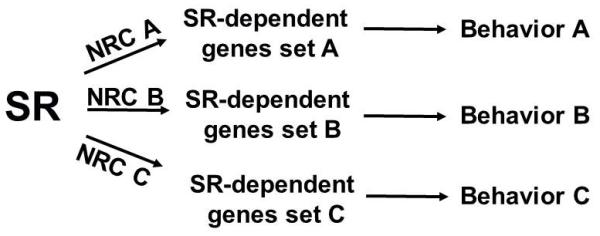
Diagram depicting the concept that steroid receptors (SR) recruit different sets of nuclear receptor coactivators (NRC) to enhance transactivation of distinct target genes and elicit different behaviors.
Acknowledgements
Studies contributed by the authors’ laboratory were supported by grants from National Institutes of Health R01 DK61935 (MJT).
References
- 1.Mangelsdorf DJ, Thummel C, Beato M, Herrlich P, Schütz G, Umesono K, Blumberg B, Kastner P, Mark M, Chambon P, Evans RM. The nuclear receptor superfamily: the second decade. Cell. 1995:83835–9. doi: 10.1016/0092-8674(95)90199-x. [DOI] [PMC free article] [PubMed] [Google Scholar]
- 2.Cornil CA, Ball GF, Balthazart J. Rapid control of male typical behaviors by brain-derived estrogens. Frontiers in neuroendocrinology. 2012;33(4):425–46. doi: 10.1016/j.yfrne.2012.08.003. [DOI] [PMC free article] [PubMed] [Google Scholar]
- 3.Vasudevan N, Pfaff DW. Membrane-initiated actions of estrogens in neuroendocrinology: emerging principles. Endocr Rev. 2007;28(1):1–19. doi: 10.1210/er.2005-0021. [DOI] [PubMed] [Google Scholar]
- 4.Tetel MJ, Lange CA. Molecular genomics of progestin actions. In: Pfaff DW, Arnold AP, Etgen AM, Fahrbach SE, Rubin RT, editors. Hormones, Brain and Behavior. Academic Press; San Diego: 2009. pp. 1439–65. [Google Scholar]
- 5.Mani SK, Mermelstein PG, Tetel MJ, Anesetti G. Convergence of multiple mechanisms of steroid hormone action. Horm Metab Res. 2012;44(8):569–76. doi: 10.1055/s-0032-1306343. [DOI] [PMC free article] [PubMed] [Google Scholar]
- 6.Kelly MJ, Ronnekleiv OK. Membrane-initiated actions of estradiol that regulate reproduction, energy balance and body temperature. Frontiers in neuroendocrinology. 2012;33(4):376–87. doi: 10.1016/j.yfrne.2012.07.002. [DOI] [PMC free article] [PubMed] [Google Scholar]
- 7.Micevych PE, Dewing P. Membrane-initiated estradiol signaling regulating sexual receptivity. Frontiers in endocrinology. 2011:21–9. doi: 10.3389/fendo.2011.00026. [DOI] [PMC free article] [PubMed] [Google Scholar]
- 8.Lonard DM, O’Malley BW. Nuclear receptor coregulators: modulators of pathology and therapeutic targets. Nat Rev Endocrinol. 2013:8598–604. doi: 10.1038/nrendo.2012.100. [DOI] [PMC free article] [PubMed] [Google Scholar]
- 9.Tsai MJ, O’Malley BW. Molecular mechanisms of action of steroid/thyroid receptor superfamily members. Annual Review of Biochemistry. 1994:63451–86. doi: 10.1146/annurev.bi.63.070194.002315. [DOI] [PubMed] [Google Scholar]
- 10.Tora L, White J, Brou C, Tasset D, Webster N, Scheer E, Chambon P. The human estrogen receptor has two independent non-acidic transcriptional activation functions. Cell. 1989:59477–87. doi: 10.1016/0092-8674(89)90031-7. [DOI] [PubMed] [Google Scholar]
- 11.Jensen EV, Suzuki T, Kawasima T, Stumpf WE, Jungblut PW, de Sombre ER. A two-step mechanism for the interaction of estradiol with rat uterus. Proceedings of the National Academy of Sciences USA. 1968:59632–8. doi: 10.1073/pnas.59.2.632. [DOI] [PMC free article] [PubMed] [Google Scholar]
- 12.Kuiper GGJM, Enmark E, Pelto-Huikko M, Nilsson S, Gustafsson J. Cloning of a novel estrogen receptor expressed in rat prostate and ovary. Proceedings of the National Academy of Sciences USA. 1996:935925–30. doi: 10.1073/pnas.93.12.5925. [DOI] [PMC free article] [PubMed] [Google Scholar]
- 13.Kuiper GGJM, Carlsson B, Grandien K, Enmark E, Häggblad J, Nilsson S, Gustafsson J. Comparison of the ligand binding specificity and transcript tissue distribution of estrogen receptors alpha and beta. Endocrinology. 1997:138863–70. doi: 10.1210/endo.138.3.4979. [DOI] [PubMed] [Google Scholar]
- 14.Delaunay F, Pettersson K, Tujague M, Gustafsson JA. Functional differences between the amino-terminal domains of estrogen receptors alpha and beta. Mol Pharmacol. 2000;58(3):584–90. doi: 10.1124/mol.58.3.584. [DOI] [PubMed] [Google Scholar]
- 15.Shughrue PJ, Lane MV, Merchenthaler I. Comparative distribution of estrogen receptor-alpha and -beta mRNA in the rat central nervous system. The Journal of comparative neurology. 1997;388(4):507–25. doi: 10.1002/(sici)1096-9861(19971201)388:4<507::aid-cne1>3.0.co;2-6. [DOI] [PubMed] [Google Scholar]
- 16.Greco B, Allegretto EA, Tetel MJ, Blaustein JD. Coexpression of ER beta with ER alpha and progestin receptor proteins in the female rat forebrain: Effects of estradiol treatment. Endocrinology. 2001;142:5172–81. doi: 10.1210/endo.142.12.8560. [DOI] [PubMed] [Google Scholar]
- 17.Osterlund M, Kuiper GG, Gustafsson JA, Hurd YL. Differential distribution and regulation of estrogen receptor-alpha and -beta mRNA within the female rat brain. Brain Res Mol Brain Res. 1998;54(1):175–80. doi: 10.1016/s0169-328x(97)00351-3. [DOI] [PubMed] [Google Scholar]
- 18.Mitra SW, Hoskin E, Yudkovitz J, Pear L, Wilkinson HA, Hayashi S, Pfaff DW, Ogawa S, Rohrer SP, Schaeffer JM, McEwen BS, Alves SE. Immunolocalization of estrogen receptor beta in the mouse brain: comparison with estrogen receptor alpha. Endocrinology. 2003:1442055–67. doi: 10.1210/en.2002-221069. [DOI] [PubMed] [Google Scholar]
- 19.Tetel MJ, Pfaff DW. Contributions of estrogen receptor-alpha and estrogen receptor-beta to the regulation of behavior. Biochim Biophys Acta. 2010:18001084–9. doi: 10.1016/j.bbagen.2010.01.008. [DOI] [PMC free article] [PubMed] [Google Scholar]
- 20.Ogawa S, Eng V, Taylor J, Lubahn DB, Korach KS, Pfaff DW. Roles of estrogen receptor-alpha gene expression in reproduction-related behaviors in female mice. Endocrinology. 1998:1395070–81. doi: 10.1210/endo.139.12.6357. [DOI] [PubMed] [Google Scholar]
- 21.Musatov S, Chen W, Pfaff DW, Kaplitt MG, Ogawa S. RNAi-mediated silencing of estrogen receptor {alpha} in the ventromedial nucleus of hypothalamus abolishes female sexual behaviors. ProcNatlAcadSciUSA. 2006:10310456–60. doi: 10.1073/pnas.0603045103. [DOI] [PMC free article] [PubMed] [Google Scholar]
- 22.Ogawa S, Chan J, Chester AE, Gustafsson JA, Korach KS, Pfaff DW. Survival of reproductive behaviors in estrogen receptor beta gene-deficient (betaERKO) male and female mice. ProcNatlAcadSciUSA. 1999:9612887–92. doi: 10.1073/pnas.96.22.12887. [DOI] [PMC free article] [PubMed] [Google Scholar]
- 23.Bodo C, Rissman EF. New roles for estrogen receptor beta in behavior and neuroendocrinology. Front Neuroendocrinol. 2006:27217–32. doi: 10.1016/j.yfrne.2006.02.004. [DOI] [PubMed] [Google Scholar]
- 24.Koehorst SGA, Cox JJ, Donker GH, Lopes da Silva S, Burbach JPH, Thijssen JHH, Blankenstein MA. Functional analysis of an alternatively spliced estrogen receptor lacking exon 4 isolated from MCF-7 breast cancer cells and meningioma tissue. MolCell Endocrinol. 1994:101237–45. doi: 10.1016/0303-7207(94)90240-2. [DOI] [PubMed] [Google Scholar]
- 25.Madsen MW, Reiter BE, Lykkesfeldt AE. Differential expression of estrogen receptor mRNA splice variants in the tamoxifen resistant human breast cancer cell line, MCF-7/TAMR-1 compared to the parental MCF-7 cell line. MolCell Endocrinol. 1995:109197–207. doi: 10.1016/0303-7207(95)03503-y. [DOI] [PubMed] [Google Scholar]
- 26.Ishunina TA, Swaab DF. Hippocampal estrogen receptor-alpha splice variant TADDI in the human brain in aging and Alzheimer’s disease. Neuroendocrinology. 2009;89(2):187–99. doi: 10.1159/000158573. [DOI] [PubMed] [Google Scholar]
- 27.Bollig A, Miksicek RJ. An estrogen receptor-alpha splicing variant mediates both positive and negative effects on gene transcription [In Process Citation] MolEndocrinol. 2000:14634–49. doi: 10.1210/mend.14.5.0460. [DOI] [PubMed] [Google Scholar]
- 28.Kastner P, Krust A, Turcotte B, Stropp U, Tora L, Gronemeyer H, Chambon P. Two distinct estrogen-regulated promoters generate transcripts encoding the two functionally different human progesterone receptor forms A and B. EMBO Journal. 1990:91603–14. doi: 10.1002/j.1460-2075.1990.tb08280.x. [DOI] [PMC free article] [PubMed] [Google Scholar]
- 29.Horwitz KB, Sheridan PL, Wei LL, Krett NL. Human progesterone receptors: synthesis, structure, and phosphorylation. Prog Clin Biol Res. 1990:32241–52. [PubMed] [Google Scholar]
- 30.Vegeto E, Shahbaz MM, Wen DX, Goldman ME, O’Malley BW, McDonnell DP. Human progesterone receptor A form is a cell-and promoter-specific repressor of human progesterone receptor B function. Molecular Endocrinology. 1993:71244–55. doi: 10.1210/mend.7.10.8264658. [DOI] [PubMed] [Google Scholar]
- 31.Tung L, Kamel Mohamed M, Hoeffler JP, Takimoto GS, Horwitz KB. Antagonist-occupied human progesterone B-receptors activate transcription without binding to progesterone response elements and are dominantly inhibited by A-receptors. Molecular Endocrinology. 1993:71256–65. doi: 10.1210/mend.7.10.8123133. [DOI] [PubMed] [Google Scholar]
- 32.Giangrande PH, Pollio G, McDonnell DP. Mapping and characterization of the functional domains responsible for the differential activity of the A and B isoforms of the human progesterone receptor. Journal of Biological Chemistry. 1997:27232889–900. doi: 10.1074/jbc.272.52.32889. [DOI] [PubMed] [Google Scholar]
- 33.Sartorius CA, Melville MY, Hovland AR, Tung L, Takimoto GS, Horwitz KB. A third transactivation function (AF3) of human progesterone receptors located in the unique N-terminal segment of the B-isoform. Molecular Endocrinology. 1994:81347–60. doi: 10.1210/mend.8.10.7854352. [DOI] [PubMed] [Google Scholar]
- 34.Hovland AR, Powell RL, Takimoto GS, Tung L, Horwitz KB. An N-terminal inhibitory function, IF, suppresses transcription by the A-isoform but not the B-isoform of human progesterone recpetors. Journal of Biological Chemistry. 1998:2735455–60. doi: 10.1074/jbc.273.10.5455. [DOI] [PubMed] [Google Scholar]
- 35.Mani SK, Reyna AM, Chen JZ, Mulac-Jericevic B, Conneely OM. Differential response of progesterone receptor isoforms in hormone-dependent and -independent facilitation of female sexual receptivity. Molecular Endocrinology. 2006:201322–32. doi: 10.1210/me.2005-0466. [DOI] [PubMed] [Google Scholar]
- 36.Mulac-Jericevic B, Conneely OM. Reproductive tissue selective actions of progesterone receptors. Reproduction. 2004:128139–46. doi: 10.1530/rep.1.00189. [DOI] [PubMed] [Google Scholar]
- 37.DeMarzo A, Beck CA, Oñate SA, Edwards DP. Dimerization of mammalian progesterone receptors occurs in the absence of DNA and is related to the release of the 90-kDa heat shock protein. Proceedings of the National Academy of Sciences USA. 1991:8872–6. doi: 10.1073/pnas.88.1.72. [DOI] [PMC free article] [PubMed] [Google Scholar]
- 38.Klein-Hitpass L, Tsai SY, Weigel NL, Allan GF, Riley D, Rodriguez R, Schrader WT, Tsai MJ, O’Malley BW. The progesterone receptor stimulates cell-free transcription by enhancing the formation of a stable preinitiation complex. Cell. 1990:60247–57. doi: 10.1016/0092-8674(90)90740-6. [DOI] [PubMed] [Google Scholar]
- 39.Kininis M, Chen BS, Diehl AG, Isaacs GD, Zhang T, Siepel AC, Clark AG, Kraus WL. Genomic analyses of transcription factor binding, histone acetylation, and gene expression reveal mechanistically distinct classes of estrogen-regulated promoters. Mol Cell Biol. 2007;27(14):5090–104. doi: 10.1128/MCB.00083-07. [DOI] [PMC free article] [PubMed] [Google Scholar]
- 40.Pfaff D. Hormone-driven mechanisms in the central nervous system facilitate the analysis of mammalian behaviours. J Endocrinol. 2005;184(3):447–53. doi: 10.1677/joe.1.05897. [DOI] [PubMed] [Google Scholar]
- 41.Blaustein JD, Mani SK. Feminine sexual behavior from neuroendocrine and molecular neurobiological perspectives. In: Blaustein JD, editor. Handbook of Neurochemistry and Molecular Neurobiology. Springer; New York: 2006. pp. 95–150. [Google Scholar]
- 42.Rosenfeld MG, Lunyak VV, Glass CK. Sensors and signals: a coactivator/corepressor/epigenetic code for integrating signal-dependent programs of transcriptional response. Genes Dev. 2006:201405–28. doi: 10.1101/gad.1424806. [DOI] [PubMed] [Google Scholar]
- 43.Johnson AB, O’Malley BW. Steroid receptor coactivators 1, 2 and 3: Critical regulators of nuclear receptor activity and steroid receptor modulator (SRM)-based cancer therapy. Molecular and cellular endocrinology. 2012:348430–9. doi: 10.1016/j.mce.2011.04.021. [DOI] [PMC free article] [PubMed] [Google Scholar]
- 44.Oñate SA, Tsai SY, Tsai MJ, O’Malley BW. Sequence and characterization of a coactivator for the steroid hormone receptor superfamily. Science. 1995:2701354–7. doi: 10.1126/science.270.5240.1354. [DOI] [PubMed] [Google Scholar]
- 45.McInerney EM, Tsai MJ, O’Malley BW, Katzenellenbogen BS. Analysis of estrogen receptor transcriptional enhancement by a nuclear hormone receptor coactivator. Proceedings of the National Academy of Sciences USA. 1996:9310069–73. doi: 10.1073/pnas.93.19.10069. [DOI] [PMC free article] [PubMed] [Google Scholar]
- 46.Tanenbaum DM, Wang Y, Williams SP, Sigler PB. Crystallographic comparison of the estrogen and progesterone receptor’s ligand binding domains. Proc Natl Acad Sci U S A. 1998;95(11):5998–6003. doi: 10.1073/pnas.95.11.5998. [DOI] [PMC free article] [PubMed] [Google Scholar]
- 47.Shiau AK, Barstad D, Loria PM, Cheng L, Kushner PJ, Agard DA, Greene GL. The structural basis of estrogen receptor/coactivator recognition and the antagonism of this interaction by tamoxifen. Cell. 1998;95(7):927–37. doi: 10.1016/s0092-8674(00)81717-1. [DOI] [PubMed] [Google Scholar]
- 48.Oñate SA, Boonyaratanakornkit V, Spencer TE, Tsai SY, Tsai MJ, Edwards DP, O’Malley BW. The steroid receptor coactivator-1 contains multiple receptor interacting and activation domains that cooperatively enhance the activation function 1 (AF1) and AF2 domains of steroid receptors. Journal of Biological Chemistry. 1998:27312101–8. doi: 10.1074/jbc.273.20.12101. [DOI] [PubMed] [Google Scholar]
- 49.Webb P, Nguyen P, Shinsako J, Anderson C, Feng W, Nguyen MP, Chen D, Huang SM, Subramanian S, McKinerney E, Katzenellenbogen BS, Stallcup MR, Kushner PJ. Estrogen receptor activation function 1 works by binding p160 coactivator proteins. Molecular Endocrinology. 1998:121605–18. doi: 10.1210/mend.12.10.0185. [DOI] [PubMed] [Google Scholar]
- 50.Dutertre M, Smith CL. Ligand-independent interactions of p160/steroid receptor coactivators and CREB-Binding Protein (CBP) with estrogen receptor-à: Regulation by phosphorylation sites in the A/B region depends on other receptor domains. Molecular Endocrinology. 2003:171296–314. doi: 10.1210/me.2001-0316. [DOI] [PubMed] [Google Scholar]
- 51.Torchia J, Rose DW, Inostroza J, Kamei Y, Westin S, Glass CK, Rosenfeld MG. The transcriptional co-activator p/CIP binds CBP and mediates nuclear-receptor function. Nature. 1997:387677–84. doi: 10.1038/42652. [DOI] [PubMed] [Google Scholar]
- 52.Misiti S, Schomburg L, Yen PM, Chin WW. Expression and hormonal regulation of coactivator and corepressor genes. Endocrinology. 1998:1392493–500. doi: 10.1210/endo.139.5.5971. [DOI] [PubMed] [Google Scholar]
- 53.Martinez de Arrieta C, Koibuchi N, Chin WW. Coactivator and corepressor gene expression in rat cerebellum during postnatal development and the effect of altered thyroid status. Endocrinology. 2000:1411693–8. doi: 10.1210/endo.141.5.7467. [DOI] [PubMed] [Google Scholar]
- 54.Meijer OC, Steenbergen PJ, de Kloet ER. Differential expression and regional distribution of steroid receptor coactivators SRC-1 and SRC-2 in brain and pituitary. Endocrinology. 2000:1412192–9. doi: 10.1210/endo.141.6.7489. [DOI] [PubMed] [Google Scholar]
- 55.Auger AP, Tetel MJ, McCarthy MM. Steroid receptor coactivator-1 mediates the development of sex specific brain morphology and behavior. Proceedings of the National Academy of Sciences USA. 2000:977551–5. doi: 10.1073/pnas.97.13.7551. [DOI] [PMC free article] [PubMed] [Google Scholar]
- 56.Molenda HA, Griffin AL, Auger AP, McCarthy MM, Tetel MJ. Nuclear receptor coactivators modulate hormone-dependent gene expression in brain and female reproductive behavior in rats. Endocrinology. 2002:143436–44. doi: 10.1210/endo.143.2.8659. [DOI] [PubMed] [Google Scholar]
- 57.Ogawa H, Nishi M, Kawata M. Localization of nuclear coactivators p300 and steroid receptor coactivator 1 in the rat hippocampus. Brain research. 2001:890197–202. doi: 10.1016/s0006-8993(00)03158-9. [DOI] [PubMed] [Google Scholar]
- 58.Nishihara E, Yoshida-Kimoya H, Chan C, Liao L, Davis RL, O’Malley BW, Xu J. SRC-1 null mice exhibit moderate motor dysfunction and delayed development of cerebellar Purkinje cells. The Journal of Neuroscience. 2003:23213–22. doi: 10.1523/JNEUROSCI.23-01-00213.2003. [DOI] [PMC free article] [PubMed] [Google Scholar]
- 59.Setiawan E, Owen D, McCabe L, Kostaki A, Andrews MH, Matthews SG. Glucocorticoids do not alter developmental expression of hippocampal or pituitary steroid receptor coactivator-1 and-2 in the late gestation fetal guinea pig. Endocrinology. 2004:1453796–803. doi: 10.1210/en.2003-1723. [DOI] [PubMed] [Google Scholar]
- 60.Zalachoras I, Grootaers G, van Weert LT, Aubert Y, de Kreij SR, Datson NA, van Roon-Mom WM, Aartsma-Rus A, Meijer OC. Antisense-mediated isoform switching of steroid receptor coactivator-1 in the central nucleus of the amygdala of the mouse brain. BMC Neurosci. 2013:145. doi: 10.1186/1471-2202-14-5. [DOI] [PMC free article] [PubMed] [Google Scholar]
- 61.Zhang D, Guo Q, Bian C, Zhang J, Lin S, Su B. Alterations of steroid receptor coactivator-1 (SRC-1) immunoreactivities in specific brain regions of young and middle-aged female Sprague-Dawley rats. Brain Res. 2011:138288–97. doi: 10.1016/j.brainres.2011.01.024. [DOI] [PubMed] [Google Scholar]
- 62.Yore MA, Im D, Webb LK, Zhao Y, Chadwick JG, Molenda-Figueira HA, Haidacher SJ, Denner L, Tetel MJ. Steroid receptor coactivator-2 expression in brain and physical associations with steriod receptors. Neuroscience. 2010:1691017–28. doi: 10.1016/j.neuroscience.2010.05.053. [DOI] [PMC free article] [PubMed] [Google Scholar]
- 63.McGinnis MY, Lumia AR, Tetel MJ, Molenda-Figuiera HA, Possidente B. Effects of anabolic androgenic steroids on the development and expression of running wheel activity and circadian rhythms in male rats. Physiology and Behavior. 2007:921010–8. doi: 10.1016/j.physbeh.2007.07.010. [DOI] [PMC free article] [PubMed] [Google Scholar]
- 64.Apostolakis EM, Ramamurphy M, Zhou D, Onate S, O’Malley B. Acute disruption of select steroid receptor coactivators prevents reproductive behavior in rats and unmasks genetic adaptation in knockout mice. Molecular Endocrinology. 2002:161511–23. doi: 10.1210/mend.16.7.0877. [DOI] [PubMed] [Google Scholar]
- 65.Nottebohm F, Arnold AP. Sexual dimorphism in vocal control areas of the songbird brain. Science. 1976;194(4261):211–3. doi: 10.1126/science.959852. [DOI] [PubMed] [Google Scholar]
- 66.Jacobs EC, Arnold AP, Campagnoni AT. Developmental regulation of the distribution of aromatase- and estrogen-receptor- mRNA-expressing cells in the zebra finch brain. Dev Neurosci. 1999;21(6):453–72. doi: 10.1159/000017413. [DOI] [PubMed] [Google Scholar]
- 67.Perlman WR, Arnold AP. Expression of estrogen receptor and aromatase mRNAs in embryonic and posthatch zebra finch brain. J Neurobiol. 2003;55(2):204–19. doi: 10.1002/neu.10190. [DOI] [PubMed] [Google Scholar]
- 68.Saldanha CJ, Coomaralingam L. Overlap and co-expression of estrogen synthetic and responsive neurons in the songbird brain--a double-label immunocytochemical study. General and comparative endocrinology. 2005;141(1):66–75. doi: 10.1016/j.ygcen.2004.11.013. [DOI] [PubMed] [Google Scholar]
- 69.Gurney ME, Konishi M. Hormone-induced sexual differentiation of brain and behavior in zebra finches. Science. 1980;208(4450):1380–3. doi: 10.1126/science.208.4450.1380. [DOI] [PubMed] [Google Scholar]
- 70.Simpson HB, Vicario DS. Early estrogen treatment of female zebra finches masculinizes the brain pathway for learned vocalizations. J Neurobiol. 1991;22(7):777–93. doi: 10.1002/neu.480220711. [DOI] [PubMed] [Google Scholar]
- 71.Grisham W, Mathews GA, Arnold AP. Local intracerebral implants of estrogen masculinize some aspects of the zebra finch song system. J Neurobiol. 1994;25(2):185–96. doi: 10.1002/neu.480250209. [DOI] [PubMed] [Google Scholar]
- 72.Adkins-Regan E, Mansukhani V, Seiwert C, Thompson R. Sexual differentiation of brain and behavior in the zebra finch: critical periods for effects of early estrogen treatment. J Neurobiol. 1994;25(7):865–77. doi: 10.1002/neu.480250710. [DOI] [PubMed] [Google Scholar]
- 73.Fusani L, Metzdorf R, Hutchison JB, Gahr M. Aromatase inhibition affects testosterone-induced masculinization of song and the neural song system in female canaries. J Neurobiol. 2003;54(2):370–9. doi: 10.1002/neu.10141. [DOI] [PubMed] [Google Scholar]
- 74.Charlier TD, Ball GF, Balthazart J. Rapid action on neuroplasticity precedes behavioral activation by testosterone. Hormones and behavior. 2008;54(4):488–95. doi: 10.1016/j.yhbeh.2008.03.001. [DOI] [PMC free article] [PubMed] [Google Scholar]
- 75.Seredynski AL, Ball GF, Balthazart J, Charlier TD. Specific activation of estrogen receptor alpha and beta enhances male sexual behavior and neuroplasticity in male Japanese quail. PLoS One. 2011;6(4):e18627. doi: 10.1371/journal.pone.0018627. [DOI] [PMC free article] [PubMed] [Google Scholar]
- 76.Balthazart J, Surlemont C. Copulatory behavior is controlled by the sexually dimorphic nucleus of the quail POA. Brain Res Bull. 1990;25(1):7–14. doi: 10.1016/0361-9230(90)90246-v. [DOI] [PubMed] [Google Scholar]
- 77.Charlier TD, Seredynski AL, Niessen NA, Balthazart J. Modulation of testosterone-dependent male sexual behavior and the associated neuroplasticity. General and comparative endocrinology. 2013 doi: 10.1016/j.ygcen.2013.03.003. [DOI] [PMC free article] [PubMed] [Google Scholar]
- 78.Balthazart J, Surlemont C. Androgen and estrogen action in the preoptic area and activation of copulatory behavior in quail. Physiol Behav. 1990;48(5):599–609. doi: 10.1016/0031-9384(90)90198-d. [DOI] [PubMed] [Google Scholar]
- 79.Charlier TD, Balthazart J, Ball GF. Sex differences in the distribution of the steroid coactivator SRC-1 in the song control nuclei of male and female canaries. Brain research. 2003;959:263–74. doi: 10.1016/s0006-8993(02)03758-7. [DOI] [PubMed] [Google Scholar]
- 80.Duncan KA, Jimenez P, Carruth LL. Distribution and sexually dimorphic expression of steroid receptor coactivator-1 (SRC-1) in the zebra finch brain. Gen Comp Endocrinol. 2011;170(2):408–14. doi: 10.1016/j.ygcen.2010.10.021. [DOI] [PubMed] [Google Scholar]
- 81.Niessen NA, Balthazart J, Ball GF, Charlier TD. Steroid receptor coactivator 2 modulates steroid-dependent male sexual behavior and neuroplasticity in Japanese quail (Coturnix japonica) Journal of neurochemistry. 2011;119(3):579–93. doi: 10.1111/j.1471-4159.2011.07438.x. [DOI] [PMC free article] [PubMed] [Google Scholar]
- 82.MacLusky NJ, McEwen BS. Oestrogen modulates progestin receptor concentrations in some rat brain regions but not in others. Nature. 1978:274276–8. doi: 10.1038/274276a0. [DOI] [PubMed] [Google Scholar]
- 83.Lauber AH, Romano GJ, Pfaff DW. Sex difference in estradiol regulation of progestin receptor messenger RNA in rat mediobasal hypothalamus as demonstrated by In situ hybridization. Neuroendocrinology. 1991:53608–13. doi: 10.1159/000125781. [DOI] [PubMed] [Google Scholar]
- 84.Blaustein JD, Turcotte JC. Estradiol-induced progestin receptor immunoreactivity is found only in estrogen receptor-immunoreactive cells in guinea pig brain. Neuroendocrinology. 1989:49454–61. doi: 10.1159/000125152. [DOI] [PubMed] [Google Scholar]
- 85.Warembourg M, Jolivet A, Milgrom E. Immunohistochemical evidence of the presence of estrogen and progesterone receptors in the same neurons of the guinea pig hypothalamus and preoptic area. Brain Res. 1989:4801–15. doi: 10.1016/0006-8993(89)91561-8. [DOI] [PubMed] [Google Scholar]
- 86.Scott REM, Wu-Peng XS, Pfaff DW. Regulation and expression of progesterone receptor mRNA isoforms A and B in the male and female rat hypothalamus and pituitary following oesterogen treatment. Journal of Neuroendocrinology. 2002:14175–83. doi: 10.1046/j.0007-1331.2001.00750.x. [DOI] [PubMed] [Google Scholar]
- 87.Brinton RD, Thompson RF, Foy MR, Baudry M, Wang J, Finch CE, Morgan TE, Pike CJ, Mack WJ, Stanczyk FZ, Nilsen J. Progesterone receptors: form and function in brain. Front Neuroendocrinol. 2008;29(2):313–39. doi: 10.1016/j.yfrne.2008.02.001. [DOI] [PMC free article] [PubMed] [Google Scholar]
- 88.Tetel MJ, Siegal NK, Murphy SD. Cells in behaviourally relevant brain regions coexpress nuclear receptor coactivators and ovarian steroid receptors. J Neuroendocrinol. 2007;19(4):262–71. doi: 10.1111/j.1365-2826.2007.01526.x. [DOI] [PMC free article] [PubMed] [Google Scholar]
- 89.Tognoni CM, Chadwick JG, Jr., Ackeifi CA, Tetel MJ. Nuclear receptor coactivators are coexpressed with steroid receptors and regulated by estradiol in mouse brain. Neuroendocrinology. 2011:9449–57. doi: 10.1159/000323780. [DOI] [PMC free article] [PubMed] [Google Scholar]
- 90.Zhu L, Yang Y, Xu P, Zou F, Yan X, Liao L, Xu J, O’Malley BW, Xu Y. Steroid receptor coactivator-1 mediates estrogenic actions to prevent body weight gain in female mice. Endocrinology. 2013;154(1):150–8. doi: 10.1210/en.2012-2007. [DOI] [PMC free article] [PubMed] [Google Scholar]
- 91.Xu J, Qiu Y, Demayo FJ, Tsai SY, Tsai MJ, O’Malley BW. Partial hormone resistance in mice with disruption of the steroid receptor coactivator-1 (SRC-1) gene. Science. 1998:2791922–5. doi: 10.1126/science.279.5358.1922. [DOI] [PubMed] [Google Scholar]
- 92.Weiss RE, Xu J, Ning G, Pohlenz J, O’Malley BW, Refetoff S. Mice deficient in the steroid receptor co-activator 1 (SRC-1) are resistant to thyroid hormone. EMBO J. 1999:181900–4. doi: 10.1093/emboj/18.7.1900. [DOI] [PMC free article] [PubMed] [Google Scholar]
- 93.Bousios S, Karandrea D, Kittas C, Kitraki E. Effects of gender and stress on the regulation of steroid receptor coactivator-1 expression in the rat brain and pituitary. The Journal of Steroid Biocheminstry and Molecular Biology. 2001:78401–7. doi: 10.1016/s0960-0760(01)00123-6. [DOI] [PubMed] [Google Scholar]
- 94.Bian C, Zhang D, Guo Q, Cai W, Zhang J. Localization and sex-difference of steroid receptor coactivator-1 immunoreactivities in the brain of adult female and male mice. Steroids. 2011;76(3):269–79. doi: 10.1016/j.steroids.2010.11.009. [DOI] [PubMed] [Google Scholar]
- 95.Mitev YA, Wolf SS, Almeida OF, Patchev VK. Developmental expression profiles and distinct regional estrogen responsiveness suggest a novel role for the steroid receptor coactivator SRC-l as a discriminative amplifier of estrogen signaling in the rat brain. The FASEB Journal. 2003:17518–9. doi: 10.1096/fj.02-0513fje. [DOI] [PubMed] [Google Scholar]
- 96.Camacho-Arroyo I, Neri-Gomez T, Gonzalez-Arenas A, Guerra-Araiza C. Changes in the content of steroid receptor coactivator-1 and silencing mediator for retinoid and thyroid hormone receptors in the rat brain during the estrous cycle. The Journal of Steroid Biocheminstry and Molecular Biology. 2005:94267–72. doi: 10.1016/j.jsbmb.2004.12.013. [DOI] [PubMed] [Google Scholar]
- 97.Zhang D, Guo Q, Bian C, Zhang J, Cai W, Su B. Expression of steroid receptor coactivator-1 was regulated by postnatal development but not ovariectomy in the hippocampus of rats. Dev Neurosci. 2011;33(1):57–63. doi: 10.1159/000322978. [DOI] [PubMed] [Google Scholar]
- 98.Maerkel K, Durrer S, Henseler M, Schlumpf M, Lichtensteiger W. Sexually dimorphic gene regulation in brain as a target for endocrine disrupters: developmental exposure of rats to 4-methylbenzylidene camphor. Toxicol Appl Pharmacol. 2007;218(2):152–65. doi: 10.1016/j.taap.2006.10.026. [DOI] [PubMed] [Google Scholar]
- 99.Faass O, Schlumpf M, Reolon S, Henseler M, Maerkel K, Durrer S, Lichtensteiger W. Female sexual behavior, estrous cycle and gene expression in sexually dimorphic brain regions after pre- and postnatal exposure to endocrine active UV filters. Neurotoxicology. 2009;30(2):249–60. doi: 10.1016/j.neuro.2008.12.008. [DOI] [PubMed] [Google Scholar]
- 100.Tetel MJ, Ungar TC, Hassan B, Bittman EL. Photoperiodic regulation of androgen receptor and steroid receptor coactivator-1 in Siberian hamster brain. Molecular Brain Research. 2004:13179–87. doi: 10.1016/j.molbrainres.2004.08.009. [DOI] [PMC free article] [PubMed] [Google Scholar]
- 101.Iannacone EA, Yan AW, Gauger KJ, Dowling ALS, Zoeller RT. Thyroid hormone exerts site-specific effects on SRC-1 and NCoR expression selectively in the neonatal rat brain. Molecular and cellular endocrinology. 2002:18649–59. doi: 10.1016/s0303-7207(01)00672-4. [DOI] [PubMed] [Google Scholar]
- 102.Ramos HE, Weiss RE. Regulation of nuclear coactivator and corepressor expression in mouse cerebellum by thyroid hormone. Thyroid. 2006;16(3):211–6. doi: 10.1089/thy.2006.16.211. [DOI] [PubMed] [Google Scholar]
- 103.Charlier TD, Ball GF, Balthazart J. Plasticity in the expression of the steroid receptor coactivator-1 in the Japanese quail brain: Effect of sex, testosterone, stress and time of the day. Neuroscience. 2006:172333–43. doi: 10.1016/j.neuroscience.2006.03.002. [DOI] [PubMed] [Google Scholar]
- 104.Kurihara I, Shibata H, Suzuki T, Ando T, Kobayashi S, Hayashi M, saito I, Saruta T. Expression and regulation of nuclear receptor coactivators in glucocorticoid action. Molecular and cellular endocrinology. 2002:189181–9. doi: 10.1016/s0303-7207(01)00717-1. [DOI] [PubMed] [Google Scholar]
- 105.Meijer OC, Kalkhoven E, van der LS, Steenbergen PJ, Houtman SH, Dijkmans TF, Pearce D, de Kloet ER. Steroid receptor coactivator-1 splice variants differentially affect corticosteroid receptor signaling. Endocrinology. 2005:1461438–48. doi: 10.1210/en.2004-0411. [DOI] [PubMed] [Google Scholar]
- 106.Bittman EL, Hegarty CM, Layden MQ, Jonassen JA. Influences of Photoperiod on Sexual Behaviour, Neuroendocrine Steroid Receptors and Adenohypophysial Hormone Secretion and Gene Expression in Female Golden Hamsters. JMolecularEndocrinol. 1990:515–25. doi: 10.1677/jme.0.0050015. [DOI] [PubMed] [Google Scholar]
- 107.Charlier TD. Importance of steroid receptor coactivators in the modulation of steroid action on brain and behavior. Psychoneuroendocrinology. 2009:34S20–S9. doi: 10.1016/j.psyneuen.2009.05.004. [DOI] [PubMed] [Google Scholar]
- 108.Nishihara E, Moriya T, Shinohara K. Expression of steroid receptor coactivator-1 is elevated during neuronal differentiation of murine neural stem cells. Brain research. 2007;1135(1):22–30. doi: 10.1016/j.brainres.2006.12.026. [DOI] [PubMed] [Google Scholar]
- 109.Han SJ, Lonard DM, O’Malley BW. Multi-modulation of nuclear receptor coactivators through posttranslational modifications. Trends in endocrinology and metabolism: TEM. 2009;20(1):8–15. doi: 10.1016/j.tem.2008.10.001. [DOI] [PMC free article] [PubMed] [Google Scholar]
- 110.Rowan BG, Weigel NL, O’Malley BW. Phosphorylation of steroid receptor coactivator-1. Identification of the phosphorylation sites and phosphorylation through the mitogen-activated protein kinase pathway. Journal of Biological Chemistry. 2000:2754475–83. doi: 10.1074/jbc.275.6.4475. [DOI] [PubMed] [Google Scholar]
- 111.Rowan BG, Garrison N, Weigel NL, O’Malley BW. 8-Bromo-cyclic AMP induces phosphorylation of two sites in SRC-1 that facilitate ligand-independent activation of the chicken progesterone receptor and are critical for functional cooperation between SRC-1 and CREB binding protein. MolCell Biol. 2000:208720–30. doi: 10.1128/mcb.20.23.8720-8730.2000. [DOI] [PMC free article] [PubMed] [Google Scholar]
- 112.Hoang T, F IS, Cook C, Børud B, Bakke M, Lien EA, Mellgren G. cAMP-dependent protein kinase regulates ubiquitin-proteasome-mediated degradation and subcellular localization of the nuclear receptor coactivator GRIP1. The Journal of biological chemistry. 2004:27949120–30. doi: 10.1074/jbc.M409746200. [DOI] [PubMed] [Google Scholar]
- 113.Narayanan R, Adigun AA, Edwards DP, Weigel NL. Cyclin-dependent kinase activity is required for progesterone receptor function: novel role for cyclin A/Cdk2 as a progesterone receptor coactivator. Mol Cell Biol. 2005;25(1):264–77. doi: 10.1128/MCB.25.1.264-277.2005. [DOI] [PMC free article] [PubMed] [Google Scholar]
- 114.Gianni M, Parrella E, Raska I, Jr., Gaillard E, Nigro EA, Gaudon C, Garattini E, Rochette-Egly C. P38MAPK-dependent phosphorylation and degradation of SRC-3/AIB1 and RARalpha-mediated transcription. EMBO J. 2006;25(4):739–51. doi: 10.1038/sj.emboj.7600981. [DOI] [PMC free article] [PubMed] [Google Scholar]
- 115.Pleim ET, Brown TJ, MacLusky NJ, Etgen AM, Barfield RJ. Dilute estradiol implants and progestin receptor induction in the ventromedial nucleus of the hypothalamus: correlation with receptive behavior in female rats. Endocrinology. 1989:1241807–12. doi: 10.1210/endo-124-4-1807. [DOI] [PubMed] [Google Scholar]
- 116.Smith CL, Oñate SA, Tsai MJ, O’Malley BW. CREB binding protein acts synergistically with steroid receptor coactivator-1 to enhance steroid receptor-dependent transcription. Proceedings of the National Academy of Sciences USA. 1996:938884–8. doi: 10.1073/pnas.93.17.8884. [DOI] [PMC free article] [PubMed] [Google Scholar]
- 117.Tetel MJ, Giangrande PH, Leonhardt SA, McDonnell DP, Edwards DP. Hormone-dependent interaction between the amino- and carboxyl-terminal domains of progesterone receptor in vitro and in vivo Molecular Endocrinology. 1999:13910–24. doi: 10.1210/mend.13.6.0300. [DOI] [PubMed] [Google Scholar]
- 118.Sharma D, Handa RJ, Uht RM. The ERbeta Ligand 5alpha-androstane, 3beta,17beta-diol (3beta-diol) Regulates Hypothalamic Oxytocin (Oxt) Gene Expression. Endocrinology. 2012;153(5):2353–61. doi: 10.1210/en.2011-1002. [DOI] [PMC free article] [PubMed] [Google Scholar]
- 119.Charlier TD, Harada N, Ball GF, Balthazart J. Targeting steroid receptor coactivator-1 expression with locked nucleic acids antisense reveals different thresholds for the hormonal regulation of male sexual behavior in relation to aromatase activity and protein expression. Behav Brain Res. 2006;172(2):333–43. doi: 10.1016/j.bbr.2006.05.023. [DOI] [PubMed] [Google Scholar]
- 120.Hemmerich P, von Mikecz A, Neumann F, Sözeri O, Wolff-Vorbeck G, Zoebelein R, Krawinkel U. Structural and functional properties of ribosomal protein L7 from humans and rodents. Nucleic Acid Res. 1993:21223–31. doi: 10.1093/nar/21.2.223. [DOI] [PMC free article] [PubMed] [Google Scholar]
- 121.Jackson TA, Richer JK, Bain DL, Takimoto GS, Tung L, Horwitz KB. The partial agonist activity of antagonist-occupied steroid receptors is controlled by a novel hinge domain-binding coactivator L7/SPA and the corepressors N-CoR or SMRT. Molecular Endocrinology. 1997:11693–705. doi: 10.1210/mend.11.6.0004. [DOI] [PubMed] [Google Scholar]
- 122.Berghöfer-Hochheimer Y, Zurek C, Wölfl S, Hemmerich P, Munder T. L7 protein is a coregulator of vitamin D receptor-retinoid X receptor-mediated transactivation. J Cell Biochem. 1998:691–12. doi: 10.1002/(sici)1097-4644(19980401)69:1<1::aid-jcb1>3.0.co;2-x. [DOI] [PubMed] [Google Scholar]
- 123.Duncan KA, Carruth LL. The sexually dimorphic expression of L7/SPA, an estrogen receptor coactivator, in zebra finch telencephalon. Dev Neurobiol. 2007:671852–66. doi: 10.1002/dneu.20539. [DOI] [PubMed] [Google Scholar]
- 124.Duncan KA, Carruth LL. The song remains the same: coactivators and sex differences in the songbird brain. Front Neuroendocrinol. 2011;32(1):84–94. doi: 10.1016/j.yfrne.2010.11.001. [DOI] [PubMed] [Google Scholar]
- 125.Duncan KA, Jimenez P, Carruth LL. The selective estrogen receptor-alpha coactivator, RPL7, and sexual differentiation of the songbird brain. Psychoneuroendocrinology. 2009:34SS30–S8. doi: 10.1016/j.psyneuen.2009.04.023. [DOI] [PubMed] [Google Scholar]
- 126.Gonzalez-Arenas A, Hansberg-Pastor V, Hernandez-Hernandez OT, Gonzalez-Garcia TK, Henderson-Villalpando J, Lemus-Hernandez D, Cruz-Barrios A, Rivas-Suarez M, Camacho-Arroyo I. Estradiol increases cell growth in human astrocytoma cell lines through ERalpha activation and its interaction with SRC-1 and SRC-3 coactivators. Biochimica et biophysica acta. 2012;1823(2):379–86. doi: 10.1016/j.bbamcr.2011.11.004. [DOI] [PubMed] [Google Scholar]
- 127.Hernandez-Hernandez OT, Gonzalez-Garcia TK, Camacho-Arroyo I. Progesterone receptor and SRC-1 participate in the regulation of VEGF, EGFR and Cyclin D1 expression in human astrocytoma cell lines. The Journal of steroid biochemistry and molecular biology. 2012;132(1-2):127–34. doi: 10.1016/j.jsbmb.2012.04.005. [DOI] [PubMed] [Google Scholar]
- 128.Molenda-Figueira HA, Williams CA, Griffin AL, Rutledge EM, Blaustein JD, Tetel MJ. Nuclear receptor coactivators function in estrogen receptor-and progestin receptor-dependent aspects of sexual behavior in female rats. Hormones and Behavior. 2006:50383–92. doi: 10.1016/j.yhbeh.2006.04.005. [DOI] [PMC free article] [PubMed] [Google Scholar]
- 129.Mazzucco CA, Walker HA, Pawluski JL, Lieblich SE, Galea LA. ERalpha, but not ERbeta, mediates the expression of sexual behavior in the female rat. Behav Brain Res. 2008;191(1):111–7. doi: 10.1016/j.bbr.2008.03.016. [DOI] [PubMed] [Google Scholar]
- 130.Hardy DF, DeBold JF. The relationship between levels of exogenous hormones and the display of lordosis by the female rat. HormsBehav. 1971:2287–97. [Google Scholar]
- 131.Erskine MS. Solicitation behavior in the estrous female rat: A review. HormsBehav. 1989:23473–502. doi: 10.1016/0018-506x(89)90037-8. [DOI] [PubMed] [Google Scholar]
- 132.Charlier TD, Ball GF, Balthazart J. Inhibition of steroid receptor coactivator-1 blocks estrogen and androgen action on male sex behavior and associated brain plasticity. Journal of Neuroscience. 2005:25906–13. doi: 10.1523/JNEUROSCI.3533-04.2005. [DOI] [PMC free article] [PubMed] [Google Scholar]
- 133.Niessen NA, Balthazart J, Ball GF, Charlier TD. Steroid receptor coactivator 2 modulates steroid-dependent male sexual behavior and neuroplasticity in Japanese quail (Coturnix japonica) J Neurochem. 2011;119(3):579–93. doi: 10.1111/j.1471-4159.2011.07438.x. [DOI] [PMC free article] [PubMed] [Google Scholar]
- 134.Molenda-Figueira HA, Murphy SD, Shea KL, Siegal NK, Zhao Y, Chadwick JG, Denner LA, Tetel MJ. Steroid receptor coactivator-1 from brain physically interacts differentially with steroid receptor subtypes. Endocrinology. 2008;149(10):5272–9. doi: 10.1210/en.2008-0048. [DOI] [PMC free article] [PubMed] [Google Scholar]
- 135.Rissman EF. Roles of oestrogen receptors alpha and beta in behavioural neuroendocrinology: beyond Yin/Yang. J Neuroendocrinol. 2008;20(6):873–9. doi: 10.1111/j.1365-2826.2008.01738.x. [DOI] [PMC free article] [PubMed] [Google Scholar]
- 136.Zhao Z, Fan L, Fortress AM, Boulware MI, Frick KM. Hippocampal Histone Acetylation Regulates Object Recognition and the Estradiol-Induced Enhancement of Object Recognition. The Journal of neuroscience: the official journal of the Society for Neuroscience. 2012;32(7):2344–51. doi: 10.1523/JNEUROSCI.5819-11.2012. [DOI] [PMC free article] [PubMed] [Google Scholar]
- 137.Fugger HN, Foster TC, Gustafsson J, Rissman EF. Novel effects of estradiol and estrogen receptor alpha and beta on cognitive function. Brain Res. 2000;883(2):258–64. doi: 10.1016/s0006-8993(00)02993-0. [DOI] [PubMed] [Google Scholar]
- 138.Isgor C, Cecchi M, Kabbaj M, Akil H, Watson SJ. Estrogen receptor beta in the paraventricular nucleus of hypothalamus regulates the neuroendocrine response to stress and is regulated by corticosterone. Neuroscience. 2003;121(4):837–45. doi: 10.1016/s0306-4522(03)00561-x. [DOI] [PubMed] [Google Scholar]
- 139.Kraichely DM, Sun J, Katzenellenbogen JA, Katzenellenbogen BS. Conformational changes and coactivator recruitment by novel ligands for estrogen receptor-alpha and estrogen receptor-beta: correlations with biological character and distinct differences among SRC coactivator family members. Endocrinology. 2000:1413534–45. doi: 10.1210/endo.141.10.7698. [DOI] [PubMed] [Google Scholar]
- 140.Zhao C, Toresson G, Xu L, Koehler KF, Gustafsson JA, Dahlman-Wright K. Mouse estrogen receptor beta isoforms exhibit differences in ligand selectivity and coactivator recruitment. Biochemistry. 2005;44(22):7936–44. doi: 10.1021/bi047691m. [DOI] [PubMed] [Google Scholar]
- 141.Cvoro A, Tatomer D, Tee MK, Zogovic T, Harris HA, Leitman DC. Selective estrogen receptor-beta agonists repress transcription of proinflammatory genes. J Immunol. 2008;180(1):630–6. doi: 10.4049/jimmunol.180.1.630. [DOI] [PubMed] [Google Scholar]
- 142.Routledge EJ, White R, Parker MG, Sumpter JP. Differential Effects of Xenoestrogens on Coactivator Recruitment by Estrogen Receptor (ER) alpha and ERbeta. Journal of Biological Chemistry. 2000:27535986–93. doi: 10.1074/jbc.M006777200. [DOI] [PubMed] [Google Scholar]
- 143.Singh M, Su C. Progesterone and neuroprotection. Hormones and behavior. 2013;63(2):284–90. doi: 10.1016/j.yhbeh.2012.06.003. [DOI] [PMC free article] [PubMed] [Google Scholar]
- 144.Sandstrom NJ, Williams CL. Memory retention is modulated by acute estradiol and progesterone replacement. Behav Neurosci. 2001;115(2):384–93. [PubMed] [Google Scholar]
- 145.Tung L, Abdel-Hafiz H, Shen T, Harvell DM, Nitao LK, Richer JK, Sartorius CA, Takimoto GS, Horwitz KB. Progesterone receptors (PR-B and -A regulate transcription by different mechanisms: AF-3 exerts regulatory control over coactivator binding to PR-B. Molecular endocrinology (Baltimore, Md. 2006;20(11):2656–70. doi: 10.1210/me.2006-0105. [DOI] [PubMed] [Google Scholar]
- 146.Giangrande PH, Kimbrel A, Edwards DP, McDonnell DP. The opposing transcriptional activities of the two isoforms of the human progesterone receptor are due to differential cofactor binding. MolCell Biol. 2000:203102–15. doi: 10.1128/mcb.20.9.3102-3115.2000. [DOI] [PMC free article] [PubMed] [Google Scholar]
- 147.Wen DX, Xu YF, Mais DE, Goldman ME, McDonnell DP. The A and B isoforms of the human progesterone receptor operate through distinct signaling pathways within target cells. Molecular and Cellular Biology. 1994:148356–64. doi: 10.1128/mcb.14.12.8356. [DOI] [PMC free article] [PubMed] [Google Scholar]
- 148.Hall JM, McDonnell DP, Korach KS. Allosteric regulation of estrogen receptor structure, function, and coactivator recruitment by different estrogen response elements. MolEndocrinol. 2002:16469–86. doi: 10.1210/mend.16.3.0814. [DOI] [PubMed] [Google Scholar]