Abstract
Background
Recently, there have been a few reports of renal cell carcinoma (RCC) cases with anaplastic lymphoma kinase (ALK) gene fusion. In this study, we screened consecutively resected RCCs from a single institution for ALK protein expression by immunohistochemistry, and then we performed fluorescence in situ hybridization to confirm the ALK gene alteration in ALK immunohistochemistry-positive cases.
Methods
We screened 829 RCCs by ALK immunohistochemistry, and performed fluorescence in situ hybridization analysis using ALK dual-color break-apart rearrangement probe. Histological review and additional immunohistochemistry analyses were done in positive cases.
Results
One ALK-positive case was found. Initial diagnosis of this case was papillary RCC type 2. This comprises 0.12% of all RCCs (1/829) and 1.9% of papillary RCCs (1/53). This patient was a 44-year-old male with RCC found during routine health check-up. He was alive without evidence of disease 12 years after surgery. The tumor showed a papillary and tubular pattern, and showed positivity for CD10 (focal), epithelial membrane antigen, cytokeratin 7, pan-cytokeratin, PAX-2, and vimentin.
Conclusions
We found the first RCC case with ALK gene rearrangement in Korean patients by ALK immunohistochemistry among 829 RCCs. This case showed similar histological and immunohistochemical features to those of previous adult cases with ALK rearrangement, and showed relatively good prognosis.
Keywords: ALK; Carcinoma, renal cell; Immunohistochemistry; In situ hybridization, fluorescence
Renal cancer is a common cancer, with an incidence of about 10.1% in Korean males in 2010.1 Renal cell carcinoma (RCC) of adults represents 90% of all renal cancers.2 The 5-year survival rate of patients with localized RCC is more than 70%, which is relatively excellent, although there are some differences according to the subtype. However, the 5-year survival rate of patients with advanced RCC is poor.3 Recently, many targeted therapies have been tried on advanced RCC patients.4
After the anaplastic lymphoma kinase (ALK) gene rearrangement was first reported in anaplastic large-cell lymphoma in 1994, it was reported in several human cancers including diffuse large B-cell lymphoma, plasmacytoma, non-small cell lung cancer, and neuroblastoma and its oncogenic role is still under research.5-9 Even though the physiologic role of the ALK gene is still being uncovered, the clinical therapeutic trial for a kinase inhibitor against ALK in non-small lung cancer patients is ongoing, and early reports indicate excellent interim result.10 The recent case report showed an excellent treatment outcome with the ALK inhibitor on an ALK-rearranged inflammatory myofibroblastic tumor patient,11 and some in vitro studies showed the effectiveness of the ALK inhibitor on ALK-positive anaplastic large cell lymphoma, diffuse large B-cell lymphoma, and neuroblastoma.12-14 Therefore, we anticipate increased use of ALK inhibitors in the treatment of human cancers in the future.
The detection methods of ALK gene rearrangement are divided into two major categories. One is the method detecting gene rearrangement indirectly via ALK immunohistochemistry, which detects protein overexpression, and the other is the direct method of in situ hybridization.15-17
Several studies have reported RCC cases with ALK gene fusion and the fusion partners of these genes have been diverse. The subtypes of these RCCs were non-clear cell type such as papillary type, medullary type, and unclassified type.18-21 But there has been no study on ALK gene rearrangement in Korean RCC patients.
In this study, we screened consecutively resected RCCs from a single institution for ALK protein expression by immunohistochemistry, and then we performed fluorescence in situ hybridization (FISH) analysis to confirm the ALK gene alteration in ALK immunohistochemistry-positive cases. Here, we report a RCC case with ALK gene rearrangement among 829 RCCs.
MATERIALS AND METHODS
Patients and tissue microarray
We examined 829 RCCs from patients who underwent radical or partial nephrectomy between 1995 and 2005 at the Seoul National University Hospital. RCCs included 689 (83.1%) clear cell RCC, 73 (8.8%) chromophobe RCC, 53 (6.4%) papillary RCC, and 14 (1.7%) other rare subtypes. Tissue microarray (TMA) blocks consisted of a representative tumor core section (2 mm in diameter) from each formalin-fixed paraffin block (SuperBioChips Laboratories, Seoul, Korea). Clinical and pathological information were collected from electronic medical records and pathologic reports. This study was approved by the Institutional Review Board (IRB) of Seoul National University Hospital.
Immunohistochemistry
Immunohistochemical staining for ALK expression was performed on 4 µm-thick sections taken from the TMA blocks. Monoclonal mouse anti-human ALK antibody (Novocastra, Newcastle upon Tyne, UK) was diluted 1:100 and immunohistochemistry was performed using the Ventana Benchmark XT automated staining system (Ventana Medical Systems, Tucson, AZ, USA).15-17 Additional immunohistochemistry for CD10 (ready-to-use, Novocastra,), cytokeratin 7 (1:300, Dako, Glostrup, Denmark), cytokeratin 20 (1:50, Dako), pan-cytokeratin (1:300, Dako), PAX-2 (1:100, Epitomics, Burlingame, CA, USA), epithelial membrane antigen (EMA; 1:300, Dako), vimentin (1:500, Dako), alpha-methylacyl-CoA racemase (AMACR; 1:300, Dako), c-kit (1:300, Dako), TFE3 (1:1,500, Santa Cruz Biotechnology Inc., Santa Cruz, CA, USA), and human melanoma black 45 (HMB45; 1:200, Dako) were also performed for ALK immunohistochemistry-positive cases.
Fluorescence in situ hybridization analysis
For analysis of the ALK gene alteration, we performed FISH analysis for ALK immunohistochemistry-positive cases. Two-micrometer-thick sections were deparaffinized, dehydrated, immersed in 0.2 N HCl, boiled in a microwave in citrate buffer (pH 6.0), and incubated in 1 M NaSCN for 35 minutes at 80℃. Sections were then immersed in pepsin solution and the tissues were fixed in 10% neutral-buffered formalin. Dual-probe hybridization was performed as described previously22 using Vysis ALK dual-color break-apart rearrangement probe (Abbott Molecular, Abbott Park, IL, USA). FISH signals for each locus-specific FISH probe were assessed under an Olympus BX51TRF microscope (Olympus, Tokyo, Japan) equipped with a triple-pass filter (DAPI/Green/Orange).
RESULTS
Identification of ALK immunohistochemical positivity and ALK gene alteration
ALK immunohistochemical staining showed only one positive case among 829 RCCs (Fig. 1A), and there were no cases showing borderline positivity. The initial diagnosis of this case was papillary RCC type 2. This case also revealed the break-apart signals in ALK FISH analysis (Fig. 1B). This comprises of 0.12% of all RCCs (1/829) and 1.9% of papillary RCCs (1/53).
Fig. 1.
Anaplastic lymphoma kinase (ALK) immunohistochemistry and fluorescence in situ hybridization. (A) Tumor cells show diffuse strong positivity for ALK in immunohistochemistry. (B) FISH analysis using dual color ALK break-apart probe shows one red, one green, and one fused signal indicating one rearranged ALK gene and one intact ALK gene.
Patient information
The patient was a 44-year-old male. The RCC was incidentally found by ultrasonography during a routine health check-up and radical nephrectomy was done. The patient had no history of sickle cell trait. He was alive, without evidence of disease, 12 years after surgery.
Pathologic findings
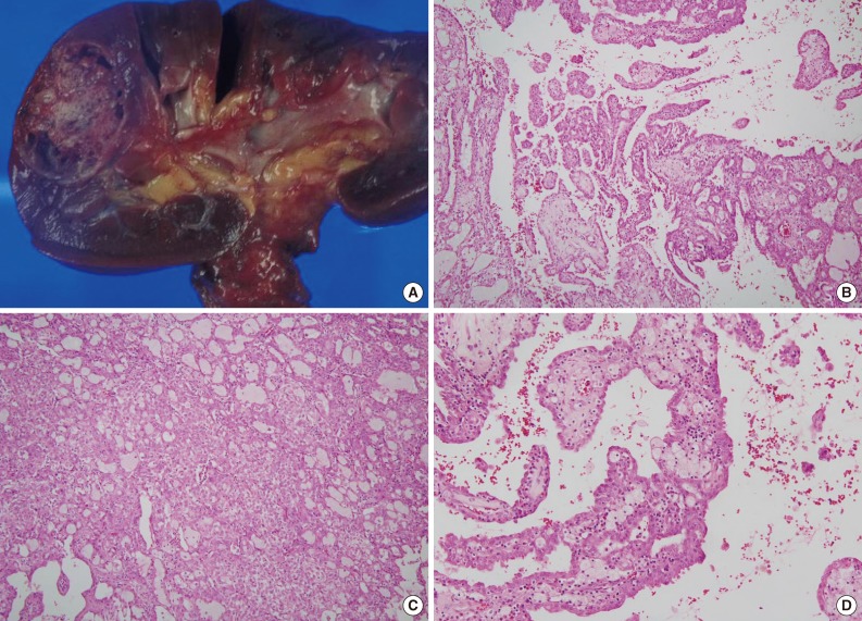
The size of tumor was 3×3×2.5 cm. The tumor was confined to the renal parenchyme and there was no regional lymph node metastases in three regional lymph nodes (pT1aN0 by American Joint Committee on Cancer [AJCC] 7th edition23). Grossly, the tumor was a well-circumscribed, partially cystic mass (Fig. 2A). Microscopically, the tumor showed a papillary, tubular or solid pattern with occasional foam cell collections. Tumor cells were columnar or cuboidal cells with abundant eosinophilic cytoplasm (Fig. 2B-D). No psammoma bodies were found. Initial diagnosis of this case was papillary RCC type 2 and the Fuhrman nuclear grade was 3. Immunohistochemical staining showed positivity for CD10 (focal), EMA, cytokeratin 7, pan-cytokeratin, PAX-2, and vimentin and negativity for cytokeratin 20, AMACR, c-kit, TFE3, and HMB45 (Fig. 3).
Fig. 2.
Pathologic features of renal cell carcinoma showing anaplastic lymphoma kinase (ALK) rearrangement. (A) Grossly, the tumor is a well-circumscribed, partially cystic mass. Microscopically, the tumor shows a papillary (B), tubular or solid pattern (C) with occasional foam cell collections (D).
Fig. 3.
Immunohistochemical findings of renal cell carcinoma showing anaplastic lymphoma kinase (ALK) rearrangement. The tumor cells are positive for cytokeratin 7 (A), PAX-2 (B), and focal positive for CD10 (C) and negative for alpha-methylacyl-CoA racemase (D).
DISCUSSION
Recently, six RCC cases showing ALK gene rearrangement were reported by 4 independent groups.18-21 Two of the reported cases were pediatric cases (6- and 16-year-old) with a history of sickle cell trait and the RCC subtypes of these cases were renal medullary carcinoma and RCC undetermined.18,19 Renal medullary carcinoma was almost exclusively associated with sickle cell trait.2 However, the following 4 cases were adult cases without history of sickle cell trait and subtypes were papillary RCC in 3 cases and one unclassified RCC.20,21 In this study, we screened 829 RCCs and found one case with ALK immunohistochemical positivity and ALK gene rearrangement. Our case is also an adult male case without history of sickle cell trait and the initial diagnosis was papillary RCC type 2. The fusion partners of ALK in RCC were variable, with VCL in two pediatric cases and TPM3 and EML4 in two adult cases.18,19,21 In this study, we did not check the ALK fusion partner (Table 1).
Table 1.
Seven ALK-positive renal cell carcinomas

ALK, anaplastic lymphoma kinase; M, male; VCL, vinculin; F, female; TPM3, tropomyosin 3; EML4, echinoderm microtubule-associated protein like 4.
Two previous adult cases showed poor prognosis,20 and the other two adult cases had a relatively short follow-up period.21 Our patient is alive without evidence of RCC for 12 years and the follow-up period for this patient was the longest of all reported cases. This case and the previous cases suggest that RCC with ALK gene rearrangement may show variable prognosis.
Histologic features of reported RCC cases with ALK rearrangement are variable. Two previous pediatric cases showed typical histologic features of renal medullary carcinoma19 or mixed features of renal medullary carcinoma, chromophobe RCC and transitional cell carcinoma.18 Three previous adult cases had the histologic features of papillary RCC and another case showed papillary, tubular or cribriform growth pattern. Psammoma bodies were found in all previous adult cases and foam cell collection was described in 3 out of 4 cases.20,21 Our case also showed the features of papillary RCC having foam cell collections but psammoma bodies were not found. Excluding the two pediatric cases with sickle cell trait, all four previous adult cases and our case showed the papillary growth pattern.20,21
Two previous studies included large-scale screenings to detect RCC with ALK gene alteration. ALK rearrangement was mainly found in papillary RCC in these two studies, and they could not find ALK rearrangement in 505 clear cell RCC and 34 chromophobe RCC.20,21 We also could not find ALK expression in 689 clear cell RCCs, 73 chromophobe RCCs, and 14 rare RCC subtypes by immunohistochemistry. Taken together, the results of previous reports and this study indicate that RCC with ALK rearrangement may be a particular subset of papillary RCC.
It is difficult to define the frequency of RCC with ALK rearrangement. A previous large-scale study showed the very low incidence of RCC with ALK rearrangement (0.56% of all RCCs), but they occupied 3.7% of non-clear cell and non-chromophobe RCC (2/32).21
Previous adult RCC cases with ALK rearrangement showed similar immunohistochemical staining results, with positivity for cytokeratin, cytokeratin 7, and focal positive or negative for CD10.20,21 These immunohistochemical results are compatible with those of typical papillary RCCs and our case also showed similar results of positivity for cytokeratin and cytokeratin 7, and focal positivity for CD10.
Many targeted therapies have been used for advanced RCC. ALK rearrangement in RCC may add another option for treatment of advanced RCC. ALK inhibition has been tried for treatment of other tumors with ALK rearrangement and some successful results has been reported.10,11 Therefore, we can try ALK inhibition in the treatment of advanced RCC with ALK rearrangement. FISH has been accepted as a standard method for detecting ALK gene rearrangement, but also has disadvantages in terms of cost and time. Instead of FISH, ALK immunohistochemistry can be a reliable screening tool.24 We would have found our case from screening based on ALK immunohistochemistry, which suggests that ALK immunohistochemistry is useful for the detection of RCC with ALK rearrangement.
In conclusion, as far as we know, we have found the first RCC case with ALK gene rearrangement in Korean patients among 829 RCCs. This case showed similar histological and immunohistochemical features to previous adult cases with ALK rearrangement, and showed a relatively good prognosis.
Acknowledgments
This work was supported by grant 03-2010-009 from the Seoul University Hospital Research Fund.
Footnotes
No potential conflict of interest relevant to this article was reported.
References
- 1.Jung KW, Won YJ, Kong HJ, Oh CM, Seo HG, Lee JS. Cancer statistics in Korea: incidence, mortality, survival and prevalence in 2010. Cancer Res Treat. 2013;45:1–14. doi: 10.4143/crt.2013.45.1.1. [DOI] [PMC free article] [PubMed] [Google Scholar]
- 2.Eble JN, Sauter G, Epstein JI, Sesterhenn IA. Pathology and genetics of tumours of the urinary system and male genital organs. Lyon: IARC Press; 2004. pp. 12–39. [Google Scholar]
- 3.Patard JJ, Leray E, Rioux-Leclercq N, et al. Prognostic value of histologic subtypes in renal cell carcinoma: a multicenter experience. J Clin Oncol. 2005;23:2763–2771. doi: 10.1200/JCO.2005.07.055. [DOI] [PubMed] [Google Scholar]
- 4.Singer EA, Gupta GN, Srinivasan R. Targeted therapeutic strategies for the management of renal cell carcinoma. Curr Opin Oncol. 2012;24:284–290. doi: 10.1097/CCO.0b013e328351c646. [DOI] [PMC free article] [PubMed] [Google Scholar]
- 5.Morris SW, Kirstein MN, Valentine MB, et al. Fusion of a kinase gene, ALK, to a nucleolar protein gene, NPM, in non-Hodgkin's lymphoma. Science. 1994;263:1281–1284. doi: 10.1126/science.8122112. [DOI] [PubMed] [Google Scholar]
- 6.Laurent C, Do C, Gascoyne RD, et al. Anaplastic lymphoma kinase-positive diffuse large B-cell lymphoma: a rare clinicopathologic entity with poor prognosis. J Clin Oncol. 2009;27:4211–4216. doi: 10.1200/JCO.2008.21.5020. [DOI] [PubMed] [Google Scholar]
- 7.Wang WY, Gu L, Liu WP, Li GD, Liu HJ, Ma ZG. ALK-positive extramedullary plasmacytoma with expression of the CLTC-ALK fusion transcript. Pathol Res Pract. 2011;207:587–591. doi: 10.1016/j.prp.2011.07.001. [DOI] [PubMed] [Google Scholar]
- 8.Cook JR, Dehner LP, Collins MH, et al. Anaplastic lymphoma kinase (ALK) expression in the inflammatory myofibroblastic tumor: a comparative immunohistochemical study. Am J Surg Pathol. 2001;25:1364–1371. doi: 10.1097/00000478-200111000-00003. [DOI] [PubMed] [Google Scholar]
- 9.Chen Y, Takita J, Choi YL, et al. Oncogenic mutations of ALK kinase in neuroblastoma. Nature. 2008;455:971–974. doi: 10.1038/nature07399. [DOI] [PubMed] [Google Scholar]
- 10.Kwak EL, Bang YJ, Camidge DR, et al. Anaplastic lymphoma kinase inhibition in non-small-cell lung cancer. N Engl J Med. 2010;363:1693–1703. doi: 10.1056/NEJMoa1006448. [DOI] [PMC free article] [PubMed] [Google Scholar]
- 11.Butrynski JE, D'Adamo DR, Hornick JL, et al. Crizotinib in ALK-rearranged inflammatory myofibroblastic tumor. N Engl J Med. 2010;363:1727–1733. doi: 10.1056/NEJMoa1007056. [DOI] [PMC free article] [PubMed] [Google Scholar]
- 12.Galkin AV, Melnick JS, Kim S, et al. Identification of NVP-TAE684, a potent, selective, and efficacious inhibitor of NPM-ALK. Proc Natl Acad Sci U S A. 2007;104:270–275. doi: 10.1073/pnas.0609412103. [DOI] [PMC free article] [PubMed] [Google Scholar]
- 13.Cerchietti L, Damm-Welk C, Vater I, et al. Inhibition of anaplastic lymphoma kinase (ALK) activity provides a therapeutic approach for CLTC-ALK-positive human diffuse large B cell lymphomas. PLoS One. 2011;6:e18436. doi: 10.1371/journal.pone.0018436. [DOI] [PMC free article] [PubMed] [Google Scholar]
- 14.Azarova AM, Gautam G, George RE. Emerging importance of ALK in neuroblastoma. Semin Cancer Biol. 2011;21:267–275. doi: 10.1016/j.semcancer.2011.09.005. [DOI] [PMC free article] [PubMed] [Google Scholar]
- 15.Kim H, Yoo SB, Choe JY, et al. Detection of ALK gene rearrangement in non-small cell lung cancer: a comparison of fluorescence in situ hybridization and chromogenic in situ hybridization with correlation of ALK protein expression. J Thorac Oncol. 2011;6:1359–1366. doi: 10.1097/JTO.0b013e31821cfc73. [DOI] [PubMed] [Google Scholar]
- 16.Paik JH, Choe G, Kim H, et al. Screening of anaplastic lymphoma kinase rearrangement by immunohistochemistry in non-small cell lung cancer: correlation with fluorescence in situ hybridization. J Thorac Oncol. 2011;6:466–472. doi: 10.1097/JTO.0b013e31820b82e8. [DOI] [PubMed] [Google Scholar]
- 17.Paik JH, Choi CM, Kim H, et al. Clinicopathologic implication of ALK rearrangement in surgically resected lung cancer: a proposal of diagnostic algorithm for ALK-rearranged adenocarcinoma. Lung Cancer. 2012;76:403–409. doi: 10.1016/j.lungcan.2011.11.008. [DOI] [PubMed] [Google Scholar]
- 18.Debelenko LV, Raimondi SC, Daw N, et al. Renal cell carcinoma with novel VCL-ALK fusion: new representative of ALK-associated tumor spectrum. Mod Pathol. 2011;24:430–442. doi: 10.1038/modpathol.2010.213. [DOI] [PubMed] [Google Scholar]
- 19.Mariño-Enríquez A, Ou WB, Weldon CB, Fletcher JA, Pérez-Atayde AR. ALK rearrangement in sickle cell trait-associated renal medullary carcinoma. Genes Chromosomes Cancer. 2011;50:146–153. doi: 10.1002/gcc.20839. [DOI] [PubMed] [Google Scholar]
- 20.Sukov WR, Hodge JC, Lohse CM, et al. ALK alterations in adult renal cell carcinoma: frequency, clinicopathologic features and outcome in a large series of consecutively treated patients. Mod Pathol. 2012;25:1516–1525. doi: 10.1038/modpathol.2012.107. [DOI] [PubMed] [Google Scholar]
- 21.Sugawara E, Togashi Y, Kuroda N, et al. Identification of anaplastic lymphoma kinase fusions in renal cancer: large-scale immunohistochemical screening by the intercalated antibody-enhanced polymer method. Cancer. 2012;118:4427–4436. doi: 10.1002/cncr.27391. [DOI] [PubMed] [Google Scholar]
- 22.Kim SH, Choi Y, Jeong HY, Lee K, Chae JY, Moon KC. Usefulness of a break-apart FISH assay in the diagnosis of Xp11.2 translocation renal cell carcinoma. Virchows Arch. 2011;459:299–306. doi: 10.1007/s00428-011-1127-5. [DOI] [PubMed] [Google Scholar]
- 23.Edge SB, Byrd DR, Compton CC, Fritz AG, Greene FL, Trotti A. AJCC cancer staging manual. 7th ed. New York: Springer; 2010. [Google Scholar]
- 24.Conklin CM, Craddock KJ, Have C, Laskin J, Couture C, Ionescu DN. Immunohistochemistry is a reliable screening tool for identification of ALK rearrangement in non-small-cell lung carcinoma and is antibody dependent. J Thorac Oncol. 2013;8:45–51. doi: 10.1097/JTO.0b013e318274a83e. [DOI] [PubMed] [Google Scholar]


