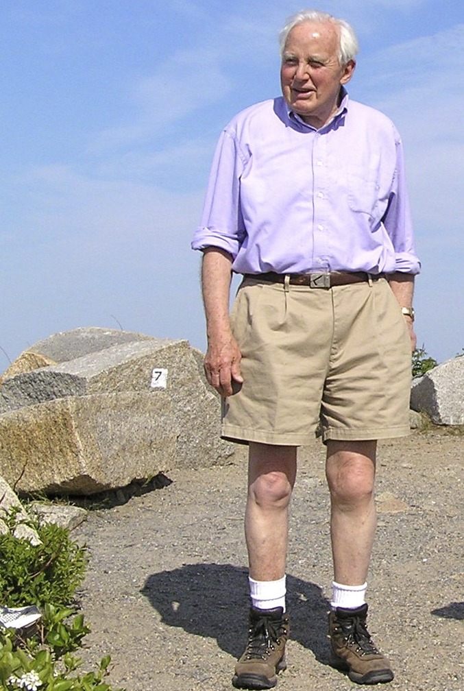
Hugh Huxley on the Boston North Shore.
Hugh Esmor Huxley was born on February 25th, 1924 and raised in Birkenhead, Cheshire, England. He gained admittance to Christ’s College Cambridge in 1941 and studied physics. Britain was at war; Hugh elected to interrupt his studies and join the Royal Air Force, working on the development of radar. He was honored for his substantial contributions by being awarded membership of the Order of the British Empire (MBE).
Hugh returned to Cambridge in 1947, his enthusiasm for nuclear physics irrevocably diminished by the horrors of Hiroshima. For his doctoral research at the Cavendish Laboratory, rather than pursue nuclear physics Hugh joined Max Perutz’s group working on the X-ray analysis of crystalline proteins, where he became John Kendrew’s first research student. Hugh had come across Dick Bear’s X-ray diffraction patterns from air-dried muscle and, moreover, was amazed to discover no one really knew how muscle works. Hugh set about building an X-ray camera and X-ray generator capable of resolving spacing of 300–400 Å to take diffraction photographs of wet “living” muscle. He saw for the first time the equatorial reflections arising from a hexagonal array of filaments and somewhat prophetically guessed that these arose from filaments of myosin on the hexagonal lattice points, with actin filaments in between. Hugh also suggested the filaments were linked by “cross bridges.”
In 1952 Hugh went to the Massachusetts Institute of Technology (MIT) as a postdoctorate scholar in Frank Schmitt’s department to learn electron microscopy. Soon, Hugh’s tranverse sections of fixed muscle showed the arrangement of filaments he had postulated from his X-ray studies, together with a hint of cross-bridges connecting the filaments. The arrival of Jean Hanson at MIT provided further impetus. Jean was experienced in phase-contrast microscopy. The units of cross-striated muscle are sarcomeres, about 2.5 μ in length, delineated by Z-lines, all in series with each other. In the phase-contrast pictures of single muscle fibers the subdivisions of the sarcomere into the H-zone, A-bands, and I-bands are clear. Examining glycerinated rabbit muscle fibers after extraction of the myosin with Hasselbach–Schneider solution, which dissolves myosin, it became clear that filaments in the I-bands were just actin, the H-zones were just myosin, and the A-bands were overlap zones for actin and myosin filaments. Carrying out these experiments at various degrees of stretch of the muscle demonstrated that the muscle consists of two sets of filaments that move past each other as muscle contracts. Hugh and Jean wrote up the results for Nature (1), but on the advice of Frank Schmitt left out the sliding filament hypothesis: Frank maintained that good data should not be spoiled with speculation. This may have been bad advice, because this groundbreaking work was never acknowledged by the Nobel committees.
While in Boston, Hugh formed a life-long friendship with Andrew Szent-Györgyi, the younger cousin of Nobel prize winner Albert Szent-Györgyi. Hugh often went down to the Marine Biological Laboratory in Woods Hole during summer weekends and stayed with Andrew and Eve Szent-Györgyi. In the summer of 1953, Hugh met his namesake (but not relative) Andrew Huxley in Woods Hole. Unlike the muscle establishment, Andrew Huxley was sympathetic to Hugh’s ideas of sliding filaments and indeed, using his own sophisticated interference microscope, was reaching similar conclusions. The next year, together with Jean Hanson and Rolf Niedergerke, the researchers published the sliding-filament hypothesis in two papers back-to-back in Nature (2, 3). This was the beginning of modern muscle research.
In the spring of 1954, Hugh returned to Cambridge, England, to a research fellowship at Christ’s College, but in 1956 he moved to a position at University College, London, in Bernard Katz’s Department. Here he made substantial advances in the fixing and sectioning of muscle. Hugh produced wonderful images of longitudinal sections of cross-striated muscle, barely 15.0-nm-thick and clearly showing the cross-bridges connecting the thick (myosin) and thin (actin) filaments. With Geoffrey Zubay, Hugh developed “negative staining.” Using negative staining, Hugh discovered that the isolated cross-bridges bound specifically and regularly to the actin filament, one cross-bridge per actin, with the symmetry of the actin filament (decorated actin). He saw with wonder that the polarity of the actin filaments reverses as you go through the Z-line.
In 1962 Hugh moved back to Cambridge, to the newly opened Medical Research Council Laboratory of Molecular Biology (LMB). At the LMB he set up a Siemens Elmiscope-1 and first used it to investigate the spontaneous assembly of myosin molecules in vitro to form bipolar thick filaments. Hugh investigated the structures of the thin actin-containing filaments. He and John Haselgrove were able to find an X-ray diffraction signal from muscle that showed how tropomyosin, one component of the thin filament, moved in response to activation by calcium ions, so as to free up the cross-bridge binding site on actin.
In February 1966 Hugh married Frances Maxon Fripp from Boston, MA. Frances brought three teenage children into the marriage. The family duly moved to Cambridge, England. In 1970 Frances gave birth to Olwen.
Hugh was still intrigued by the low-angle X-ray diffraction pattern of muscle that he had uncovered in his doctoral research. He wished to follow the changes when a resting muscle is stimulated and allowed to contract. According to the “swinging cross-bridge” hypothesis that he first presented in a Scientific American article in 1958 (4), the cross-bridges bind in an initial conformation and “row” the actin filaments past the myosin filaments by going over into a second more angled conformation, followed by release from actin and rebinding in the first conformation, but for many years the swinging cross-bridge hypothesis remained unsubstantiated. A change in orientation of the cross-bridge would manifest itself as a change in the low-angle X-ray scattering pattern, if you could get enough intensity to measure this in a few milliseconds. Conventional X-ray sources had reached their limit but Synchrotron Radiation was in principle a very bright X-ray source. With Hugh’s support, the newly founded European Molecular Biology Laboratory in Heidelberg opened an outstation on the DORIS (Doppel-Ring-Speicher) electron storage ring at Deutsches Elektronen-Synchrotron Hamburg for the exploitation of synchrotron radiation as an X-ray source. Here in 1981 Hugh and his team of collaborators were finally able to demonstrate that swinging cross-bridges actually swung (5).
Together with Aaron Klug, Hugh became joint head of the Division of Structural Studies. Later he became deputy Director of the LMB, but enforced retirement at 65 was imminent. Hugh was offered a professorial appointment at the Rosenstiel Basic Medical Sciences Research Center at Brandeis, which he accepted. In 1987 the family moved to Concord, MA. In 1988 Hugh was appointed Director of the Rosenstiel Center, a position he held for six years.
Hugh and Frances built a house in Woods Hole, where Frances spent her summers directing local drama groups and sailing. Hugh came at weekends. After Gordon Conferences on muscle Hugh would invite friends and expostdoctorate scholars to visit Woods Hole to enjoy a not always uneventful trip to Martha’s Vineyard on his sloop Saraband.
In 1997 the Advanced Photon Source at Argonne became available as the world’s brightest X-ray source. The Biophysics Collaborative Access Team (BioCAT) team led by Tom Irving set up excellent beam lines that provided Hugh with the opportunity to investigate a phenomenon first explored with single muscle fibers by Vincenzo Lombardi and his collaborators: the 14.5-nm meridional reflection is actually split into two closely spaced reflections that arise from interference between the two halves of the sarcomere. Because it is an interference phenomenon, this process allows the average position of the cross-bridges attached to actin to be measured with high precision. Massimo Reconditi and Hugh were able to work out the distribution of cross-bridges during a contraction. This work was reported in Hugh’s two last papers, which demonstrate his characteristic rigor and analytical ability (6); they are a fitting conclusion to his scientific career and provide an elegant vindication of the swinging cross-bridge.
Hugh died unexpectedly on July 25th, 2013 in Woods Hole. Active to the last, he combined a wonderful experimental ability with a very analytical mind. He was also a gentle humanist and a man of great integrity. Hugh’s originality and creativity led to his being elected to the Royal Society of London at the young age of 36. He was a member of the National Academy of Sciences. In 1971 Hugh was awarded the Louisa Gross Horwitz Prize. In 1975 he received the Gairdner Award. In 1997 he was awarded the prestigious Royal Society Copley Medal. The citation reads:
In recognition of his pioneering work on the structure of muscle and on the molecular mechanisms of muscle contraction, providing solutions to one of the great problems in physiology.
Footnotes
This article is an abridged version of the Obituary of Hugh Huxley published in J Muscle Res Cell Motil, 10.1007/s10974-013-9365-6.
References
- 1. Hanson J, Huxley HE (1953) Nature 172:530–532. [DOI] [PubMed]
- 2. Huxley HE, Hanson J (1954) Nature 173:973–976. [DOI] [PubMed]
- 3. Huxley AF, Niedergerke R (1954) Nature 173:971–973. [DOI] [PubMed]
- 4. Huxley HE (1958) Scientific American 199:58–67. [PubMed]
- 5. Huxley HE, Simmons RM, Faruqi AR, Kress M, Bordas j, and Koch MHJ (1981) Proc Natl Acad Sci USA 78:2297–2301. [DOI] [PMC free article] [PubMed]
- 6. Huxley H, Reconditi M, Stewart A, Irving T (2006) J Molec Biol 383:743–761, 762–772. [DOI] [PubMed]


