

Abstract
The emergence of Nipah virus (NiV) infection into the pig population and subsequently into the human population is believed to be due to changes in ecological conditions. In Malaysia, A major NiV outbreak occurred in pigs and humans from September 1998 to April 1999 that resulted in infection of 265 and death of 105 persons. About 1.1 million pigs had to be destroyed to control the outbreak. The disease was recorded in the form of a major outbreak in India in 2001 and then a small incidence in 2007, both the outbreaks in West Bengal only in humans without any involvement of pigs. There were series of human Nipah incidences in Bangladesh from 2001 till 2013 almost every year with mortality exceeding 70 %. The disease transmission from pigs acting as an intermediate host during Malaysian and Singapore outbreaks has changed in NIV outbreaks in India and Bangladesh, transmitting the disease directly from bats to human followed by human to human. The drinking of raw date palm sap contaminated with fruit bat urine or saliva containing NiV is the only known cause of outbreak of the disease in Bangladesh outbreaks. The virus is now known to exist in various fruit bats of Pteropus as well as bats of other genera in a wider belt from Asia to Africa.
Keywords: Nipah virus, NiV, Henipavirus, Fruit bats, Pigs, Encephalitis
Introduction
The recent pandemic threat posed by the viral pathogens such as Coronovirus, Influenza virus etc. implies that disease emergence and spread are not limited by geographical boundaries. In many cases the animals are found to be the source of infection for human infection [95]. Only 87 out of 1,399 human pathogen have been first reported in humans in the years since 1980 [95]. India’s fast-growing human population and resulting increasing animal-human interactions, combined with changing environmental conditions and inadequate sanitation and regulation, have made India one of the world’s top hotspots for livestock diseases, including zoonotic diseases—those that pass from animals to humans and which make up 75 % of all human diseases. Controlling zoonoses is particularly important in developing countries, where the absolute burden of these diseases is up to 130 times greater than in developed countries [31]. Emerging zoonoses are the product of socioeconomic and anthropogenic environmental changes. Expansion of road networks, development of agricultural land, and intensification of wildlife trade have caused novel pathogens to emerge from wildlife, Nipah virus (NiV) is one of the best examples of the emerging zoonoses [19].
Nipah is a viral zoonotic disease caused by NiV of the Henipavirus genus of Paramyxoviridae family. Pteropus bats (fruit eating species, popularly known as flying foxes) are supposed to be the natural hosts of the virus.
The emergence of NiV into the pig population and subsequently into the human population is believed to be due to changes in ecological conditions. Urbanization, deforestation and drought resulting in a shortage of resources for bat populations could have compelled bats to move from their natural habitats to agricultural areas. Among the factors that contributed to the disease emergence in Malaysia is the establishment of pig farms within the range of natural host that led to the initial introduction into the pig population; the maintenance of high densities of pigs led to the rapid dissemination of the infection within local pig populations; and the transport of pigs to other geographic areas for commerce led to the rapid spread of disease in pigs in southern Malaysia and Singapore. The presence of high density, amplifying host population facilitated transmission of the virus to human [48]. The same may be true for the NiV outbreaks in Bangladesh and India and it is postulated that the outbreak in humans may be due to direct contact with bats or indirectly by contact with material contaminated by bats. It is apparent from the presence of the virus and antibodies in the fruit bats of the region and 13 years of continuous NiV outbreaks in humans in Bangladesh that it is the potential threat to the Indian subcontinent. The medical and veterinary professionals should also increase the awareness of the disease particularly hosts and mode of transmission of the virus. Therefore this brief account of the virus has been presented in this paper.
Historical background
The first incidence of the NiV was recorded simultaneously in humans and pigs in Malaysia in 1998–1999 [66]. Chua et al. [13, 14] has presented a detailed account of the initial Nipah outbreak in Malaysia, where several human cases of viral encephalitis occurred over a 35-week period from September 1998 to May 1999. On the basis of clinical signs and association of pigs, the disease was considered to be already circulating Japanese encephalitis among pig farmers, creating considerable anxiety and fear throughout the country. Total 265 cases were reported of which 105 died. The outbreak started in the pig farmers near Ipoh in the Kinta District of Perak, some 200 km north of Kuala Lumpur and spread to three other major pig-rearing areas (the largest in Southeast Asia) in Negeri Sembilan and Sungei Buloh in Selangor. The disease was named after Kampung Sungai Nipah (Nipah River Village), where the first viral isolate was obtained and therefore named as NiV. Retrospective investigations conducted later had suggested that NiV was responsible for sporadic disease in pigs in Peninsular Malaysia since late 1996, but was not recognized because the clinical signs were not markedly different from those of several endemic pig diseases, and because morbidity and mortality were not remarkable [2]. Singapore recorded the subsequent outbreak in 1999 in slaughterhouse persons due to infected pigs brought from Malaysia [76]. In India, the disease was recorded in humans without any involvement of pigs. There were two outbreaks, major one occurred in 2001 in Siliguri in West Bengal and an isolated incidence in Nadia also in West Bengal in 2007. Bangladesh recorded several Nipah outbreaks in human beings covering several districts almost every year during 2001–2013 [98].
The disease was eradicated from Malaysia in 1999 and the last of the two recognized outbreaks from India was in 2007 but the clinical expression of the disease in human being with very high case fatality has been continued in Bangladesh. Nevertheless, in addition to these countries, the virus has been detected in fruit bats or the bats were seropsitive to NiV antibodies from Cambodia [79], Thailand [90], Indonesia [81], India [23, 99], Madagascar in Southern Africa [41] and Ghana in West Africa [22, 37]. P. giganteus is the only Pteropus species present in Bangladesh [59]. In the Naogaon (Bangladesh) investigation, 2 of 19 P. giganteus specimens had antibody against NiV. None of 31 other animals from various species had Nipah antibodies [39].
In the initial Malaysian outbreaks, concurrent respiratory ailment, encephalitis with mortality in pigs was recorded. It was also revealed that majority of the human cases comprised of occupationally related to the pigs and having the history of direct contact with live pigs [77]. It was also revealed that the disease spread to Negeri Sembilan through the sale and movement of infected pigs. Preliminary characterization of an isolate from a human case at the Centers for Disease Control and Prevention (CDC) in Fort Collins and Atlanta, USA, showed the primary causative agent in the outbreak to be a previously undescribed virus of the family Paramyxoviridae. These investigations showed the new virus, named NiV, was closely related to (already known since 1994) Hendra virus [13].
During the Malaysian NiV outbreak, two Indonesian farm workers who returned home to Indonesia from Malaysian pig farm were also succumbed to infection [27]. The outbreak also spread to neighboring Singapore during March 1999 where 11 abattoir workers handling pigs from infected farms in Malaysia developed the disease with one fatality. In addition to these 11 cases in Singapore, there were two apparently asymptomatic case patients among workers in this abattoir [8, 77, 78].
NiV in Malaysia emerged in 1998 during an outbreak of infectious respiratory and neurologic disease in commercially farmed pigs, presumably after virus spillover from Malaysian flying foxes [14]. Pigs were the source of infection for farm and abattoir workers, resulting in a widespread outbreak of severe febrile encephalitic disease among humans [77]. NiV infection has not been detected in Malaysia or Singapore after 1999, but recurring (almost annual) human cases of Nipah encephalitis with very high case fatalities in Bangladesh and sporadic outbreaks in India since 2001 (Table 1) [43, 44, 46, 47, 98].
Table 1.
| Year/month | Location | No. cases | No. deaths | Case fatality (%) |
|---|---|---|---|---|
| Jan–Feb 2001 | Siliguri (India) | 66 | 45 | 68 |
| Apr–May 2001 | Meherpur (Bangladesh) | 13 | 9 | 69 |
| Jan 2003 | Naogaon (Bangladesh) | 12 | 8 | 67 |
| Jan 2004 | Rajbari(Bangladesh) | 31 | 23 | 74 |
| Apr 2004 | Faridpur (Bangladesh) | 36 | 27 | 75 |
| Jan–Mar 2005 | Tangail (Bangladesh) | 12 | 11 | 92 |
| Jan–Feb 2007 | Thakurgaon (Bangladesh) | 7 | 3 | 43 |
| Mar 2007 | Kushtia, Pabna, Natore (Bangladesh) | 8 | 5 | 63 |
| Apr 2007 | Naogaon (Bangladesh) | 3 | 1 | 33 |
| April 2007 | Nadia (India) | 5 | 5 | 101 |
| Feb 2008 | Manikgonj (Bangladesh) | 4 | 4 | 101 |
| Apr 2008 | Rajbari and Faridpur (Bangladesh) | 7 | 5 | 71 |
| Jan 2009 | Gaibandha, Rangpur and Nilphamari (Bangladesh) | 3 | 0 | 0 |
| Jan 2009 | Rajbari (Bangladesh) | 1 | 1 | 101 |
| Feb–Mar 2010 | Faridpur, Rajbari,Gopalganj,Madaripur (Bangladesh) | 16 | 14 | 87.5 |
| Jan–Feb 2011 | Lalmohirhat, Dinajpur, Comilla, Nilphamari and Rangpur (Bangladesh) | 44 | 40 | 91 |
| Feb 2012 | Joypurhat, Rajshahi, Natore, Rajbari and Gopalganj (Bangladesh) | 12 | 10 | 83 |
| Jan–Feb 2013 | Gaibandha, Natore, Rajshahi, Naogaon, Rajbari, Pabna, Jhenaidah, Mymensingh (Bangladesh) | 12 | 10 | 83 |
| Total | 292 | 221 | 75.7 |
The detailed account of the first NiV outbreak in India (Siliguri, West Bengal) during 2001 has been published in two reports [5, 36]. An outbreak of acute encephalitis occurred in Siliguri (West Bengal) town of India between January 31 and February 23, 2001. A total of 66 probable human cases and 45 deaths were reported. Later, the clinical material obtained during the Siliguri outbreak was retrospectively analyzed for evidence of NiV infection [97]. Analysis of the limited sequence data suggested that the NiV strains associated with the outbreak were more closely related to NiV isolated in Bangladesh than to NiV isolated in Malaysia [5].
In the Malaysian outbreak, pigs were the intermediate hosts. NiV was isolated from fruit bats in Malaysia [16] Fruit bats with antibodies to NiV were captured in the outbreak areas of Bangladesh but no intermediate animal host was identified. In Bangladesh, NiV might have been transmitted to humans by direct contact with bats or indirectly by contact with material contaminated by bats. Person-to-person spread was also noted during the 2004 NiV outbreak in Faridpur, Bangladesh [45, 96]. The range of Pteropus giganteus, one of the flying foxes commonly found in South Asia [72] includes West Bengal. Therefore, the range of the proposed natural reservoir for NiV extends into northeastern India. Since the geographical features of West Bengal are similar to those of Bangladesh, environmental circumstances that favor transmission of NiV to humans would also likely be same in West Bengal. Many of the epidemiologic features of the outbreak in Siliguri were similar to those of the recent NiV outbreaks in Bangladesh.
Analysis of the limited sequence data suggested that the NiV strains associated with the outbreak were more closely related to NiV isolated in Bangladesh than to NiV isolated in Malaysia. These data extend the previous observation that viruses circulating in different areas have unique genetic signatures and suggest that these strains may have co-evolved within local natural reservoirs [35, 79].
After 2001, the second NiV outbreak was reported in West Bengal in India in 2007. Between 11 and 28 April 2007, Krishnan [53] reported 30 cases of fever with acute respiratory distress and/or neurological symptoms from Nadia district of West Bengal in India. The cases presented mainly with fever, headache and bodyache with a few cases having episodes of vomiting, disorientation, respiratory distress. Five cases ended fatally within 3–10 days of onset. However, Arankalle et al. [1] investigated the same outbreak and stated that similar other cases had not been reported from the village or the surrounding area, thus showed case fatality (5/5) as 100 %. They also amplified full-genome sequence of NiV (18252 nt) from lung tissue that showed 99.2 % nucleotide and 99.8 % amino acid identity with the Bangladesh-2004 isolate, suggesting a common source of the virus.
Chong et al. [10] discussed the relationship between Henipaviruses and fruit bats. Epidemiological studies have shown that the virus could be transmitted from bat to human and from human to human. Wildlife studies have also shown that the virus was widely distributed in at least 10 genera and 23 species of bats in a large part of Asia and Africa. As bats are long distant flying, gregarious animals living in large colonies which could exchange novel viruses from one species to another, it is not unexpected that the seroprevalence of Henipavirus among bat colonies are relatively high. Chong et al. [10] also commented that due to widespread distribution of both the Henipavirus and its hosts, the virus will remain an important cause of zoonotic disease.
Aetiology
The NiV is classified under the subfamily Paramyxovirinae in the family Paramyxoviridae that comprises the five genera Respiro-, Morbilli-, Rubula-, Avula-,and Henipavirus, as well as a group of yet unclassified viruses [55]. The genus Henipavirus contains two of the most pathogenic viruses known in humans, Hendra- and NiV, which were discovered only in 1994 and 1998, respectively [14, 56, 91] Due to its high pathogenicity, Biosafety level-4 containment is required to work with live NiV in laboratories. [58].
The genome consists of six genes (N, P, M, F, G and L) yielding nucleoprotein, phosphoprotein, matrix, fusion, glycoprotein and large RNA polymerase [6]. The genome of Henipa group of viruses is large (around 18250 nucleotides) as compared to other members of paramyxoviruses due to an extended open reading frame of the P gene and to longer non-coding regions for all the genes except L gene. NiV does not have the hemagglutinin and neuraminidase properties as commonly found in many Paramyxoviruses. The G and F proteins of NiV are required in mediating the viral entry into the cell as well as for inducing neutralizing antibodies, [83]. The P gene of Henipaviruses encodes at least three nonstructural proteins (C, V, and W) in addition to the P protein [33, 34]; however, P protein is the only essential gene product for genome replication. The additional gene products are usually not required for virus replication in vitro, though they often serve as virulence factors in vivo [20, 21].
The studies conducted by Tanimura et al. [88] indicated that NiV is highly pathogenic to chicken embryos and that the chicken embryo represented a useful model for studying the vascular and neuronal tropisms of NiV. The virus also grows well in many of the mammalian cells but the rate of growth and patterns of cytopathic effect (CPE) produced in all culture varies with the type of mammalian cells used. With Vero cells, CPE could be visible by 5th–7th day post-inoculation of clinical samples; in subsequent passages, a complete CPE is observed by 24 h post-inoculation. It produces high viral titre (108 infectious particles per mL) in Vero cells at full CPE. The progression of the CPE also shows inclusions of viral nucleocapsids within the cytoplasm, budding of the nucleocapsid at the plasma membrane and pleomorphic extracellular enveloped virus particles filled with collection of tangled viral nucleocapsids [30, 40]. In electron microscopic studies, negative stain preparations revealed nucleocapsids with the typical ‘herringbone’ appearance that is characteristic for paramyxoviruses [11, 30]. Virus particles vary in size from 120 to 500 nm. Thin-section EM studies of infected cells revealed filamentous nucleocapsids within cytoplasmic inclusions incorporated into virions budding from the plasma membrane. Pleomorphic extracellular virus particles, with an average diameter of 500 nm, and fine surface projections, were sporadically seen [12, 54].
The host range
Yob et al. [101] and Chua [12] have confirmed that fruit bats (popularly known as flying foxes) found in Malaysia, P. vampyrus and P. hypomelanus, are the natural reservoir hosts of NiV. The virus is assumed to have been introduced into the swine population from bat species of Pteropus. Malaysia has diverse bat fauna, with at least13 species of fruit bats, including two species of flying foxes (Pteropid bats) and more than 60 species of insectivorous bats [63]. A serological survey during the 1999 outbreak demonstrated neutralizing antibodies in 5 species of bats, 4 species of fruit bats and one species of insectivorous bats. This included 31 % positive antibody status to Pteropus hypomelanus (island flying foxes) and 17 % positive antibody status to Pteropus vampyrus (Malayan flying foxes) in Peninsular Malaysia [100]. Serologic and RT-PCR testing of blood and urine from Pteropid fruit bats in Malaysia and Cambodia showed NiV infection, which suggested that these animals were reservoir hosts [76, 100]. The geographic range of Pteropus includes all of southeast and south Asia and several other species have been found with antibodies to NiV. The virus has been found in excretions of Pteropid bats, Chua et al. [15] isolated NiV from urine of bats in Malaysia.
Pigs play the part of amplifying host. It has been proved that the human outbreaks in Malaysia and Singapore with encephalitis and ~40 % case fatality occurred after contact with infected swine. The outbreak in Singapore ended when the importation of pigs from Malaysia was prohibited, and the outbreak in Malaysia ceased when over 1 million pigs (45 % of all pigs in Malaysia) were culled from the outbreak area and immediately surrounding areas [77]. Serological studies indicated that NiV occurred in dogs, cats, horses and goats in the outbreak areas of Malaysia but all were effectively dead-end hosts [84]. Apart from pigs, experimental infection has been produced in cats [64], golden hamsters [94] and squirrel monkeys (Saimiri sciureus) [62]. Experimental studies have shown that infected cats shed NiV through the nasopharynx and in urine during viremic phase and of 2 cats infected, one recovered from experimental NiV infection with a high neutralizing antibody titer within 21 days [64]. In a pregnant cat experimentally infected with NiV, the placenta and uterine fluid contained high levels of recoverable virus. The high levels of viral shedding combined with viral replication in fetal tissues suggested that both vertical and horizontal transmission of NiV could play a role in spillover events, an essential element in the epidemiology of Henipavirus infection [69].
Dogs in the outbreak area have also been shown to suffer from NiV infection [73]. NiV infection was confirmed by immunohistochemical examination of one dead and one dying dog from the epidemic area in Malaysia. Both showed histologic evidence of severe disease [38]. Many dogs sampled from the Bukit Pelanduk and Sepang epidemic area around the time of the pig culling had antibodies to NiV [28]. However, Mills et al. [65] analysed the serologic status in the dogs carefully and concluded that in the absence of infected pigs, dogs were not a secondary reservoir for NiV.
The transmission route of Nipah virus
The direct contact with pigs or fresh pig products was responsible for NiV transmission to humans, confirming preliminary observations by health workers [13]. The Malaysian experience showed that the disease spread rapidly among pigs in the infected farm that was attributed to direct contact with excretions and secretions such as urine, saliva, pharyngeal and respiratory secretions of infected pigs. The findings in the pig’s respiratory system could explain the severe pulmonary symptoms in these animals and provide support for the suggestion that aerosol spread of NiV from pig to human represents an important mode of transmission [66]. Widespread surveillance of pig populations to detect infected pigs, and culling of sick pigs stopped the epidemic [67]. Urine exposure may also be associated with transmission as NiV antigen has been demonstrated in the renal tubules of pigs and a concurrent outbreak of NiV among abbatoir workers in Singapore also showed the associatedness of infected pig urine and exposure to the workers [67, 68].The possible mechanical transmission by repetitive use of same needles or equipment without further sterilization after each use for health intervention and artificial insemination and sharing of boar semen within a farm were also implicated. The possible role of transmission by infected dogs and cats found in the affected farm could not be excluded [12].
Several outbreaks of NiV have been reported in human beings in Bangladesh from 2001 to 2013 [43, 97] and two outbreaks in India [1, 5], but none had showed any involvement of pigs. Investigation of different Nipah outbreaks in Bangladesh have identified different routes of transmission including climbing trees (probably contaminated with infected date palm sap), contact with sick persons, and contact with sick animals [39, 44–46, 68]. Another way of NiV getting transmitted from P. giganteus to humans recorded in Bangladesh is food-borne. Fruit bats (P. giganteus) are a nuisance to date palm sap collectors because the bats drink the sap at night from the clay pots used to collect the sap. The investigations by Luby et al. [59] suggested that NiV was transmitted from P. giganteus to persons through drinking fresh date palm sap. Date palm sap is a national delicacy that is enjoyed by millions of Bangladeshis each winter. However, the sap is occasionally contaminated with NiV-infected bat urine or saliva that contains a sufficient dose of NiV to be fatal to humans. In India, in a bat sample survey, NiV RNA was detected in a liver homogenate of P. giganteus captured in Myanaguri, West Bengal [100] (Figs. 1, 2, 3)
Fig. 1.
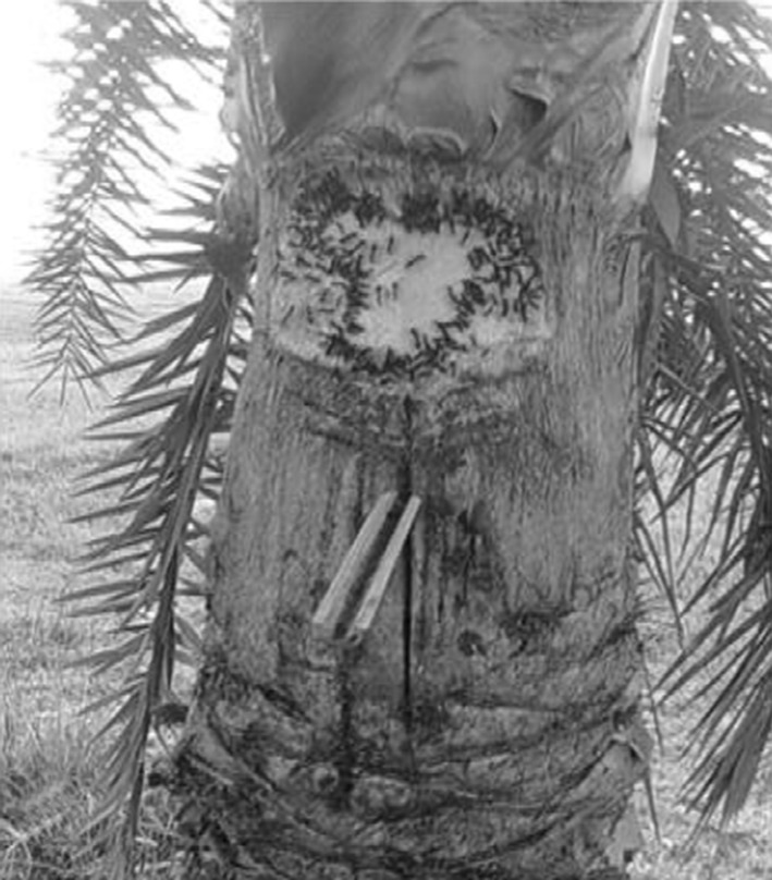
The date palm trees are scratched to collect sap through a bamboo channel
Fig. 2.
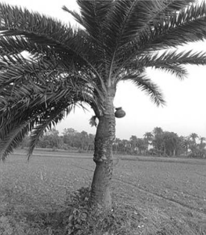
An earthen pot is tied for collection of the sap
Fig. 3.

A bat (P. giganteus) colony in Malda town in West Bengal
In Siliguri, India, transmission of the virus was also reported within a health-care setting, where 75 % of cases occurred among hospital staff or visitors [5]. Nipah cases tend to occur in a cluster or as an outbreak, although 18 % of cases in Bangladesh were isolated. Strong evidence indicative of human-to-human transmission of NiV was found in Siliguri (India) in 2001 and in Bangladesh in 2004 [97]. Approximately one-half of recognized Nipah case patients in Bangladesh developed their disease following person-to-person transmission of the virus [60]. Sazzad et al. [80] further confirmed human-to-human transmission during Bangladesh outbreak in 2010 while studying 16 case-patients.
The clinical signs in animals
Pigs: NiV infection in pigs is known as porcine respiratory and neurologic syndrome, porcine respiratory and encephalitic syndrome, and barking pig syndrome [75]. The only experience of the disease in pigs is available from Malaysia during 1998–1999; the disease was generally associated with low (<5 %) mortality but high (~80 %) morbidity.
An account of NiV in pigs has been given by Nor [71]. The incubation period was estimated to be 7–14 days. NiV caused illness with both CNS and respiratory manifestations though the clinical patterns of the disease varied according to the age of the pigs. Most pigs developed a febrile respiratory disease with a severe cough that led to the local names for the disease, “barking pig syndrome” and “one-mile cough.” It was associated with neurological disease in sows and boars, while weanling and feeder pigs more commonly manifest respiratory signs.
Pigs less than 6 months old usually presented with an acute febrile illness with respiratory signs ranging from rapid and labored respiration to a harsh, non-productive cough and open mouth breathing with severe cough and haemoptysis occurred in severe cases. Neurological signs such as trembling, muscular twitching, spasm and myoclonus, hind-leg weakness with varying degrees of spastic or flaccid paresis and incoordinate gait accompanied respiratory signs in some affected pigs. In boars and sows, acute febrile illness with labored respiration, increased salivation and nasal discharge accompanied neurological signs such as agitation, head pressing or knocking, clamping of mouth, nystagmus, tetanus-like spasm and seizures. Early abortions might have occurred in pregnant sows [12]. Pulmonary inflammation, glomerular and tubular necrosis with syncytia formation in kidneys was reported in infected dogs. Cats are also susceptible to infection with vasculopathy, endothelial syncytia in many organs. There is severe pulmonary inflammation with prominent involvement of bronchial epithelium. Several other animals have been experimentally infected include the Guinea pig, hamster, ferret, non human primates (Squirrel monkey and African green monkey) and chick embryo with extensive vasculopathy and parenchymal lesions in CNS and other organs viz., lungs, kidneys, liver, urinary bladder, female genital tract, muscles, lymphoid organs. Surprisingly, Mouse and rat do not apparently develop clinical disease for reasons yet to be known [92].
In post-mortem finding of affected pigs, lungs showed varying degrees of consolidation and petechiae to ecchymotic hemorrhages. The bronchi and trachea were filled with frothy and in some cases blood stained fluids. The cut surface showed exudate of varying consistency in the bronchi. The brain and kidneys showed generalized congestion and oedema [71]. Histologically, the major lesion was moderate to severe interstitial pneumonia with widespread hemorrhages and syncytial-cell formations in the endothelial cells of blood vessels of the lung [14, 71]. Generalized vasculitis with fibrinoid necrosis, hemorrhages, and infiltration of mononuclear cells sometimes associated with thrombosis, were observed notably in the lung, kidney and brain tissue. Immunohistology showed a high concentration of the viral antigens in the endothelium of the blood vessels, particularly in the lung. A lesser number of pigs showed prominent meningeal inflammatory infiltrates. This picture suggested that respiratory secretions from infected pigs were likely to be a rich source of infectious virus.
Dogs: Necropsy of dogs presenting clinical signs revealed severe hemorrhage and congestion of the kidneys and exudates present in the trachea and bronchi [70].
Disease in humans
NiV caused severe, rapidly progressive encephalitis that carried a high mortality rate [29]. Based on the time interval between last exposure to pigs and subsequent onset of illness, the incubation period ranged from 4 days to 2 months with more than 90 % of patients giving a history of 2 weeks or less. In NiV, the rate of subclinical infection ranged from 8 to 15 % [77, 85]. The majority of patients showed symptoms related to the central nervous system, but respiratory system involvement was also seen in many patients in Singapore. About 40 % patients presented were accompanied with respiratory disease [14, 93]. The clinical signs were fever, headache, dizziness and vomiting. More than 50 % of the patients had a reduced level of consciousness and prominent brain-stem dysfunction. Older patients, especially those having diabetes mellitus and those with severe brain-stem involvement carried a poorer prognosis [29]. The symptoms observed in patients during Siliguri outbreak were fever, headache and myalgia, vomiting, altered sensorium, respiratory symptoms (tachypnea to acute respiratory distress) and involuntary movements or convulsions. Patients were normotensive at admission but became hypertensive before death [5]. The case fatality in clinical cases was around 40 % in the Malaysian outbreaks but in Bangladesh and India, it was on as average 75 % (Table 1).
Diagnostic tests and facilities
Infections by NiV in humans and animals are confirmed by virus isolation, nucleic acid amplification tests and serologic tests [18]. For isolation and propagation of NiV, biosafety level-4 (BSL-4) facilities are needed commensurate with its classification. However, Primary virus isolation from suspected samples may be conducted under BSL3 conditions under stringent guidelines to ensure operator safety. The culture fluid has to be immediately transferred to a BSL-4 lab, if the virus is confirmed by immunofluorescent detection in acetone fixed infected cell [74].
Although the disease is noticed only in Bangladesh, almost all developed countries have continued the advanced research and preparedness plans. The OIE Reference Laboratory for Henipaviruses in Asia–Pacific region is located at Australian Animal Health Laboratory, Geelong. Bangladesh NiV outbreaks are handled by ICDDRB and IEDCR in collaboration with CDC, USA. In India, recently established full-fledged BSL4 lab at National Institute of Virology (ICMR), Pune has got all the preparedness for diagnosis of NiV that takes care of any eventuality in the country. High Security Animal Disease Laboratory, Bhopal with BSL3 + facility caters the need for exotic animal disease diagnosis.
Viral antigen capture ELISAs would also provide a high throughput format at relatively low cost for screening of the samples. Chiang et al. [9] have reported the development of monoclonal antibody based antigen capture ELISAs for virus detection and for differentiation between NiV and HeV. Polyclonal antibody derived from the immunization of rabbits with NiV-G protein DNA vaccine construct was used for development for a novel antigen-capture sandwich ELISA system. It is suggested that considering the recent emergence of genetic variants of Henipaviruses and the resultant problems that arise for PCR-based detection, this system could serve as an alternative rapid diagnostic and detection assay [50].
Infections by NiV in humans and animals can also be confirmed by serological tests. The most commonly used serologic assays are ELISAs. They use infected cell lysate antigens for coating the plates and are available with AAHL, Geelong and CDC, Atlanta. However, its use is limited to BSL4 laboratories. To overcome this problem, recombinant proteins have been developed and used as an alternative antigen for serological detections of Henipaviruses [7, 24, 102]. A recombinant N protein ELISA was developed and used for screening the pig serum samples at HSADL. A novel serum neutralization test using pseudotyped particles has also been reported [51]. The NiV infection can be detected by molecular diagnostic tests like RT-PCR [14], Real time RT-PCR (Taqman) [32] and Duplex nested RT-PCR [89] which can be confirmed by sequencing of amplified products. The diagnostic techniques or reagents developed in various laboratories are presented in Table 2.
Table 2.
Brief summary of lab diagnostic tests developed in various laboratories
| Technique/product developed | Place | Reference |
|---|---|---|
| 1. Rapid immune plaque assay for the detection of Nipah viruse and anti-virus antibodies | CSIRO, Australia | [17] |
| 2. Solid-phase blocking ELISA for detection of antibodies to Nipah virus. | DVS, Malaysia | [52] |
| 3. Real-time RT-PCR (TaqMan) | Institut Pasteur, France | [32] |
| 4. MAb against formalin-inactivated NiV | National Institute of Animal Health Japan. | [42] |
| 5. Recombinant nucleocapsid protein produced in Escherichia coli | University Putra Malaysia | [86] |
| 6. MAb-based immunohistochemical diagnosis NiV | National Institute of Animal Health, Japan | [87] |
| 7. Recombinant glycoprotein produced in insect cells | University Putra Malaysia, | [24] |
| 8. Recombinant nucleocapsid protein produced in insect cells | University Putra Malaysia, | [26] |
| 9. Recombinant glycoprotein produced in E. coli | University Putra Malaysia | [25] |
| 10. Monoclonal antibodies against NiV (4 MAbs against “N” protein and 1 against “M” protein | NCFAD, Canada | [3] |
| 11. Indirect ELISA for the detection of Henipavirus antibodies based on a recombinant nucleocapsid protein expressed in E. coli | Chinese National Diagnostic Center for Exotic Animal Diseases, | [7] |
| 12. Indirect IgG ELISA for human and swine sera and an IgM capture-ELISA for human sera using the recombinant NiV-N protein as an antigen | Institute of Tropical Medicine, Japan | [102] |
| 13. Neutralization assays for differential Henipavirus serology using Bio-Plex Protein Array Systems | CSIRO, Australia | [4] |
| 14. Duplex nested RT-PCR for detection of Nipah virus RNA from urine specimens of bats | Chulalongkorn University Hospital, Thailand | [89] |
| 15. Monoclonal antibodies against the nucleocapsid proteins | Institute of Veterinary Sciences, China | [99] |
| 16. Neutralization test for specific detection of NiV antibodies using pseudotyped VSV | National Inst. Inf. Diseases, Japan | [49] |
| 17. Recombinant matrix protein produced in E. coli | University Putra Malaysia Malaysia | [82] |
| 18. Neutralization assay using VSV pseudotype particles expressing the F and G proteins of NiV (pVSV-NiV-F/G) as target antigens | CDC, Atlanta | [84] |
| 19. MAb based antigen capture ELISAs for virus detection and differentiation between NiV and HeV | CDC, Atlanta | [9] |
| 20. Antigen capture ELISA using polyclonal antibodies obtained by DNA immunization | National Institute Inf. Diseases, Japan | [50] |
| 21. Second generation of pseudotype-based serum neutralization assay for NiV antibodies | National Institute Inf Diseases, Japan | [51] |
Concluding comments and lessons to be learned from Nipah experience
The disease pattern during Malaysian and Indo-Bangladesh Nipah outbreaks presented different pictures. The virus is now known to exist in various fruit bats of even other than Pteropus genus from Asia to Africa. Continuous outbreaks in Bangladesh have underlined the risk to human population due to food-borne infection contaminated by fruit bats carrying the virus but it should also be noted that more number of human patients could also be infected by human-to-human transmission. This also poses more risk to the medical professionals as evidenced from Siliguri outbreak in India. In view of fruit bats in Bangladesh contaminating food sources and proved human-to-human transmission method, India cannot sit back idle even though there are not many outbreaks occurring. Moreover bat population from Northeast to North-west states like Haryana has NiV antibodies means there might be active NiV infection among Indian bats too. Preparedness, surveillance and constant vigil would have to be mounted continuously in the country.
Ling [57] published the editorial after Nipah outbreak in Singapore, in which he has stated that for the prevention of NiV and other zoonoses, there is a need to inspect all the imported livestock at the point of origin and when they arrive. All the facilities for their slaughter are maintained at the highest level of hygiene possible. It is also essential to take proper precautions for workers For those who come down with these zoonoses, we must ensure that facilities for the care of patients with high level (dangerous) organisms are available, that all health care and veterinary care personnel have adequate protection, and that laboratories are equipped with P3 or P4 facilities to handle these dangerous organisms. A breakdown or neglect in the use of safety precautions because one is not aware that one could be exposed to dangerous organisms can lead to an outbreak of tragic consequences. After Bangladesh and Indian Nipah experience, Mackenzie [61] in his presentation from the Global Outbreak Alert and Response Network (GOARN) platform, underlined the importance of communication between medical and veterinary health, and of the ‘One Health’ approach. He also warned about the need of improved communication between human and animal health in many countries. The key to controlling the outbreak and reducing mortalities is early detection of the outbreak and installing preventive measures as soon as possible. There is a need to have active inter-institutional and international coordination among human-animal virologists as well as virologists and ecologists to fully understand how and when the bats excrete the virus. Simultaneously there is also a need for educating the common people about personal and food hygiene. There is a need to have active inter-institutional and international coordination among human-animal virologists as well as virologists and ecologists to fully understand how and when the bats excrete the virus. Simultaneously there is a need for educating the common people about personal and food hygiene.
Acknowledgments
The work on Nipah virus was carried out under ICMR-sponsored Extramural Project (2007-05080) entitled ‘Development of diagnostics and zoo-epidemiology of Nipah virus infection in India’. The authors are thankful to ICMR and ICAR for providing facilities for Nipah virus work at HSADL, Bhopal.
References
- 1.Arankalle VA, Bandyopadhyay BT, Ramdasi AY, Jadi R, Patil DR, Rahman M, Majumdar M, Banerjee PS, Hati AK, Goswami RP, Neogi DK, Mishra AC. Genomic characterization of Nipah virus, West Bengal, India. Emerg Infect Dis. 2011;17:907–909. doi: 10.3201/eid1705.100968. [DOI] [PMC free article] [PubMed] [Google Scholar]
- 2.Aziz J, Olson J, Lee OB, Daniels P, Adzhar AB, Bunning M, Field H, Johra MY, Hooper P. NiV of animals in Malaysia. In: Abstracts of the XI International Congress of Virology, Sydney, Australia, Aug 9–13, 1999.
- 3.Berhane Y, Berry JD, Ranadheera C, Marszal P, Nicolas B, Yuan X, Czub M, Weingartl H. Production and characterization of monoclonal antibodies against binary ethylenimine inactivated Nipah virus. J Virol Methods. 2006;132(1–2):59–68. doi: 10.1016/j.jviromet.2005.09.005. [DOI] [PubMed] [Google Scholar]
- 4.Bossart KN, McEachern JA, Hickey AC, Choudhry V, Dimitrov DS, Eaton BT, Wang LF. Neutralization assays for differential Henipavirus serology using Bio-Plex protein array systems. J Virol Methods. 2007;142(1–2):29–40. doi: 10.1016/j.jviromet.2007.01.003. [DOI] [PubMed] [Google Scholar]
- 5.Chadha MS, Comer JA, Lowe L, Rota PA, Rollin PE, Bellini WJ, Ksiazek TG, Mishra A. Nipah virus-associated encephalitis outbreak, Siliguri, India. Emerg Infect Dis. 2006;12(2):235–240. doi: 10.3201/eid1202.051247. [DOI] [PMC free article] [PubMed] [Google Scholar]
- 6.Chan YP, Chua KB, Koh CL, Lim ME, Lam SK. Complete nucleotide sequences of Nipah virus isolates from Malaysia. J Gen Virol. 2001;82:2151–2155. doi: 10.1099/0022-1317-82-9-2151. [DOI] [PubMed] [Google Scholar]
- 7.Chen JM, Yu M, Morrissy C, Zhao YG, Meehan G, Sun YX, Wang QH, Zhang W, Wang LF, Wang ZL. A comparative indirect ELISA for the detection of Henipavirus antibodies based on a recombinant nucleocapsid protein expressed in Escherichia coli. J Virol Methods. 2006;136(1–2):273–276. doi: 10.1016/j.jviromet.2006.05.003. [DOI] [PubMed] [Google Scholar]
- 8.Chew MH, Arguin PM, Shay DK, Goh KT, Rollin PE, Shieh WJ, Zaki SR, Rota PA, Ling AE, Ksiazek TG, Chew SK, Anderson LJ. Risk factors for Nipah virus infection among abattoir workers in Singapore. J Infect Dis. 2000;181:1760–1763. doi: 10.1086/315443. [DOI] [PubMed] [Google Scholar]
- 9.Chiang CF, Lo MK, Rota PA, Spiropoulou CF, Rollin PE. Use of monoclonal antibodies against Hendra and Nipah viruses in an antigen capture-ELISA. Virol J. 2010;7:115. doi: 10.1186/1743-422X-7-115. [DOI] [PMC free article] [PubMed] [Google Scholar]
- 10.Chong HT, Abdullah S, Tan CT. Nipah virus and bats. Neurol Asia. 2009;14:73–76. [Google Scholar]
- 11.Chow VT, Tambyan PA, Yeo WM, Phoon MC, Howe J. Diagnosis of Nipah virus encephalitis by electron microscopy of cerebrospinal fluid. J Clin Virol. 2000;19:143–147. doi: 10.1016/S1386-6532(00)00094-9. [DOI] [PubMed] [Google Scholar]
- 12.Chua KB. Nipah virus outbreak in Malaysia. J Clin Virol. 2003;26(3):265–275. doi: 10.1016/S1386-6532(02)00268-8. [DOI] [PubMed] [Google Scholar]
- 13.Chua KB, Goh KJ, Wong KT, Kamarulzaman A, Tan PSK, Ksiazek TG, Zaki SR, Paul G, Lam SK, Tan CT. Fatal encephalitis due to Nipah virus among pig farmers in Malaysia. Lancet. 1999;354:1257–1259. doi: 10.1016/S0140-6736(99)04299-3. [DOI] [PubMed] [Google Scholar]
- 14.Chua KB, Bellini WJ, Rota PA, Harcourt BH, Lam SK, Ksiazek TG, Rollin PE, Zaki SR, Shieh WJ, Goldsmith CS, Gubler DJ, Roehrig JT, Eaton BT, Gould A, Olson J, Field H, Daniels P, Ling AE, Peters CJ, Anderson LJ, Mahy WJ. Nipah virus: a recently emergent deadly paramyxovirus. Science. 2000;288:1432–1435. doi: 10.1126/science.288.5470.1432. [DOI] [PubMed] [Google Scholar]
- 15.Chua KB, Chua BH, Wang CW. Anthropogenic deforestation, El Niño and the emergence of Nipah virus in Malaysia. Malays J Pathol. 2002;24:15–21. [PubMed] [Google Scholar]
- 16.Chua KB, Koh CL, Hooi PS, Wee KF, Khong JH, Chua BH, Chan YP, Lim ME, Lam SK. Isolation of Nipah virus from Malaysian Island flying-foxes. Microbes Infect. 2002;4:145–151. doi: 10.1016/S1286-4579(01)01522-2. [DOI] [PubMed] [Google Scholar]
- 17.Crameri G, Wang LF, Morrissy C, White J, Eaton BT. A rapid immune plaque assay for the detection of Hendra and Nipah viruses and anti-virus antibodies. J Virol Methods. 2002;99(1–2):41–51. doi: 10.1016/S0166-0934(01)00377-9. [DOI] [PubMed] [Google Scholar]
- 18.Daniels P, Ksiazek T, Eaton BT. Laboratory diagnosis of Nipah and Hendra virus infections. Microbes Infect. 2001;3:289–295. doi: 10.1016/S1286-4579(01)01382-X. [DOI] [PubMed] [Google Scholar]
- 19.Daszak P. Anatomy of a pandemic. Lancet. 2012;380(9857):1883–1884. doi: 10.1016/S0140-6736(12)61887-X. [DOI] [PMC free article] [PubMed] [Google Scholar]
- 20.Devaux P, Cattaneo R. Measles virus phosphoprotein gene products: conformational flexibility of the P/V protein amino-terminal domain and C protein infectivity factor function. J Virol. 2004;78(21):11632–11640. doi: 10.1128/JVI.78.21.11632-11640.2004. [DOI] [PMC free article] [PubMed] [Google Scholar]
- 21.Devaux P, Hodge G, McChesney MB, Cattaneo R. Attenuation of V- or C defective measles viruses: infection control by the inflammatory and interferon responses of rhesus monkeys. J Virol. 2008;82(11):5359–5367. doi: 10.1128/JVI.00169-08. [DOI] [PMC free article] [PubMed] [Google Scholar]
- 22.Drexler JF, Corman VM, Gloza-Rausch F, Seebens A, Annan A, Ipsen A, Kruppa T, Müller MA, Kalko EK, Adu-Sarkodie Y, Oppong S, Drosten C. Henipavirus RNA in African bats. PLoS ONE. 2009;4(7):e6367. doi: 10.1371/journal.pone.0006367. [DOI] [PMC free article] [PubMed] [Google Scholar]
- 23.Epstein JH, Prakash V, Smith CS, Daszak P, McLaughlin AB. Meehan, Henipavirus infection in Fruit bats (Pteropusgiganteusgiganteus), India. Emerg Infect Dis. 2008;14(8):1309–1311. doi: 10.3201/eid1408.071492. [DOI] [PMC free article] [PubMed] [Google Scholar]
- 24.Eshaghi M, Tan WS, Mohidin TB, Yusoff K. Nipah virus glycoprotein: production in baculovirus and application in diagnosis. Virus Res. 2004;106(1):71–76. doi: 10.1016/j.virusres.2004.06.011. [DOI] [PubMed] [Google Scholar]
- 25.Eshaghi M, Tan WS, Chin WK, Yusoff K. Purification of the extra-cellular domain of Nipah virus glycoprotein produced in Escherichia coli and possible application in diagnosis. J Biotechnol. 2005;116(3):221–226. doi: 10.1016/j.jbiotec.2004.10.020. [DOI] [PMC free article] [PubMed] [Google Scholar]
- 26.Eshaghi M, Tan WS, Ong ST, Yusoff K. Purification and characterization of Nipah virus nucleocapsid protein produced in insect cells. J Clin Microbiol. 2005;43(7):3172–3177. doi: 10.1128/JCM.43.7.3172-3177.2005. [DOI] [PMC free article] [PubMed] [Google Scholar]
- 27.FAO Manual on the Diagnosis of Nipah Virus Infection in Animals. 2002; ISBN 974-680-208-9. http://www.fao.org/docrep/005/AC449E/ac449e04.htm. Accessed 28 Oct 2011.
- 28.Field H, Young P, Yob JM, Mills J, Hall L, Mackenzie J. The natural history of Hendra and Nipah viruses. Microbes Infect. 2001;3:307–314. doi: 10.1016/S1286-4579(01)01384-3. [DOI] [PubMed] [Google Scholar]
- 29.Goh KJ, Tan CT, Chew NK, Tan PSK, Kamarulzaman A, Sarji SA, Wong KT, Abdullah BJ, Chua KB, Lam SK. Clinical features of Nipah virus encephalitis among pig farmers in Malaysia. New Engl J Med. 2000;342:1229–1235. doi: 10.1056/NEJM200004273421701. [DOI] [PubMed] [Google Scholar]
- 30.Goldsmith CS, Whistler T, Rollin PE, Chua KB, Bellini W, Rota P, Wong KT, Daszak P, Ksiazek TG, Zaki SR. Ultrastructural studies of Nipah virus, a newly emergent paramyxovirus, using thin section, negative stain, immunogold, and in situ hybridization electron microscopy. Microsc Microanal. 2000;6:644–645. [Google Scholar]
- 31.Grace D, Mutua F, Ochungo P, Kruska R, Jones K, Brierley L, Lapar L, Said M, Herrero M, Phuc PM, Thao NB, Akuku I and Ogutu F. Mapping of poverty and likely zoonoses hotspots. Zoonoses Project 4. Report to the UK Department for International Development. Nairobi, Kenya: ILRI 2012. (http://hdl.handle.net/10568/21161). Accessed 13 Jan 2013.
- 32.Guillaume V, Lefeuvre A, Faure C, Marianneau P, Buckland R, Lam SK, Wild TF, Deubel V. Specific detection of Nipah virus using real-time RT-PCR (TaqMan) J Virol Methods. 2004;120(2):229–237. doi: 10.1016/j.jviromet.2004.05.018. [DOI] [PubMed] [Google Scholar]
- 33.Harcourt BH, Tamin A, Ksiazek TG, Rollin PE, Anderson LJ, Bellini WJ, Rota PA. Molecular characterization of Nipah virus, a newly emergent paramyxovirus. Virology. 2000;271:334–349. doi: 10.1006/viro.2000.0340. [DOI] [PubMed] [Google Scholar]
- 34.Harcourt BH, Tamin A, Halpin K, Ksiazek TG, Rollin PE, Bellini WJ, Rota PA. Molecular characterization of the polymerase gene and genomic termini of Nipah virus. Virology. 2001;287:192–201. doi: 10.1006/viro.2001.1026. [DOI] [PubMed] [Google Scholar]
- 35.Harcourt BH, Lowe L, Tamin A, Liu X, Bankamp B, Bowden N, Rollin PE, Comer JA, Ksiazek TG, Hossain MJ, Gurley ES, Breiman RS, Bellini WJ, Rota PA. Genetic characterization of Nipah viruses isolated during two outbreaks in Bangladesh in 2004. Emerg Infect Dis. 2005;11:1594–1597. doi: 10.3201/eid1110.050513. [DOI] [PMC free article] [PubMed] [Google Scholar]
- 36.Harit AK, Ichhpujani RL, Gupta S, Gill KS, Lal S, Ganguly NK, Agarwal SP. Nipah/Hendra virus outbreak in Siliguri, West Bengal, India in 2001. Indian J Med Res. 2006;123:553–560. [PubMed] [Google Scholar]
- 37.Hayman DTS, Suu-Ire R, Breed AC, McEachern JA, Wang L, et al. Evidence of Henipavirus infection in West African fruit bats. PLoS ONE. 2008;3(7):e2739. doi: 10.1371/journal.pone.0002739. [DOI] [PMC free article] [PubMed] [Google Scholar]
- 38.Hooper P, Zaki S, Daniels P, Middleton DA. Comparative pathology of the diseases caused by Hendra and Nipah viruses. Microbes Infect. 2001;3:315–322. doi: 10.1016/S1286-4579(01)01385-5. [DOI] [PubMed] [Google Scholar]
- 39.Hsu VP, Hossain MH, Parashar UD, Ali MM, Ksiazek TG, Kuzmin I, Niezgoda M, Rupprecht C, Bresee J, Breiman RF. Nipah virus encephalitis reemergence, Bangladesh. Emerg Infect Dis. 2004;10:2082–2087. doi: 10.3201/eid1012.040701. [DOI] [PMC free article] [PubMed] [Google Scholar]
- 40.Hyatt AD, Zaki SR, Goldsmith CS, Wise TG, Hengstberger SG. Ultrastructure of Hendra virus and Nipah virus within cultured cells and host animals. Microbes Infect. 2001;3:297–306. doi: 10.1016/S1286-4579(01)01383-1. [DOI] [PubMed] [Google Scholar]
- 41.Iehle C, Razafitrimo G, Razainirina J, Andriaholinirina N, Goodman SM, Faure C, Courbot MCG, Rousset D, Reynes JM. Henipavirus and Tioman virus antibodies in pteropodid bats, Madagascar. Emerg Infect Dis. 2007;13:159–161. doi: 10.3201/eid1301.060791. [DOI] [PMC free article] [PubMed] [Google Scholar]
- 42.Imada T, Abdul Rahman MA, Kashiwazaki Y, Tanimura N, Syed Hassan S, Jamaluddin A. Production and characterization of monoclonal antibodies against formalin-inactivated Nipah virus isolated from the lungs of a pig. J Vet Med Sci. 2004;66(1):81–83. doi: 10.1292/jvms.66.81. [DOI] [PubMed] [Google Scholar]
- 43.Institute of Epidemiology, Disease Control and Research (IEDCR). Nipah Infection in 2013—Update on 5 Feb, 2013.
- 44.International Centre for Diarrheal Disease Research, Bangladesh (ICDDRB) Outbreaks of encephalitis due to Nipah/Hendra-like viruses. Western Bangladesh. Health Sci Bull. 2003;1:1–9. [Google Scholar]
- 45.International Centre for Diarrheal Disease Research, Bangladesh (ICDDRB) Person-to-person transmission of Nipah virus during outbreak in Faridpur District. Health Sci Bull. 2004;2:5–9. [Google Scholar]
- 46.International Centre for Diarrheal Disease Research, Bangladesh (ICDDRB) Nipah encephalitis outbreak over wide area of western Bangladesh. Health Sci Bull. 2004;2:7–11. [Google Scholar]
- 47.International Centre for Diarrheal Disease Research, Bangladesh (ICDDRB) Nipah outbreak in Faridpur District, Bangladesh. Health Sci Bull. 2010;8:6–11. [Google Scholar]
- 48.Jamaluddin BAA, Adzhar BA. Case story 2: Nipah virus: experience from Malaysia 1998–1999. OIE Global Conference on wildlife animal health and biodiversity: Preparing for the future. Paris (France), 23–25 Feb, 2011.
- 49.Kaku Y, Noguchi A, Marsh GA, McEachern JA, Okutani A, Hotta K, Bazartseren B, Fukushi S, Broder CC, Yamada A, Inoue S, Wang LF. A neutralization test for specific detection of Nipah virus antibodies using pseudotyped vesicular stomatitis virus expressing green fluorescent protein. J Virol Methods. 2009;160(1–2):7–13. doi: 10.1016/j.jviromet.2009.04.037. [DOI] [PMC free article] [PubMed] [Google Scholar]
- 50.Kaku Y, Noguchi A, Marsh GA, Barr JA, Okutani A, Hotta K, Bazartseren B, Broder CC, Yamada A, Inoue S, Wang LF. Antigens capture ELISA system for Henipaviruses using polyclonal antibodies obtained by DNA immunization. Arch Virol. 2012;157(8):1605–1609. doi: 10.1007/s00705-012-1338-3. [DOI] [PubMed] [Google Scholar]
- 51.Kaku Y, Noguchi A, Marsh GA, Barr JA, Okutani A, Hotta K, Bazartseren B, Fukushi S, Broder CC, Yamada A, Inoue S, Wang LF. Second generation of pseudotype-based serum neutralization assay for Nipah virus antibodies: sensitive and high-throughput analysis utilizing secreted alkaline phosphatase. J Virol Methods. 2012;179(1):226–232. doi: 10.1016/j.jviromet.2011.11.003. [DOI] [PubMed] [Google Scholar]
- 52.Kashiwazaki Y, Na YN, Tanimura N, Imada T. A solid-phase blocking ELISA for detection of antibodies to Nipah virus. J Virol Methods. 2004;121(2):259–261. doi: 10.1016/j.jviromet.2004.06.015. [DOI] [PubMed] [Google Scholar]
- 53.Krishnan S. Nipah outbreak in India and Bangladesh, WHO Communicable Disease Department Newsletter. 2007; 4(2).
- 54.Lam SK. Nipah virus: a potential agent for bioterrorism. Antiviral Res. 2003;57(1/2):113–119. doi: 10.1016/S0166-3542(02)00204-8. [DOI] [PubMed] [Google Scholar]
- 55.Lamb RA, Collins PL, Kolakofsky D, Melero JA, Nagai Y, Oldstone MBA, Pringle CR, Rima BK. Family paramyxoviridae. In: Fauquet CM, editor. Virus taxonomy: the classification and nomenclature of viruses. The eighth report of the International Committee in Taxonomy of Viruses; 2005. p 655–668.
- 56.Lamb RA, Parks GD. Paramyxoviridae: the viruses and their replication. In: Knipe DM, Griffin DE, Lamb RA, Straus SE, Howley PM, editors. Fields virology. Philadelphia: Lippincott; 2007. p 1449–1496.
- 57.Ling AE. Lesson to be learnt from the Nipah virus outbreak in Singapore. Singapore Med J. 1999;40(05):3. [PubMed] [Google Scholar]
- 58.Lo MK, Rota PA. Emergence of Nipah virus, a highly pathogenic Paramyxovirus. J Clin Virol. 2008;43:396–400. doi: 10.1016/j.jcv.2008.08.007. [DOI] [PubMed] [Google Scholar]
- 59.Luby SP, Rahman M, Hossain MJ, Blum LS, Husain MM, Gurley E, Khan R, Ahmed B, Rahman S, Nahar N, Kenah E, Comer JA, Ksiazek KT. Foodborne Transmission of Nipah Virus, Bangladesh. Emerg Inf Dis. 2006;12:1888–1894. doi: 10.3201/eid1212.060732. [DOI] [PMC free article] [PubMed] [Google Scholar]
- 60.Luby SP, Hossain MJ, Gurley ES, Ahmed B-N, Banu S, Khan SU, Homaira N, Rota PA, Rollin PE, Comer JA, Kenah E, Ksiazek TG, Rahman M. Recurrent zoonotic transmission of Nipah virus into humans Bangladesh 2001–2007. Emerg Infect Dis. 2009;15:1229–1235. doi: 10.3201/eid1508.081237. [DOI] [PMC free article] [PubMed] [Google Scholar]
- 61.Mackenzie JS. Global Outbreak Alert and Response Network (GOARN) and One Health: Nipah virus as a source of lessons learnt—CDC Presentation; March 16, 2009 http://www.cdc.gov/onehealth/archived.../march2009-may2010.html. Accessed 16 Dec 2010.
- 62.Marianneau P, Guillaume V, Wong KT, Badmanathan M, Looi RY, Murri S, Loth P, Tordo N, Wild TF, Horvat B, Contamin H. Experimental infection of squirrel monkeys with Nipah virus. Emerg Inf Dis. 2010;16(3):507–510. doi: 10.3201/eid1603.091346. [DOI] [PMC free article] [PubMed] [Google Scholar]
- 63.Medway L. The wild mammals of Malaya (Peninsular Malaysia) and Singapore. 2. Kuala Lumpur: Oxford University Press; 1978. [Google Scholar]
- 64.Middleton DJ, Westbury HA, Morrissy CJ, van der Heide BM, Russell GM, Braun MA, Hyatt AD. Experimental Nipah virus infection in pigs and cats. J Comp Pathol. 2002;126:124–136. doi: 10.1053/jcpa.2001.0532. [DOI] [PubMed] [Google Scholar]
- 65.Mills JN, Alim ANM, Bunning ML, Lee OB, Wagoner KD, Amman BR, Stockton PC, Ksiazek TG. Nipah virus infection in dogs, Malaysia, 1999. Emerg Infect Dis. 2009;15(6):950–952. doi: 10.3201/eid1506.080453. [DOI] [PMC free article] [PubMed] [Google Scholar]
- 66.MMWR. Outbreak of Hendra-like virus-Malaysia & Singapore 1998–1999. Morb Mortal Weekly Report. 1999; 48: 265–269. [PubMed]
- 67.MMWR Update: Outbreak of Nipah virus—Malaysia and Singapore, 1999 Morb Mortal Wkly Rep 48: 335–337. [PubMed]
- 68.Montgomery J, Hossain MJ, Gurley E, Carroll DS, Croisier A, Bertherat E, Asgari N, Formenty P, Keeler N, Comer J, Bell MR, Akram K, Molla AR, Zaman K, Islam M, Wagoner RK, Mills JN, Rollin PE, Ksiazek TG, Breiman RF. Risk factors for Nipah virus infection in Bangladesh. Emerg Infect Dis. 2008;14(10):1526–1532. doi: 10.3201/eid1410.060507. [DOI] [PMC free article] [PubMed] [Google Scholar]
- 69.Mungall BA, Middleton D, Crameri G, Halpin K, Bingham J, Eaton BT, Broder CC. Vertical transmission and fetal replication of Nipah virus in an experimentally infected cat. J Infect Dis. 2007;196(6):812–816. doi: 10.1086/520818. [DOI] [PubMed] [Google Scholar]
- 70.Nor MMN. Emergency Report by the DG, Malaysian Vet Services, OIE May 28, 1999; 12(20). Available as Animal Health Fact Sheet of California Dept of Food & Agriculture (CDFA). http://www.cdfa.ca.gov/ahfss/animal_health/pdfs/nipah.pdf. Accessed 28 July 2007.
- 71.Nor MMN, Gan CH, Ong BL. Nipah virus infection of pigs in peninsular Malaysia. Rev Scin tech Off Int Epiz. 2000;19(1):160–165. doi: 10.20506/rst.19.1.1202. [DOI] [PubMed] [Google Scholar]
- 72.Nowak RM. Walker’s bats of the world. Baltimore: The Johns Hopkins University Press; 1994. [Google Scholar]
- 73.OIE. Nipah Disease in Malaysia. OIE Disease Information, May 28, 1999;12(20)(Animal Health Fact Sheet, CDFA).
- 74.OIE. Nipah and Hendra virus diseases. Manual of diagnostic tests and vaccines for terrestrial animals. 2010; Chapter 2.9.6.
- 75.OIE. Technical Disease Card. Nipah virus http://www.oie.int/fileadmin/Home/eng/Media_Center/docs/pdf/Disease_cards/NIPAH-EN.pdf.
- 76.Olson J, Rupprecht CE, Rollin PE, An US, Niezgoda M, Clemins T, Walston J, Ksiazek TG. Antibodies to Nipah-Like virus in Bats (Pteropus lylei), Cambodia. Emerg Inf Dis. 2002;8(9):987–988. doi: 10.3201/eid0809.010515. [DOI] [PMC free article] [PubMed] [Google Scholar]
- 77.Parashar UD, Sunn LM, Ong F, Mounts AW, Arif MT, Ksiazek TG, Kamaluddin MA, Mustafa AN, Kaur H, Ding LM, Othman G, Radzi HM, Kitsutani PT, Stockton PC, Arokiasamy J, Gary HE, Jr, Anderson LJ. Case–control study of risk factors for human infection with a new zoonotic paramyxovirus, Nipah virus in Malaysia. J Infect Dis. 2000;181:1755–1759. doi: 10.1086/315457. [DOI] [PubMed] [Google Scholar]
- 78.Paton NI, Leo YS, Zaki SR, Auchus AP, Lee KE, Ling AE, Chew SK, Ang BSP, Rollin PE, Umapathi T, Sng I, Lee CC, Lim E, Ksiazek TG. Outbreak of Nipah virus infection among abattoir workers in Singapore. Lancet. 1999;354:1253–1256. doi: 10.1016/S0140-6736(99)04379-2. [DOI] [PubMed] [Google Scholar]
- 79.Reynes JM, Counor D, Ong S, Faure C, Seng V, Molia S, Walston J, Georges-Courbot MC, Deubel V, Sarthou JL. Nipah virus in Lyle’s flying foxes, Cambodia. Emerg Infect Dis. 2005;11:1042–1047. doi: 10.3201/eid1107.041350. [DOI] [PMC free article] [PubMed] [Google Scholar]
- 80.Sazzad HMS, Hossain MJ, Gurley ES, Ameen KMH, Parveen S, Islam MS, et al. Nipah virus infection outbreak with nosocomial and corpse-to-human transmission, Bangladesh. Emerg Infect Dis. 2013;19(2):210–217. doi: 10.3201/eid1902.120971. [DOI] [PMC free article] [PubMed] [Google Scholar]
- 81.Sendow I, Field HE, Darminto CJ, Morrissy C, Meehan G, Buick T, Daniels PW. Henipavirus in Pteropus vampyrus Bats, Indonesia. Emerg Infect Dis. 2006;12:711–712. doi: 10.3201/eid1204.051181. [DOI] [PMC free article] [PubMed] [Google Scholar]
- 82.Subramanian SK, Tey BT, Hamid M, Tan WS. Production of the matrix protein of Nipah virus in Escherichia coli: virus-like particles and possible application for diagnosis. J Virol Methods. 2009;162(1–2):179–183. doi: 10.1016/j.jviromet.2009.07.034. [DOI] [PubMed] [Google Scholar]
- 83.Tamin A, Harcourt BH, Ksiazek TG, Rollin PE, Bellini WJ, Rota PA. Functional properties of the fusion and attachment glycoproteins of Nipah virus. Virology. 2002;296(1):190–200. doi: 10.1006/viro.2002.1418. [DOI] [PubMed] [Google Scholar]
- 84.Tamin A, Harcourt BH, Lo MK, Roth JA, Wolf MC, Lee B, Weingartl H, Audonnet JC, Bellini WJ, Rota PA. Development of a neutralization assay for Nipah virus using pseudotype particles. J Virol Methods. 2009;160(1–2):1–6. doi: 10.1016/j.jviromet.2009.02.025. [DOI] [PMC free article] [PubMed] [Google Scholar]
- 85.Tan KS, Tan CT, Goh KJ. Epidemiological aspects of Nipah virus infection. Neurol J SE Asia. 1999;4:77–81. http://www.asean-neurology.org/pdf/1999-12-077.pdf. Accessed 28 July 2007.
- 86.Tan WS, Ong ST, Eshaghi M, Foo SS, Yusoff K. Solubility, immunogenicity and physical properties of the nucleocapsid protein of Nipah virus produced in Escherichia coli. J Med Virol. 2004;73(1):105–112. doi: 10.1002/jmv.20052. [DOI] [PubMed] [Google Scholar]
- 87.Tanimura N, Imada T, Kashiwazaki Y, Shahirudin S, Sharifah SH, Aziz AJ. Monoclonal antibody-based immunohistochemical diagnosis of Malaysian Nipah virus infection in pigs. J Comp Pathol. 2004;131(2–3):199–206. doi: 10.1016/j.jcpa.2004.03.006. [DOI] [PubMed] [Google Scholar]
- 88.Tanimura N, Imada T, Kashiwazaki Y, Sharifah SH. Distribution of viral antigens and development of lesions in chicken embryos inoculated with Nipah virus. J Comp Pathol. 2006;135:74–82. doi: 10.1016/j.jcpa.2006.05.001. [DOI] [PubMed] [Google Scholar]
- 89.Wacharapluesadee S, Hemachudha T. Duplex nested RT-PCR for detection of Nipah virus RNA from urine specimens of bats. J Virol Methods. 2007;141(1):97–101. doi: 10.1016/j.jviromet.2006.11.023. [DOI] [PubMed] [Google Scholar]
- 90.Wacharapluesadee S, Lumlertdacha B, Boongird K, Wanghongsa S, Chanhome L, Rollin P, Stockton P, Rupprecht CE, Ksiazek TG, Hemachudha T. Bat Nipah virus, Thailand. Emerg Infect Dis. 2005;11:1949–1951. doi: 10.3201/eid1112.050613. [DOI] [PMC free article] [PubMed] [Google Scholar]
- 91.Wang LF, Chua KB, Yu M, Eaton BT. Genome diversity of emerging Paramyxoviruses. Curr Genomics. 2003;4:263–273. doi: 10.2174/1389202033490385. [DOI] [Google Scholar]
- 92.Wong KT, Ong KC. Pathology of acute Henipavirus infection in humans and animals. Pathol Res Int. 2011;2011:567248. doi: 10.4061/2011/567248. [DOI] [PMC free article] [PubMed] [Google Scholar]
- 93.Wong KT, Shieh WJ, Kumar S, Norain K, Abdullah W, Guarner J, Goldsmith CS, Chua KB, Lam SK, Tan CT, Goh KJ, Chong HT, Jusoh R, Rollin PE, Ksiazek TG, Zaki SR, The Nipah Virus Pathology Working Group Nipah virus infection: an emerging paramyxoviral zoonosis. Am J Pathol. 2002;161:215–228. doi: 10.1016/S0002-9440(10)64493-8. [DOI] [PMC free article] [PubMed] [Google Scholar]
- 94.Wong KT, Grosjean I, Brisson C, Blanquier B, Fevre-Montagne M, Bernard A, Loth P, Georges-Courbot MC, Chevallier M, Akaoka H, Marianneau P, Lam SK, Wild TF, Deubel V. A golden hamster model for human acute Nipah virus infection. Am J Pathol. 2003;163(5):2127–2137. doi: 10.1016/S0002-9440(10)63569-9. [DOI] [PMC free article] [PubMed] [Google Scholar]
- 95.Woolhouse M, Gaunt E. Ecological origins of novel human pathogens. Crit Rev Microbiol. 2007;33:231–242. doi: 10.1080/10408410701647560. [DOI] [PubMed] [Google Scholar]
- 96.World Health Organization. Nipah virus outbreak(s) in Bangladesh, January–April 2004. Wkly Epidemiol Rec. 2004;17:168–71. [PubMed]
- 97.World Health Organization Global Early Warning System for Major Animal Diseases, including Zoonoses. Zoonoses & Vet Pub Hlth. 2007 (http://www.who.int/zoonoses/outbreaks/glews/en/index.html).
- 98.World Health Organization Nipah virus outbreaks in the WHO South-East Asia Region, Communicable Diseases Department, Disease Surveillance and Epidemiology. Update 1 May 2012.
- 99.Xiao C, Liu Y, Jiang Y, Magoffin DE, Guo H, Xuan H, Wang G, Wang LF, Tu C. Monoclonal antibodies against the nucleocapsid proteins of Henipaviruses: production, epitope mapping and application in immunohistochemistry. Arch Virol. 2008;153(2):273–281. doi: 10.1007/s00705-007-1079-x. [DOI] [PubMed] [Google Scholar]
- 100.Yadav PD, Raut CG, Shete AM, Mishra AC, Towner JS, Nichol ST, Mourya DT. Detection of Nipah virus RNA in fruit bat (Pteropus giganteus) from India. Am J Trop Med Hyg. 2012;87(3):576–578. doi: 10.4269/ajtmh.2012.11-0416. [DOI] [PMC free article] [PubMed] [Google Scholar]
- 101.Yob JM, Field H, Rashdi AM, Morrissy C, van der Heide B, Rota P, Adzhar A, White J, Daniels P, Jamaluddin A, Ksiazek T. Nipah virus infection in bats (order Chiroptera) in Peninsular Malaysia. Emerg Infect Dis. 2001;7:439–441. doi: 10.3201/eid0703.010312. [DOI] [PMC free article] [PubMed] [Google Scholar]
- 102.Yu F, Khairullah NS, Inoue S, Balasubramaniam V, Berendam SJ, Teh LK, Ibrahim NS, Abdul Rahman S, Hassan SS, Hasebe F, Sinniah M, Morita K. Serodiagnosis using recombinant Nipah virus nucleocapsid protein expressed in Escherichia coli. J Clin Microbiol. 2006;44:3134–3138. doi: 10.1128/JCM.00693-06. [DOI] [PMC free article] [PubMed] [Google Scholar]