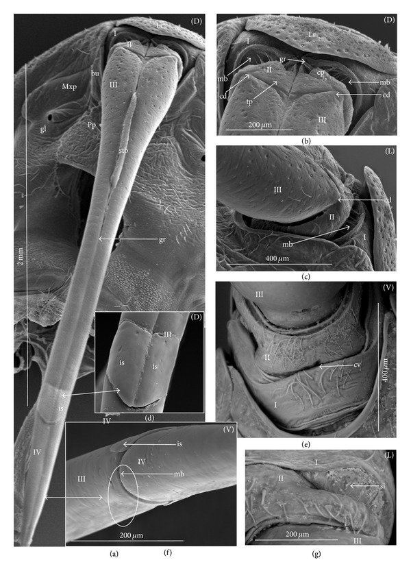
Figure 19.

Shapes of the labial segments of the Aphelocheiridae. (a)–(g) Aphelocheirus aestivalis (a) Dorsal view (D) of the labium, all segments (I, II, III, IV) are visible, (b) the shape of the first segment (I), the stylet groove (gr) is opened in the proximal part, the dorsal surface of the second segment (II) is divided into the triangular plate (tp) and the convex plate (cp), the second segment (II)—the stylet groove is opened, the articulation of the dorsal condyle (cd) between the II and III segments on the dorsal side, (c) lateral view, the proximal part of the third segment, the intersegmental membrane (mb) is visible, the second segment, ventral (V) view, (d) shapes of the intercalary sclerites (is), (e) the midventral condyle (cv) on the distal edge of the first segment, ventral view (V), the intersegmental membrane is distinctly visible, the shape of the first (I) and second (II) segments, viewed from the ventral side, (f) the connection between third and fourth segments on the ventral side, the midventral condyle (cv) on the third segment is not visible (circle), only the membrane (mb) is present, and (g) an intersegmental sclerit (si) between the first and second segment, lateral view. bu: buccula of the posterior plate, gl: orifice of the maxillary gland, Lr: labrum, Mxp: maxillary plate, Pp: posterior plate of the cranium, stb: stylet bundle.
