Abstract
Extract of Taraxacum platycarpum (AF-343) has been reported to have several biological properties such as skin hydration and anti-inflammatory effects. Although clinical evidences of skin hydration and antiinflammatory effect were proven in clinical trial, precise mechanism of skin hydration was not fully understood yet. In this study, we have focused skin hydration mechanism related filaggrin, collagen, and matrix metalloproteinase (MMP) in vitro and animal study. Herein, skin hydration mechanism of AF-343 is due to recovery of filaggrin in mice model and increased production of collagen with suppression of matrix MMP in vitro fibroblast cell line.
Keywords: Taraxacum platycarpum, Type I collagen, MMP-1, Smad2/3
INTRODUCTION
Several positive effects of dandelion extract were reported in the literature, which were related with skin hydration, anti-inflammatory effect, and anti-oxidative effect (Schtz et al., 2006; Cheong et al., 1998). In our previous clinical trial, we have revealed that dandelion extract of Taraxacum platycarpum (AF-343) have skin hydration and anti-inflammatory effect (Kim et al., 2010). However, we have not fully understood how AF-343 plays a role for skin hydration. To have a moisturizing effect in human skin, several factors are required such as recovery of natural moisturizing factors (NMF), increased expression of collagen, and suppression of matrix metalloproteinase (MMP) (Seo et al., 2005; Hu and Kitts, 2005). In addition to skin hydration, these factors are also known to increase skin elasticity, antiwrinkle effect, and smoothing of skin texture (Homebeck, 2003). Among NMFs, ceramide, filaggrin, and loricrin are representative and filaggrin mutation is one of the important precipitating factor in human atopic dermatitis (AD) skin (Irvine et al., 2011). AD is characterized by repetitive chronic skin inflammation and significant dry skin which has lower amount of water content when compared with those of normal healthy control.
Filaggrin gene (FLG) loss-of-function mutations underlie ichthyosis vulgaris, a semi-dominant disorder of keratinization, and are the strongest and most widely replicated genetic risk factor for AD (Sandilands et al., 2009). We have recently shown using Raman spectroscopy that FLG genotype is a major determinant of NMF in the stratum corneum (Kezic et al., 2008). FLG codes for profilaggrin, a large protein precursor comprised of 10~12 filaggrin repeats. The most abundant amino acid residues in filaggrin repeats are basic amino acids histidine and arginine and the polar residue glutamine. The protein is also significantly rich in glycine and serine, consistent with the structural similarity of filaggrin and related proteins such as loricrin to the variable domains of keratins, which largely consist of glycine/serine loop structures (Korge et al., 1992).
Here, we compared the levels of filaggrin degradation products in NC/Nga mice after oral administration of AF-343. And we evaluated the increased production of collagen with suppression of MMP in vitro fibroblast cell line.
MATERIALS AND METHODS
Animals. Fifteen female 5-week-old NC/Nga mice were purchased from Charles River Japan (Yokohama, Japan), and maintained under conventional conditions: a 12 h light/ 12 h dark cycle with freely available food and water. The colony room temperature was kept at 22~23℃ with a humidity of 55 ± 15%.
Induction of AD-like skin lesion. Mice were anesthetized with ether, after which the animal’s back hair was shaved by clipper 1 day prior to sensitization. Next, 100 mg of mite cream (Biostir Inc., Kobe, Japan) impregnated with Dermatophagoides farina crude extract was applied to the dorsal skin of these animals and secured by a patch every second day for fifteen days. Each gram of mite cream contained 234 μg of Der f 1, 7 μg of Der f 2 and 136.4 mg of proteins.
Systemic application of AF-343. After the induction of the AD-like skin lesions, the animals were divided into three groups, each containing five mice. These groups were then treated systemically for 5 days; AF-343 (experimental), hydrocortisone (positive control), or placebo only (normal control). The relative dermatitis severity was assessed macroscopically using the following scoring procedure. The total skin severity score was defined as the sum of the individual scores for each of the following seven signs: erythema, hemorrhage, edema, excoriation, erosion, scaling and dryness (Kunz et al., 1997). In this system, 0 was defined as exhibiting no symptoms, 1 as mild symptoms, 2 as moderate symptoms, and 3 as severe symptoms. Additionally, the mice were photographed after 5 days.
Cell culture. HS68 Cells (human skin fibroblasts) were cultured in 48-well plate at 37℃ in a humidified atmosphere containing an atmosphere of 5% (v/v) CO2 in air. The medium was changed every third day. Subconfluent cell cultures were enzymatically split by 100 nM TPA, 0.5% DMSO then propagated in culture medium as described above.
AF-343 application of the HS68 cells. Human skin fibroblasts were treated by various concentrations of AF-343 (0, 0.1, 0.5, 1, 2 mg/ml). The expressions of type I collagen, MMP-1, Smad2/3, and TIMP-1 proteins were analyzed by Western blot analysis. We used quantified proteins extracted from mixture of cultured cell with lysis buffer [10 mM Tris-Cl (pH 7.4), 5 mM EDTA (pH 8.0), 130 mM NaCl, 1% TritonX-100], 0.2 M PMSF (phenyl-methyl-sulfonyl- fluoride) and proteinase inhibitors (0.02 mM aprotinin, 2 mM leupeptin, 5 mM phenanthroline, 28 mM benzamidine- HCl). In addition, level of type I collagen mRNA was analyzed by CAT assay.
RESULTS
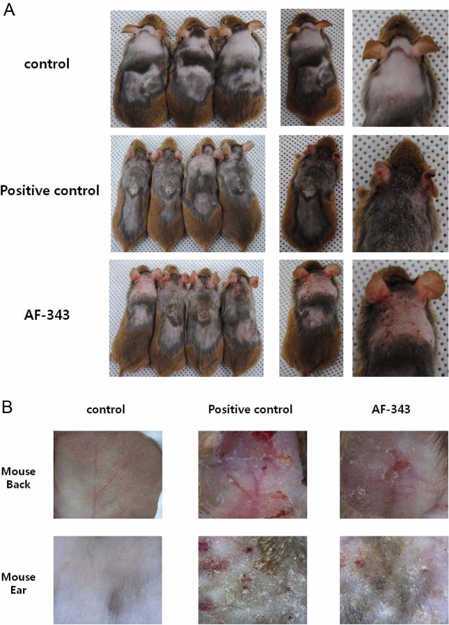
Anti-inflammatory effect of AF-343 on the AD-like skin lesion. To determine the clinical severity of each group, seven major clinical signs and symptoms of AD - erythema, hemorrhage, edema, excoriation, erosion, scaling and dryness - were scored using the criteria defined above (Kunz et al., 1997). These major clinical signs and symptoms developed shortly after the Dermatophagoides farina crude extract was applied to the backs and ears of the mice, and the AD-like skin lesions progressively worsened until 15 days after the initial induction. However, animals receiving systemic treatment with AF-343 exhibited a significantly lower degree of clinical skin severity comparable to those treated with hydrocortisone (the positive control). Conversely, no change was observed in the normal control group during the experimental period (Fig. 1). Such results indicate that AF-343 is able to effectively decrease the skin severity of mite-induced AD-like skin lesions in NC/Nga mice.
Fig. 1. Effect of AF-343 on clinical observations. AD-like skin lesions induced by attachment of mite patches on the backs of mice for 15 days, and AF-343 treated orally for 5 days. (Positive control, hydrocortisone).
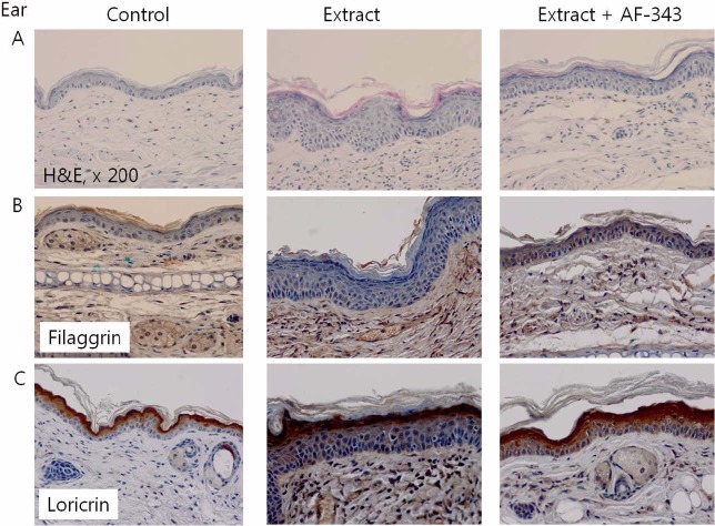
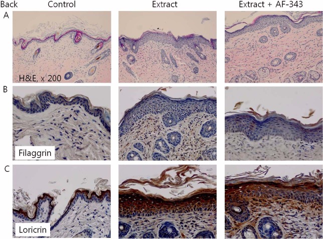
Increased expression of filaggrin in NC/Nga mice. We obtained skin tissues from backs and ears of AD induced NC/Nga mice. Histopathologically, epidermis became thicker when compared to that of normal epidermal tissue, while infiltration of inflammatory cells increased in the upper dermis (Fig. 2A). AF-343 is orally administrated, normalization of epidermal thickness and decrease of inflammatory cell infiltration were observed. These showed more prominent on the ear tissues than back samples (Fig. 3A).
Fig. 2. Immunohistochemical staining of filaggrin and loricrin in control, extract treated, or extract/AF-343 treated mice ear tissues.
Fig. 3. Immunohistochemical staining of filaggrin and loricrin in control, extract treated, or extract/AF-343 treated mice back tissues.
For normal skin tissue, immunohistochemistry revealed that filaggrin expression was evenly distributed in many horny layers with the increase of expression mostly in granular layers. Meanwhile, for skin tissue with AD, remarkable decrease of expression was observed in some granular layers and this seems to also affect the distribution of expression for filaggrin in horny layers (Fig. 2B). AF-343 is orally administrated, we could observed normalized distribution of filaggrin expression (Fig. 3B). In the case of loricrin, such results were less obvious than in the case of filaggrin (Fig. 2C, 3C).
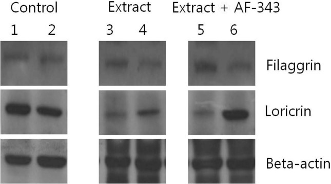
According to evaluation of filaggrin and loricrin expression by using Western blotting, filaggrin expression in AD-like skin lesion was slightly decreased. After administration of oral AF-343, the results showed increase of filaggrin expression (Fig. 4).
Fig. 4. Expressions of filaggrin and loricrin in control, extract treated, or extract/AF-343 treated mice ear and back tissues.
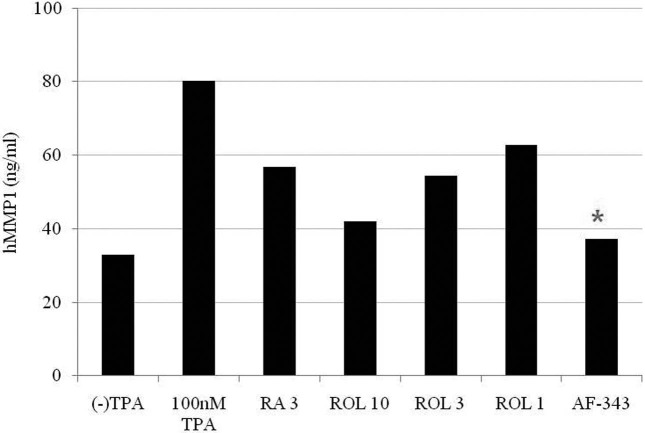
Expression of type I collagen and MMP-1. In order to evaluate a possible role of AF-343 on the collagen neogenesis, we focused on the type I collagen protein and MMP-1 expression after treatment with AF-343 in a concentration range from 0.1 to 2 mg/ml. AF-343 exerted different effects on type I collagen protein and MMP-1 expression depending on the concentration administered (Fig. 5). Expression of type I collagen protein was increased in AF-343-treated human skin fibroblasts by dose and time-dependent manners. Consistent with this result, the expressions of phosphor-Smad2/3 in skin fibroblasts were increased and MMP-1 expression was decreased by AF- 343 treatment (data not shown). TIMP-1 expression was not significantly changed in AF-343 treated skin fibroblasts.
Fig. 5. MMP-1 expression was decreased by AF-343 treatment. TPA: 12-O-tetradecanoylphorbol-13-acetate, RA: retinoic acid, ROL: retinol.
DISCCUSION
This study showed that when AD is induced on NC/Nga mice, epidermis becomes thicker when compared to that of normal epidermal tissue while infiltration of inflammatory cells increases and distribution of filaggrin expression changes. For normal skin tissue, immunohistochemistry revealed that filaggrin expression was evenly distributed in many horny layers with the increase of expression mostly in granular layers. Meanwhile, for skin tissue with AD, remarkable decrease of expression was observed in some granular layers and this seems to also affect the distribution of expression for filaggrin in horny layers (Sandilands et al., 2009). The study unveiled an important fact that when AF-343 is orally administrated, anti-inflammatory effects improved AD and at the same time, normalized distribution of filaggrin expression. In the case of loricrin, a differentiation marker, such results were less obvious than in the case of filaggrin so it seems that it would be more desirable to use filaggrin, rather than loricrin, as a marker when evaluating the efficacy of treatment for AD on animals.
Therefore, the study demonstrated that oral administration of AF-343 increased the thickness of epidermis and normalized the distribution of filaggrin expression in AD induced animals, which means that AF-343 relates to antiinflammatory effect and normalization of skin NMF expression. It appears that further study is necessary to look into what mechanism makes AF-343 relate to normalization process of filaggrin expression.
In conclusion, we have focused skin hydration mechanism related filaggrin, collagen, and MMP in vitro and animal study. Herein, skin hydration mechanism of AF-343 is due to recovery of filaggrin in mice model which means that AF-343 relates to anti-inflammatory effect and normalization of skin NMF expression. This study can also provide that AF-343 plays a role in increased production of collagen with suppression of MMP in vitro fibroblast cell line.
Acknowledgments
This study was carried out with the support of “Research Program for Agricultural Science & Technology Development (Project No. PJ006753012011)”, National Academy of Agricultural Science, Rural Development Administration, Republic of Korea.
References
- 1.Schütz K., Carle R., Schieber A. Taraxacum--a review on its phytochemical and pharmacological profile. J. Ethnopharmacol. (2006);107:313–323. doi: 10.1016/j.jep.2006.07.021. [DOI] [PubMed] [Google Scholar]
- 2.Cheong H., Choi E.J., Yoo G.S., Kim K.M., Ryu S.Y. Desacetylmatricarin, an anti-allergic component from Taraxacum platycarpum. Planta. Med. (1998);64:577–578. doi: 10.1055/s-2006-957520. [DOI] [PubMed] [Google Scholar]
- 3.Kim J.H., Lee H.I., Park J.H., Lim Y.Y., Kim B.J., Lim I.S., Kim M.N., Kim H.S., Kim J.K., Han S.H., Cho S.M., Kim J.H., Park K.M. The Effect of the Extracts of Taraxacum platycarpum (AF-343) in Atopic Dermatitis. Korean. J. Asthma. Allergy. Clin. Immunol. (2010);30:36–42. [Google Scholar]
- 4.Seo S.W., Koo H.N., An H.J., Kwon K.B., Lim B.C., Seo E.A., Ryu D.G., Moon G., Kim H.Y., Kim H.M., Hong S.H. Taraxacum officinale protects against cholecystokinininduced acute pancreatitis in rats. World J. Gastroenterol. (2005);11:597–599. doi: 10.3748/wjg.v11.i4.597. [DOI] [PMC free article] [PubMed] [Google Scholar]
- 5.Hu C., Kitts D.D. Dandelion (Taraxacum officinale) flower extract suppresses both reactive oxygen species and nitric oxide and prevents lipid oxidation in vitro. Phytomedicine. (2005);12:588–597. doi: 10.1016/j.phymed.2003.12.012. [DOI] [PubMed] [Google Scholar]
- 6.Homebeck W. Down-regulation of tissue inhibitor of matrix metalloprotease-1 (TIMP-1) in aged human skin contributes to matrix degradation and impaired cell growth and survival. Pathol. Biol. (2003);51:569–573. doi: 10.1016/j.patbio.2003.09.003. [DOI] [PubMed] [Google Scholar]
- 7.Irvine A.D., McLean W.H., Leung D.Y. Filaggrin mutations associated with skin and allergic diseases. N. Engl. J. Med. (2011);365:1315–1327. doi: 10.1056/NEJMra1011040. [DOI] [PubMed] [Google Scholar]
- 8.Sandilands A., Sutherland C., Irvine A.D., McLean W.H. Filaggrin in the frontline: role in skin barrier function and disease. J. Cell Sci. (2009);122:1285–1294. doi: 10.1242/jcs.033969. [DOI] [PMC free article] [PubMed] [Google Scholar]
- 9.Kezic S., Kemperman P.M., Koster E.S., de Jongh C.M., Thio H.B., Campbell L.E., Irvine A.D., McLean W.H., Puppels G.J., Caspers P.J. Loss-of-function mutations in the filaggrin gene lead to reduced level of natural moisturizing factor in the stratum corneum. J. Invest. Dermatol. (2008);128:2117–2119. doi: 10.1038/jid.2008.29. [DOI] [PubMed] [Google Scholar]
- 10.Korge B.P., Gan S.Q., McBride O.W., Mischke D., Steinert P.M. Extensive size polymorphism of the human keratin 10 chain resides in the C-terminal V2 subdomain due to variable numbers and sizes of glycine loops. Proc. Natl. Acad. Sci., U.S.A. (1992);89:910–914. doi: 10.1073/pnas.89.3.910. [DOI] [PMC free article] [PubMed] [Google Scholar]
- 11.Kunz B., Oranje A.P., Labrze L., Stalder J.F., Ring J., Taeb A. Clinical validation and guidelines for the SCORAD index: consensus report of the European Task Force on Atopic Dermatitis. Dermatology. (1997);195:10–19. doi: 10.1159/000245677. [DOI] [PubMed] [Google Scholar]
- 12.Bhogal R.K., Stoica C.M., McGaha T.L., Bona C.A. Molecular aspects of regulation of collagen gene expression in fibrosis. J. Clin. Immunol. (2005);25:592–603. doi: 10.1007/s10875-005-7827-3. [DOI] [PubMed] [Google Scholar]
- 13.Davis G.E., Saunders W.B. Molecular balance of capillary tube formation versus regression in wound repair: role of matrix metalloproteinases and their inhibitors. J. Investig. Dermatol. Symp. Proc. (2006);11:44–56. doi: 10.1038/sj.jidsymp.5650008. [DOI] [PubMed] [Google Scholar]


