Fig. 1.
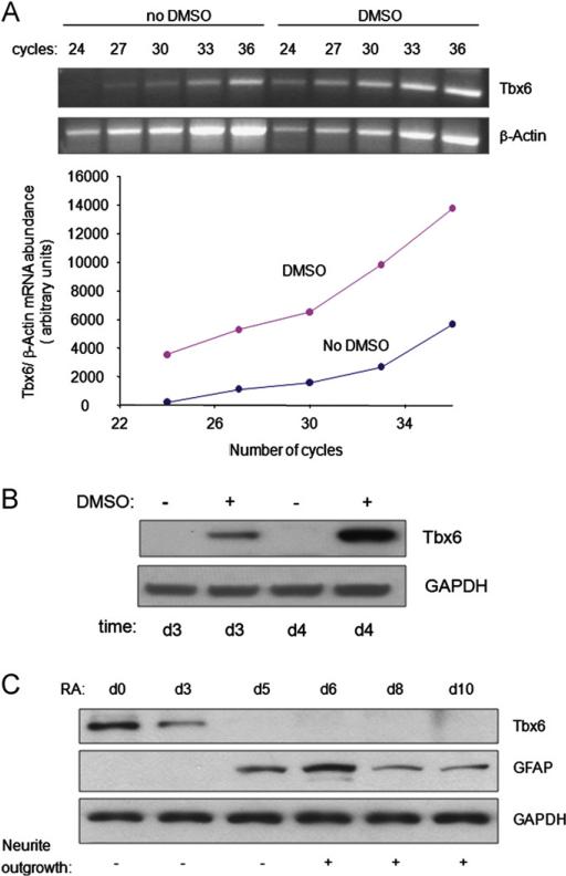
The abundance of Tbx6 mRNA and protein during differentiation of P19CL6 cells into cardiac myocytes or neural cells. (A) Tbx6 mRNA levels increase during DMSO-induced cardiac myocyte differentiation. Cells were cultured with or without DMSO for 6 days and Tbx6 and β-actin mRNA levels were assessed by semi-quantitative RT-PCR following the indicated number of amplification cycles. The abundance of Tbx6 mRNA has been normalized to that of β-actin mRNA. Similar results were obtained from three independent experiments. (B) Tbx6 protein levels increase during DMSO-induced cardiac myocyte differentiation. Cells were cultured with or without DMSO for 3 or 4 days (d) as indicated. Tbx6 protein levels were assessed by Western blot. Results are the representative of three independent experiments. (C) Tbx6 protein levels decrease during RA-induced neural differentiation. Cells were treated with RA for the times indicated. Levels of Tbx6 and GFAP, a glial marker, were assessed by Western blot. Neurite outgrowth was observed by microscopy. Western blot is the representative of three independent differentiation assays.

