Abstract
Background
Compared with invasive breast cancer, breast cancer in situ (BCIS) is seldom life threatening. However, an increasing incidence has been observed in recent years over the world. The purpose of our study is to investigate the epidemiological, clinical and pathological profiles of BCIS in Chinese women from 1999-2008.
Methods
Four thousand and two hundred-eleven female breast cancer (BC) patients were enrolled in this hospital-based nation-wide and multi-center retrospective study. Patients were randomly selected from seven hospitals in seven representative geographical regions of China between 1999 and 2008. The epidemiological, clinical and pathological data were collected based on the designed case report form (CRF).
Results
There were one hundred and forty-three BCIS cases in four thousand and two hundred-eleven BC patients (3.4%). The mean age at diagnosis was 48.3 years and BCIS peaked in age group 40-49 yrs (39.9%). The most common subtype was ductal carcinoma in situ (DCIS) (88.0%). 53.8% were positive for estrogen receptor (ER). Human epidermal growth factor receptor 2 (HER2) positive status was observed in 23.8% of patients. All patients underwent surgeries and 14.7% of them had breast conservation therapies (BCT) (21/143), but 41.9% accepted chemotherapy (64/143). Much less patients underwent radiotherapy (16.0%, 23/143) and among patients who had BCT, 67% accepted radiotherapy (14/21). Endocrine therapy was taken in 44.1% patients (63/143).
Conclusions
The younger age of BCIS among Chinese women than Western countries and increasing number of cases pose a great challenge. BCT and endocrine therapy are under great needs.
Introduction
Cancer In situ (CIS) is an early form of cancer, which is characteristically localized within the epithelium with the basement membrane intact and without any signs of invasion [1]. CIS, when compared with invasive cancer, often corresponds to better prognosis and is seldom life threatening. Invasive breast cancer develops from normal epithelium of the terminal duct or lobular unit through a series of events morphologically recognized as hyperplasia, atypical hyperplasia, in situ carcinoma, and finally malignant and invasive neoplasia [2,3]. Although most invasive cancers develop in this course, this process may be discontinuous in some other invasive cancers and is certainly not inviolate or universal. In addition, there has been evidence showing that many forms of invasive carcinoma originated from the progression of a CIS lesion [4,5]. Therefore, CIS is considered a precursor or incipient form of cancer that may, if left untreated long enough, transform into a malignant neoplasm.
Importantly, incidence rate of breast cancer in situ (BCIS) has risen dramatically over the last two decades, largely because of increased diagnosis as a result of increases in mammography screening. According to the Breast Cancer Statistics, there were about 57,650 new diagnoses of BCIS among US women in 2011, accounting for approximately 20% of all 288,130 new breast cancer cases the same year [6]. In China, due to limited cancer registries, only a few studies focus on the epidemiology of BCIS and most of them were based on small populations. So far, no nation-wide representative data of BCIS in China is available. Moreover, in recent years, great progress has been made in early detections and treatment options for BCIS. However, current data which describes these improvements and changes are almost all based on Western population [7-11]. Due to the difference of religion, race, circumstance and economic status, these data can not represent all patients over the world. Therefore, studies that could give a description of epidemiological, pathological and clinical characteristics of BCIS in other populations are of great need.
Here, we performed a nation-wide multi-center 10-year (1999-2008) retrospective clinical epidemiological study of females with BCIS in China in which patients were confirmed BCIS with pathology from 7 geographic regions across China (North, North-East, Central, South, East, North-West, and South-West). The aims of this study were to document (1) the sociodemographic characteristics and the distribution of some risk factors among Chinese female BCIS cases; (2) the clinical characteristics of female BCIS in China and (3) current treatment options for Chinese female BCIS.
Patients and Methods
Ethics Statement
This study was approved by the Institutional Review Board, Cancer Foundation of China. The written consent was given by the patients for their information to be stored in the hospital database and used for research.
Study Design
This study was part of a hospital-based multi-center 10-year retrospective study of randomly selected females whose medical chart review showed primary breast cancer [12]. To study the epidemiological, pathological, clinical characteristics and treatment options of BCIS, we selected cases that were pathologically confirmed BCIS out of the data pool in these Chinese women between 1999 and 2008.
Selection of Hospitals and Patients
A total of 7 hospitals selected from all seven traditional China districts were involved in this study. The seven districts were north, northeast, northwest, central, east, south and southwest of China, which extend over the majority of the country and represent different levels of breast cancer burden. Each selected hospital was one of the best leading hospitals at the tertiary level and had regional referral centers providing pathology diagnosis, surgery, radiotherapy, medical oncology, and routine follow-up care for patients with breast cancer.
Females who were pathologically confirmed primary breast cancer inpatients in one randomly selected month each from year 1999 to 2008 were enrolled. Every hospital collected patients randomly for no fewer than 50 cases in any month from March to December by an enrollment scheme. All cases collected were reviewed and patients’ information was collected based on the designed case report form (CRF) with quality control. In each selected month, if the number of inpatients were fewer than 50 in that year, additional cases from the following months were reviewed until the total number in that year reached 50. But if the number of patients exceeded 50, all cases were reviewed. To ensure that the national study was geographically representative, it was designed to include patients enrolled at sites from all seven traditional regions across China.
Pathology Diagnostic Criteria
Histological subtype was based on the 1981 and 2003 WHO histological classification criteria [13,14]. Staging of breast cancer was done according to the AJCC TNM staging system of year 1997 and after [15,16].
Data Collection and Quality Control
The data were systematically collected for all enrolled patients by medical chart review as described above [12]. Briefly, it included the general information, demographic characteristics, breast cancer risk factors, clinical breast examination, imaging, pathology and treatments of the patients. All of the above information was extracted from medical charts to the designed CRF by local trained-clerks. The data were finally transferred from paper to a database (FoxPro) and double-checked to ensure accuracy.
Sample Size
There were a total number of no fewer than 500 cases per hospital over 10 years (between 1999 and 2008) due to a minimum of 50 patients per site per year. Therefore, pooling of data across all sites in China was employed to adequately describe disease and treatment characteristics across the country.
Data Analysis
Due to the loss of some data or too many unknowns, not all of the information in CRF was analyzed and only data that focused on the epidemiological, pathological, clinical characteristics, and treatment options were used. All data were presented based on the original CRF.
Results
Trends of both primary breast cancer and BCIS cases over ten years across China
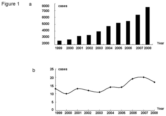
All seven selected hospitals provided the number of all their breast cancer inpatients each year, and there were a total of 45,200 patients with breast cancer during 1999- 2008. In 1999, 2,590 cases were diagnosed and under treatment, and by 2008, this number had reached 7,512 with a 2.9-fold increase (Figure 1a). Though we did not acquire all BCIS cases in the seven hospitals over ten years, cases in selected months were collected. A steady increase was observed in number of BCIS inpatients from 1999 to 2008 (Figure 1b).
Figure 1. Trends of the number of inpatients of BC and BCIS over ten years.

a. Numbers of Chinese breast cancer inpatients in each year between 1999-2008. b. The trend of Chinese breast cancer in situ cases in 1999-2008.
Patients Characteristics
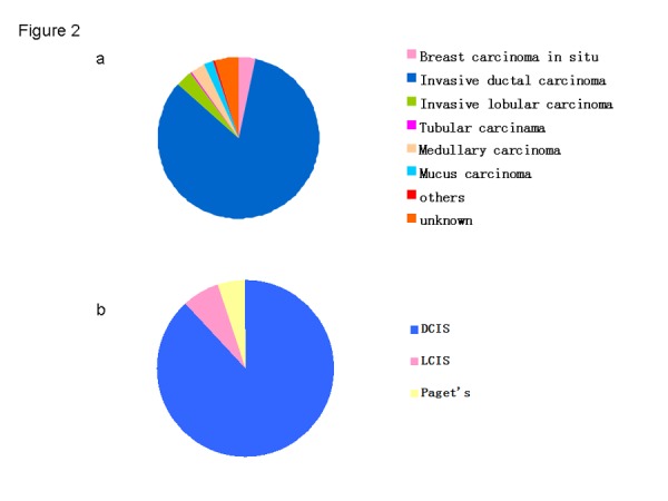
There were 143 patients diagnosed with BCIS, representing 3.4% of all 4,211 selected patients (143/4211) (Figure 2). Table 1 summarizes the general information of BCIS from seven districts during 1999-2008. The mean age at diagnosis and age range for all breast cancer patients was 48.3 years (s.d. = 11.2 yrs) and 24-80 years, respectively. The majority (69.1%) of BCIS patients were of normal Body Mass Index (BMI). Most BCIS patients were either manual or mental workers, accounting for 74.8% of all 143 patients, and housewives only presenting 3.5% of all occupations. Except for unknowns, middle school education is most common in BCIS cases. Approximately 68.5% of the cases were pre-menopausal and 31.5% were post-menopausal. The majority had been married and only two women were single. There were 94 BCIS cases with breast feeding history, accounting for 65.7% of all patients. Only 5 women had a breast cancer family history, representing only 3.5% of all cases.
Figure 2. Pathological type of BC and BCIS over ten years.

a. The percentage of each pathological type of BC in 19999-2008. b. The percentage of each Pathological type of BCIS in 19999-2008.
Table 1. Epidemiological characteristics of breast carcinoma in situ in China.
| Characteristics | N | % |
|---|---|---|
| Age (years) | ||
| Mean±SD | 48.28±11.24 | - |
| Range | 24~80 | - |
| ≤29 | 3 | 2.10 |
| 30-39 | 29 | 20.28 |
| 40-49 | 57 | 39.86 |
| 50-59 | 29 | 20.28 |
| ≥60 | 25 | 17.48 |
| BMI | ||
| Mean±SD | 23.38±3.15 | - |
| Range | 16.82~31.63 | - |
| Underweight(≤18.49) | 3 | 2.44 |
| Normal(18.50~24.99) | 85 | 69.11 |
| Overweight(25.00~29.99) | 30 | 24.39 |
| Obesity(≥30.00) | 5 | 4.07 |
| Occupation | ||
| Housewife | 5 | 3.50 |
| Manual worker | 56 | 39.16 |
| Mental worker | 51 | 35.66 |
| Others | 31 | 21.68 |
| Education | ||
| None | 3 | 2.10 |
| Primary school | 8 | 5.59 |
| Middle school | 16 | 11.19 |
| High school | 9 | 6.29 |
| University and above | 12 | 8.39 |
| Unknown | 95 | 66.43 |
| Menopausal status | ||
| Pre-menopause | 98 | 68.53 |
| Post-menopause | 45 | 31.47 |
| Marital status | ||
| Single | 2 | 1.40 |
| Married | 138 | 96.50 |
| Widowed/divorced | 3 | 2.10 |
| Breast feeding history | ||
| Yes | 94 | 65.73 |
| No | 11 | 7.69 |
| Unkown | 38 | 26.57 |
| Breast cancer family history | ||
| Yes | 5 | 3.50 |
| No | 133 | 93.01 |
| Unknown | 5 | 3.50 |
BMI, body mass index
The clinical and pathological characteristics of patients
Table 2 illustrates the clinical and pathologic characteristics of patients. Among all 143 BCIS cases, tumors had almost the same probability of being located on either the left or right. However, it should be noted that 8 cases were non-palpable, indicating that besides clinical physical examination, mammography and ultrasound were required for screening. Most tumors happened in the upper outer quadrant, accounting for almost half of all cases. About 55.4% of the patients had a tumor of which the size was less than 20 mm, tumors that were larger than 50 mm only present 10.7%. Over the 10 years, ductal cancer in situ (DCIS) remained the dominant pathologic subtype (88.1%). Among the 140 patients that had estrogen receptor (ER) and progesterone receptor (PR) information, fewer than 10% (8.2%) were ER positive and PR negative; 12.3% were positive with PR but negative with ER; more than half (52.5%) were both ER and PR positive and 24.6% were negative with both. Human epidermal growth factor receptor 2(HER2) information was available for 92 patients and the majority (63.0%) of them were HER2 negative.
Table 2. Clinical and pathological characteristics of breast carcinoma in situ in China.
| Characteristics | N | % |
|---|---|---|
| Tumor location | ||
| left | 70 | 48.95 |
| right | 65 | 45.45 |
| Non-palpable | 8 | 5.59 |
| Quadrant | ||
| upper inner | 21 | 14.69 |
| upper outer | 60 | 41.96 |
| lower inner | 6 | 4.20 |
| lower outer | 9 | 6.29 |
| center | 21 | 14.69 |
| others | 13 | 9.09 |
| Tumor size | ||
| <20mm | 62 | 55.36 |
| 20~49mm | 38 | 33.93 |
| ≥50mm | 12 | 10.71 |
| Pathology | ||
| DCIS | 126 | 88.11 |
| LCIS | 10 | 6.99 |
| Paget's disease | 7 | 4.90 |
| ER/PR status | ||
| ER+&PR+ | 64 | 52.46 |
| ER+&PR- | 10 | 8.20 |
| ER-&PR+ | 15 | 12.30 |
| ER-&PR- | 30 | 24.59 |
| unknown | 3 | 2.46 |
| HER2 status | ||
| HER2+ | 34 | 23.78 |
| HER2- | 58 | 40.56 |
| uncertain | 17 | 11.89 |
| unknown | 5 | 3.50 |
ER, estrogen receptor; PR, progesterone receptor; HER2, human epidermal growth factor receptor-2.
Treatment options
Treatment options for all patients were listed in Table 3. Among all BCIS cases, the majority (95.8%, 137/143) had undergone surgical procedures, with modified radical mastectomy being the predominant option (67.8%, 97/143). A minority of women (9.8%, 14/143) received breast conservative surgery. Hormonal therapy was the second most important treatment option (61.8%, 55/143) for BCIS patients in which ER/PR was positive. There were a total of 62 patients having chemotherapy, including neoadjuvant and adjuvant chemotherapy; these patients accounted for 43.4% of all cases. Only 15.4% of the patients accepted radiotherapy, and 66.7% of all those who had undergone breast conserving therapy (BCT) had radiotherapies.
Table 3. Treatment options for breast carcinoma in situ in China.
| Options | N | % |
|---|---|---|
| Surgery | ||
| Radical mastectomy | 7 | 4.90 |
| Modified radical mastectomy | 97 | 67.83 |
| Breast conservative surgery | 14 | 9.79 |
| Simple mastectomy | 12 | 8.39 |
| BCT+SLN biopsy | 7 | 4.90 |
| others | 6 | 4.20 |
| Radiotherapy | ||
| No | 117 | 81.25 |
| RT after BCT | 14 | 9.72 |
| RT after Radical/Modified radical mastectomy | 8 | 5.56 |
| unknown | 5 | 3.47 |
| Chemotherapy | ||
| No | 73 | 49.32 |
| Neoadjuvant chemotherapy | 7 | 4.73 |
| Adjuvant chemotherapy | 55 | 37.16 |
| Unknown | 13 | 8.79 |
| Endocrine therapy | ||
| Total | ||
| No | 64 | 44.76 |
| Yes | 63 | 44.06 |
| Unknown | 16 | 11.19 |
| Among ER/PR positive | ||
| No | 24 | 26.97 |
| Yes | 55 | 61.80 |
| Unknown | 10 | 11.24 |
| Anong ER&PR negative | ||
| No | 24 | 80.00 |
| Yes | 2 | 6.67 |
| Unkown | 4 | 13.33 |
| Among Pre-menopausal | ||
| SERM | 42 | 95.46 |
| AI | 2 | 4.54 |
| Among Post-menopausal | ||
| SERM | 12 | 54.54 |
| AI | 10 | 45.45 |
| Target-HER2 therapy | ||
| No | 122 | 85.31 |
| Unknown | 21 | 14.69 |
BCT, breast conservation therapy; SLNB, sentinel lymph node biopsy; RT, radiotherapy; SERM, selective estrogen receptor modular; AI, aromatase inhibitor.
Discussion
So far, ours is the first geographically-representative epidemiological study of BCIS in China, with 143 patients selected from a data pool of 4211 primary breast cancer cases. This study covered a large number of sites across all seven traditional regions of China [12], making it possible for us to thoroughly access the epidemiological, clinical, pathological characteristics and managements of BCIS across the entire country. A retrospect of ten years’ time shows the trends of the number of BCIS inpatients from 1999 to 2008. It also helps us to analyze the current limitation in treatments and how to improve treatment options in China, the biggest developing country. From 1999 to 2008, both the numbers of breast cancer and BCIS inpatients have increased in China, a trend similar to the data in Western countries [6,10,17-21]. According to the statistics from the SEER program, incidence rates of BCIS in Western countries rose rapidly in the 1980s and 1990s, largely because of increased diagnosis as a result of increases in mammography screening [22-24], which may be the same reason for the current increase in China. It is important to note that since 1999, incidence rates of BCIS have stabilized in women aged 50 years and older, but continue to increase in younger women [6]. However, due to the limited data and lack of age-control groups, such results could not be acquired in our study. All patients enrolled in this study were ethnically Chinese, and their clinical characteristics were significantly different from the women in Western countries. The mean age at diagnosis was 48.3 years, and this was consistent with the findings from studies in other regions of China [25-33]. There was no difference in the mean age between breast cancer (48.7 years) and BCIS in China, both of which were around the mid-40s. The mean age was about ten years younger than the reports in Western countries, in which breast cancer clusters peak around 60-69 years [10,19]. The reasons for this difference are still unclear, but several reasons could explain it: (1) mammography screening are more frequently used among older women in Western countries. (2) older Chinese women have been less exposed to estrogen-related risk factors. (3) younger women are more aware of breast cancer due to inefficient mammography screening in China. (4) younger women were more genetically predisposed to breast cancer [12].
Of all BCIS cases, DCIS accounted for 88.1% and remained the dominant pathological subtype. However, in this study, DCIS had only 2.7% (113/4211) of all primary breast cancer cases, differing drastically from the data of other studies performed in Western countries. There have been reports that, before 1980s, the incidence of DCIS was low and represented ~7% of all breast cancer cases, but now, DCIS represents 15-25% of cases in countries with efficient mammography screening programs [8,34]. The increase of incidence in Western countries can easily be explained by the advent of screening mammography. Patients presented with DCIS until they had become clinically symptomatic in earlier times. There may be two reasons for low proportions observed in China. First, several studies reported the incidence of DCIS by race or ethnicity and there may be some difference between Asian and Caucasian women. Secondly, the use of mammography screening in China was not as popular as in Western countries because of lower economic levels. In recent years, there have been reports on the effectiveness of screening mammography on breast-cancer incidence especially early stage breast cancer including DCIS, suggesting that there is substantial over-diagnosis, accounting for nearly a third of all newly diagnosed breast cancers [23,35-37]. Thus, due to inefficient mammography screening programs, there may be fewer occurrences of over-diagnosis caused by mammography screening in China.
Surgery was the most common treatment in Chinese female BCIS patients, followed by hormonal therapy. Ninety-five percent of all BCIS patients underwent surgeries. Overall, modified radical mastectomy was still the most common form of surgery, accounting for 67.8% of the 143 BCIS cases. Only 15.6% of women were treated with BCT, including 4.9 percent who accepted sentinel lymph node biopsy (SLNB). This is inconsistent with studies in the USA, suggesting that a large proportion are still treated with mastectomy [19,34], in some cases combined with SLNB.
However, there was no obvious increase observed in the percentage of BCIS patients who underwent BCT in China over the ten years, which is not consistent with the data from a study showing that an increasing percentage of patients were being treated with BCT (from 10% to 70%) between 1981 to 2001 in California [34]. The less widespread use of BCT in China in that time period may be caused by three reasons: (1) Although there was no difference in overall survival (OS ) between mastectomy and BCT plus radiotherapy, most patients still choose mastectomy as the “safer option”; 2) radiotherapy was required after BCT, but due to their economic statuses, some patients could not afford the cost; (3) BCT is not only a form of surgery; it requires advanced techniques in imaging diagnosis such as MRI and radiotherapy design. Due to limited medical resources in China, it is risky to carry BCT in patients. Therefore, with more recognition of BCT by patients and development in medicine, more BCT could be performed in China, in order to increase the living quality of future patients.
In our study, 62.2% (89/143) of patients were ER/PR positive and among these patients, 61.8% (55/89) accepted hormonal therapy. Since 1998, when U.S. Food and Drug Administration approved the use of tamoxifen to decrease the rate of recurrence of invasive breast cancer, which had been proved by several studies [38,39], the percentage of patients with DCIS treated with hormonal therapy have increased. The proportion of patients using hormonal therapy in China is much higher than that in the USA, which was reported at 14.1% [19]. Though this data was acquired in DCIS patients, due to the smaller proportion of LCIS in all BCIS, it still could demonstrate that hormonal therapy is more widespread in China than that in Western countries. However, about 40% of ER positive patients did not accept hormonal therapy, and that percentage of patients maybe expected to reduce the recurrence rate if they receive hormonal treatment. About 23.8% of all BCIS patients were HER2 positive and a similar percentage was observed in all breast cancer patients. Yet, there have been studies in Western countries reporting that pure DCIS overexpressed HER2 in approximately 45% [40]. The difference between Chinese and Caucasian population was obscure. Considering the fact that HER2 targeted therapy was not necessary for DCIS according to NCCN guidelines, this variety may not be so important for treatment choices. Compared with surgery and hormonal therapy, options for radiotherapy and chemotherapy were relatively fewer. Though there have been data demonstrating that radiotherapy after BCT could reduce the local recurrence of breast cancer [5,41,42], not all patients who underwent BCT accepted radiotherapy, showing that standard procedure should be strengthened. About 37.2% of patients had chemotherapy, which may reflect over-treatment on BCIS in China, because for in situ cases, chemotherapy is not the necessary choice.
This study provided a detailed description of BCIS across China. However, there are still some limitations: (1) selection bias may exist in the selected hospitals and months; (2) the number of BCIS cases was not large enough(3); There is no comparison between BCIS and invasive breast cancer. Overall, this is a representative study of BCIS in China to understand epidemiological, pathological ,clinical characteristics and therapies by over ten years’ retrospective study. The younger age of BCIS onset among Chinese women and increased number of cases pose a great challenge for the Chinese government regarding incidence control. BCT, as a surgical option safe and effective, is of great need. It is also necessary for hormonal therapy to be used more widely, in order to decrease the recurrence of invasive breast cancer. One should note that the application of chemotherapy should adhere strictly to the guidelines and its abuse should be prevented. A complete consideration of social, economic and medical factors by both government and doctors will ensure that better decisions are made regarding prevention and treatment for patients.
Acknowledgments
We thank all the patients who participate in this study.
Funding Statement
Key Program of National Natural Science Foundation of China (31030061); Natural Science Foundation of Guangdong Province, China (9151008901000124); Science and Technology Planning Project of Guangzhou, China (10C32060205); China Postdoctoral Science Foundation (2012M520075). The funders had no role in study design, data collection and analysis, decision to publish, or preparation of the manuscript.
References
- 1.(1997) Consensus Conference on the classification of ductal carcinoma in situ. The Consensus Conference Committee. Cancer 80: 1798-1802. doi: 10.1002/(SICI)1097-0142(19971101)80:9. PubMed: 9351550. [DOI] [PubMed] [Google Scholar]
- 2. Dupont WD, Parl FF, Hartmann WH, Brinton LA, Winfield AC et al. (1993) Breast cancer risk associated with proliferative breast disease and atypical hyperplasia. Cancer 71: 1258-1265. doi: 10.1002/1097-0142(19930215)71:4. PubMed: 8435803. [DOI] [PubMed] [Google Scholar]
- 3. London SJ, Connolly JL, Schnitt SJ, Colditz GA (1992) A prospective study of benign breast disease and the risk of breast cancer. JAMA 267: 941-944. doi: 10.1001/jama.267.7.941. PubMed: 1734106. [DOI] [PubMed] [Google Scholar]
- 4. Clark SE, Warwick J, Carpenter R, Bowen RL, Duffy SW et al. (2011) Molecular subtyping of DCIS: heterogeneity of breast cancer reflected in pre-invasive disease. Br J Cancer 104: 120-127. doi: 10.1038/sj.bjc.6606021. PubMed: 21139586. [DOI] [PMC free article] [PubMed] [Google Scholar]
- 5. Scribner KC, Behbod F, Porter WW (2013) Regulation of DCIS to invasive breast cancer progression by Singleminded-2s (SIM2s). Oncogene 32: 2631-2639. doi: 10.1038/onc.2012.286. PubMed: 22777354. [DOI] [PMC free article] [PubMed] [Google Scholar]
- 6. DeSantis C, Siegel R, Bandi P, Jemal A (2011) Breast cancer statistics, 2011. CA Cancer J Clin 61: 409-418. PubMed: 21969133. [DOI] [PubMed] [Google Scholar]
- 7. Alvarado R, Lari SA, Roses RE, Smith BD, Yang W et al. (2012) Biology, treatment, and outcome in very young and older women with DCIS. Ann Surg Oncol 19: 3777-3784. doi: 10.1245/s10434-012-2413-4. PubMed: 22622473. [DOI] [PMC free article] [PubMed] [Google Scholar]
- 8. Cuzick J, Sestak I, Pinder SE, Ellis IO, Forsyth S et al. (2011) Effect of tamoxifen and radiotherapy in women with locally excised ductal carcinoma in situ: long-term results from the UK/ANZ DCIS trial. Lancet Oncol 12: 21-29. doi: 10.1016/S1470-2045(10)70266-7. PubMed: 21145284. [DOI] [PMC free article] [PubMed] [Google Scholar]
- 9. Di Saverio S, Catena F, Santini D, Ansaloni L, Fogacci T et al. (2008) 259 Patients with DCIS of the breast applying USC/Van Nuys prognostic index: a retrospective review with long term follow up. Breast Cancer Res Treat 109: 405-416. doi: 10.1007/s10549-007-9668-7. PubMed: 17687650. [DOI] [PubMed] [Google Scholar]
- 10. Virnig BA, Shamliyan T, Tuttle TM, Kane RL, Wilt TJ (2009) Diagnosis and management of ductal carcinoma in situ (DCIS). Evid Rep Technol Assess (Full Rep): 1-549. [PMC free article] [PubMed]
- 11. Wallis MG, Clements K, Kearins O, Ball G, Macartney J et al. (2012) The effect of DCIS grade on rate, type and time to recurrence after 15 years of follow-up of screen-detected DCIS. Br J Cancer 106: 1611-1617. doi: 10.1038/bjc.2012.151. PubMed: 22516949. [DOI] [PMC free article] [PubMed] [Google Scholar]
- 12. Li J, Zhang BN, Fan JH, Pang Y, Zhang P et al. (2011) A nation-wide multicenter 10-year (1999-2008) retrospective clinical epidemiological study of female breast cancer in China. BMC Cancer 11: 364. doi: 10.1186/1471-2407-11-364. PubMed: 21859480. [DOI] [PMC free article] [PubMed] [Google Scholar]
- 13.(1982) Histological typing of breast tumors. Second edition. World Health Organization; Geneva, 1981. Ann Pathol 2: 91-105. [PubMed] [Google Scholar]
- 14. Böcker W (2002) WHO classification of breast tumors and tumors of the female genital organs: pathology and genetics. Verh Dtsch Ges Pathol 86: 116-119. PubMed: 12647359. [PubMed] [Google Scholar]
- 15. Brenin DR, Morrow M (1998) Accuracy of AJCC staging for breast cancer patients undergoing re-excision for positive margins. American Joint Committee on Cancer. Ann Surg Oncol 5: 719-723. doi: 10.1007/BF02303483. PubMed: 9869519. [DOI] [PubMed] [Google Scholar]
- 16. Edge SB, Compton CC (2010) The American Joint Committee on Cancer: the 7th edition of the AJCC cancer staging manual and the future of TNM. Ann Surg Oncol 17: 1471-1474. doi: 10.1245/s10434-010-0985-4. PubMed: 20180029. [DOI] [PubMed] [Google Scholar]
- 17. Clarke CA, Keegan TH, Yang J, Press DJ, Kurian AW et al. (2012) Age-specific incidence of breast cancer subtypes: understanding the black-white crossover. J Natl Cancer Inst 104: 1094-1101. doi: 10.1093/jnci/djs264. PubMed: 22773826. [DOI] [PMC free article] [PubMed] [Google Scholar]
- 18. Johnson RH, Chien FL, Bleyer A (2013) Incidence of breast cancer with distant involvement among women in the United States, 1976 to 2009. JAMA 309: 800-805. doi: 10.1001/jama.2013.776. PubMed: 23443443. [DOI] [PubMed] [Google Scholar]
- 19. Sumner WR, Koniaris LG, Snell SE, Spector S, Powell J et al. (2007) Results of 23,810 cases of ductal carcinoma-in-situ. Ann Surg Oncol 14: 1638-1643. doi: 10.1245/s10434-006-9316-1. PubMed: 17245612. [DOI] [PubMed] [Google Scholar]
- 20. Virnig BA, Tuttle TM, Shamliyan T, Kane RL (2010) Ductal carcinoma in situ of the breast: a systematic review of incidence, treatment, and outcomes. J Natl Cancer Inst 102: 170-178. doi: 10.1093/jnci/djp482. PubMed: 20071685. [DOI] [PubMed] [Google Scholar]
- 21. Youlden DR, Cramb SM, Dunn NA, Muller JM, Pyke CM et al. (2012) The descriptive epidemiology of female breast cancer: an international comparison of screening, incidence, survival and mortality. Cancer Epidemiol 36: 237-248. doi: 10.1016/j.canep.2012.02.007. PubMed: 22459198. [DOI] [PubMed] [Google Scholar]
- 22. Autier P, Koechlin A, Smans M, Vatten L, Boniol M (2012) Mammography screening and breast cancer mortality in Sweden. J Natl Cancer Inst 104: 1080-1093. doi: 10.1093/jnci/djs272. PubMed: 22811439. [DOI] [PubMed] [Google Scholar]
- 23. Bleyer A, Welch HG (2013) Effect of screening mammography on breast cancer incidence. N Engl J Med 368: 679 PubMed: 23406034. [DOI] [PubMed] [Google Scholar]
- 24. Nekhlyudov L, Habel LA, Achacoso N, Jung I, Haque R et al. (2012) Ten-year risk of diagnostic mammograms and invasive breast procedures after breast-conserving surgery for DCIS. J Natl Cancer Inst 104: 614-621. doi: 10.1093/jnci/djs167. PubMed: 22491230. [DOI] [PMC free article] [PubMed] [Google Scholar]
- 25. Li Jun (2009) Diagnosis and therapy of 74 cases of ductal carcinoma of breast. Clinical Medicine of China. 25: 971-973. [Google Scholar]
- 26. Li W, Wang Q, Zhang A, Xu J, Lai R (2003) Daignosis and Treatment of Ductal Carcinoma in Situ (A report of 41 cases). Lingnan Modern Clinics in Surgery 3: 288-290. [Google Scholar]
- 27. Qian X (2006) Diagnosis and management of 22 cases of DCIS. SUZHOU UNIVERSITY. Journal of Medical Sciences 26: 861-862. [Google Scholar]
- 28. Yu Y, Fang Z, Liu J (2003) Clinical characters and long-term treatment effect on 123 cases of dutal carcinoma in situ of breast. Journal of Tianjin Medical University 9: 180-182. [Google Scholar]
- 29. Zhang X, Gu Y, Wang J, Shi L, Chen Q (2011) Diagnosis and treatments of 36 cases of breast carcinoma in situ. Journal Clinical Surgery. 19: 142-143. [Google Scholar]
- 30. Zhao W (2004) Clinical analysis of 112 cases of breast ductal carcinoma . Journal Surgical Concepts and Practice 9: 428-430. [Google Scholar]
- 31. Huang J, Tang Y, Hu J (2006) A study on 8 DCIS and a literature of DCIS. Chinese Journal Difficult and Complicated Cases. 5: 139-140. [Google Scholar]
- 32. Jiang Y (2008) A clinical analysis of 123 cases of CIS: China. Medical University. [Google Scholar]
- 33. Le X (2005) A clinical analysis of 11 cases of DCIS. JIANGXI MEDICAL. Journal. 40: 541-542. [Google Scholar]
- 34. Verkooijen HM, Fioretta G, De Wolf C, Vlastos G, Kurtz J et al. (2002) Management of women with ductal carcinoma in situ of the breast: a population-based study. Ann Oncol 13: 1236-1245. doi: 10.1093/annonc/mdf194. PubMed: 12181247. [DOI] [PubMed] [Google Scholar]
- 35. Falk RS, Hofvind S, Skaane P, Haldorsen T (2013) Overdiagnosis among women attending a population-based mammography screening program. Int J Cancer 133: 705-712. doi: 10.1002/ijc.28052. PubMed: 23355313. [DOI] [PMC free article] [PubMed] [Google Scholar]
- 36. Kalager M, Adami HO, Bretthauer M, Tamimi RM (2012) Overdiagnosis of invasive breast cancer due to mammography screening: results from the Norwegian screening program. Ann Intern Med 156: 491-499. doi: 10.7326/0003-4819-156-7-201204030-00005. PubMed: 22473436. [DOI] [PubMed] [Google Scholar]
- 37. Smith RA, Kerlikowske K, Miglioretti DL, Kalager M (2012) Clinical decisions. Mammography screening for breast cancer. N Engl J Med 367: e31. doi: 10.1056/NEJMclde1212888. PubMed: 23171121. [DOI] [PubMed] [Google Scholar]
- 38. Cunnick GH, Mokbel K (2003) Radiotherapy and tamoxifen in women with completely excised ductal carcinoma in situ. Lancet 362: 1154: 1155-1156. PubMed: 14550707. [DOI] [PubMed] [Google Scholar]
- 39. Houghton J, George WD, Cuzick J, Duggan C, Fentiman IS et al. (2003) Radiotherapy and tamoxifen in women with completely excised ductal carcinoma in situ of the breast in the UK, Australia, and New Zealand: randomised controlled trial. Lancet 362: 95-102. doi: 10.1016/S0140-6736(03)13859-7. PubMed: 12867108. [DOI] [PubMed] [Google Scholar]
- 40. von Minckwitz G, Darb-Esfahani S, Loibl S, Huober J, Tesch H et al. (2012) Responsiveness of adjacent ductal carcinoma in situ and changes in HER2 status after neoadjuvant chemotherapy/trastuzumab treatment in early breast cancer--results from the GeparQuattro study (GBG 40). Breast Cancer Res Treat 132: 863-870. doi: 10.1007/s10549-011-1621-0. PubMed: 21667238. [DOI] [PubMed] [Google Scholar]
- 41. Bijker N, Meijnen P, Peterse JL, Bogaerts J, Van Hoorebeeck I et al. (2006) Breast-conserving treatment with or without radiotherapy in ductal carcinoma-in-situ: ten-year results of European Organisation for Research and Treatment of Cancer randomized phase III trial 10853--a study by the EORTC Breast Cancer Cooperative Group and EORTC Radiotherapy Group. J Clin Oncol 24: 3381-3387. doi: 10.1200/JCO.2006.06.1366. PubMed: 16801628. [DOI] [PubMed] [Google Scholar]
- 42. Punglia RS, Burstein HJ, Weeks JC (2012) Radiation therapy for ductal carcinoma in situ: a decision analysis. Cancer 118: 603-611. doi: 10.1002/cncr.26293. PubMed: 21720992. [DOI] [PMC free article] [PubMed] [Google Scholar]


