Abstract
Bovine tuberculosis has been tackled for decades by costly eradication programs in most developed countries, involving the laboratory testing of tissue samples from allegedly infected animals for detection of Mycobacterium tuberculosis complex (MTC) members, namely Mycobacterium bovis. Definitive diagnosis is usually achieved by bacteriological culture, which may take up to 6–12 weeks, during which the suspect animal carcass and herd are under sanitary arrest. In this work, a user-friendly DNA extraction protocol adapted for tissues was coupled with an IS6110-targeted semi-nested duplex real-time PCR assay to enhance the direct detection of MTC bacteria in animal specimens, reducing the time to achieve a diagnosis and, thus, potentially limiting the herd restriction period. The duplex use of a novel β-actin gene targeted probe, with complementary targets in most mammals, allowed the assessment of amplification inhibitors in the tissue samples. The assay was evaluated with a group of 128 fresh tissue specimens collected from bovines, wild boars, deer and foxes. Mycobacterium bovis was cultured from 57 of these samples. Overall, the full test performance corresponds to a diagnostic sensitivity and specificity of 98.2% (CIP95% 89.4–99.9%) and 88.7% (CIP95% 78.5–94.7%), respectively. An observed kappa coefficient was estimated in 0.859 (CIP95% 0.771–0.948) for the overall agreement between the semi-nested PCR assay and the bacteriological culture. Considering only bovine samples (n = 69), the diagnostic sensitivity and specificity were estimated in 100% (CIP95% 84.0–100%) and 97.7% (CIP95% 86.2–99.9%), respectively. Eight negative culture samples exhibiting TB-like lesions were detected by the semi-nested real-time PCR, thus emphasizing the increased potential of this molecular approach to detect MTC-infected animal tissues. This novel IS6110-targeted assay allows the fast detection of tuberculous mycobacteria in animal specimens with very high sensitivity and specificity, being amenable and cost effective for use in the routine veterinary diagnostic laboratory with further automation possibilities.
Introduction
Tuberculosis (TB) is a leading cause of morbidity and mortality in the world, also affecting a wide range of animal species, particularly livestock, in both developed and developing countries. The disease is caused by tuberculous mycobacteria belonging to the Mycobacterium tuberculosis complex (MTC). This complex consists of several closely-related pathogenic species, namely M. tuberculosis, the main agent of human TB, and, amongst others, M. bovis and M. caprae that are primary agents of bovine and caprine TB, respectively [1]. These species are genetically very similar but may differ in host preference and epidemiological characteristics [2]. Mycobacterium bovis and M. caprae also represent a high potential for zoonotic transmission to humans [3–5], with evidence of possible person-to-person transmission [6]. However, the main routes of transmission are the contact with infected animals and ingestion of unpasteurized dairy products. These zoonotic MTC species may be responsible for up to 7.2% and 15% of human TB cases in industrialized and developing countries, respectively [7]. Rapid and reliable laboratory tests for the direct detection of tuberculous mycobacteria in biological samples are in high demand, in both human health and veterinary settings, and are crucial for an improved TB control.
In most developed countries, bovine tuberculosis has been tackled during the last decades by costly eradication programs, involving the culling of reactor animals and laboratory testing of suspect samples for the definitive confirmation of the presence of MTC. Presently, the detection of MTC bacteria in animal tissues is mainly based in lengthy and cumbersome conventional methods, involving the examination of Ziehl-Neelsen stained smears, histopathology and culture in selective media, followed by biochemical or molecular identification of typical mycobacteria colonies. The microscopic identification of acid-fast mycobacteria is non-specific and highly insensitive, particularly in the case of paucibacillary forms of TB. Culture remains the gold-standard method to confirm TB infection but requires several weeks to obtain positive results due to the extremely fastidious growth of tuberculous mycobacteria. In spite of the significant advances in the development of novel molecular diagnostic assays towards a faster and accurate detection of MTC in human samples, only a few assays have been described for detecting these agents in animal tissues, particularly in fresh tissues from livestock [8-12]. Most of these molecular approaches are PCR-based and target specific polymorphisms, insertion sequences, and the so-called regions of difference in the genome of MTC members [9,10,13-18]. Nevertheless, most of the amplification-based assays described for detecting MTC nucleic acids directly in fresh or formalin-fixed paraffin-embedded tissues only yield a moderate sensitivity, usually up to 75%, particularly when testing tissues without the characteristic lesions or detectable acid-fast bacilli [8-10,15,19]. This limitation is mostly related to the inefficiency of mycobacterial DNA extraction procedures from affected animal tissues, especially those exhibiting strong fibrosis, calcification, and with no histological evidence of acid-fast bacteria [8,19]. The use of immunomagnetic separation approaches to concentrate mycobacteria cells from animal tissues prior to DNA extraction may enhance PCR sensitivities [20,21]. Nevertheless, these approaches usually involve more experimental steps and expensive equipment and consumables not readily available in veterinary diagnostic settings.
In the present work we have developed a novel and simple Taqman-based semi-nested real-time PCR assay yielding extremely high sensitivity and specificity for the direct detection of tuberculous mycobacteria in fresh animal tissues, namely of bovine origin, capable of being introduced in routine diagnostic veterinary laboratories.
Materials and Methods
Bacterial strains
Reference strains and clinical isolates of MTC, non-MTC mycobacteria and non-mycobacterial species, maintained at the Portuguese reference laboratory for animal diseases (INIAV, IP), were used for optimization of PCR assays (Table 1). The identification of each isolate was based on standard methodologies [22]. MTC isolates were identified to the species level by PCR-restriction endonuclease analysis of the gyrB gene [23–25] and hybridization with species-specific probes [26].
Table 1. Bacterial reference strains and clinical isolates whose cultures were used in the present study for the evaluation of specificity of the amplification assays and respective results.
| Species | Reference strains/Isolates | Presence of IS61101 |
|---|---|---|
| Mycobacterium tuberculosis | ATCC 25177; LNIV 9605 | + |
| M. bovis | AN5; ATCC 27291 (BCG); LNIV 13027; 5530/0/05; 11265; 7230/4; 14421/2; 24497/6; 8855; 5889; 10044; 14577; 13280/6; 13280/4; 34875; and 20564 | + |
| M. caprae | LNIV 17320; 4958/0/05; 8403; 15244; and 20752 | + |
| M. avium subsp. avium | ATCC 25291 | - |
| M. avium subsp. hominissuis | LNIV 23063/4 | - |
| M. avium subsp. paratuberculosis | LNIV 39888 | - |
| M. scrofulaceum | LNIV 31389 | - |
| Acinetobacter baumannii | LNIV 1628/12 | - |
| Arcanobacterium pyogenes | VLA 1884 | - |
| Bacillus licheniformis | VLA 1831 | - |
| Corynebacterium striatum | LNIV 12352 | - |
| Enterobacter amnigenus | LNIV 6050/II | - |
| Klebsiella pneumonia | VLA 1643 | - |
| Listeria monocytogenes | VLA 1774 | - |
| Proteus mirabilis | LNIV 2269/II | - |
| Pseudomonas aeruginosa | VLA 67 | - |
| Salmonella Dublin | VLA 1272 | - |
| Staphylococcus aureus | VLA 1032 | - |
| Streptococcus agalactiae | VLA 33 | - |
| Yersinia enterocolitica | VLA 1884 | - |
ATCC, American Type Culture Collection, USA; LNIV, Laboratório Nacional de Investigação Veterinária (currently INIAV, IP), Lisbon, Portugal; VLA, Veterinary Laboratory Agency, UK; 1Amplification (+) or no amplification (-) of IS6110 element using the real-time PCR assay with P_IS6110 TaqMan probe and respective flanking primers F_IS6110 and R_IS6110.
Tissue samples
One hundred and twenty eight animal lymph nodes, liver, spleen or lung tissue samples (69 bovines, 35 wild boars, 15 deer and 9 foxes) were used in this work (Table 2). No animals were sacrificed for the purposes of this specific study. None of the authors were responsible for the death of any animals and samples were originally collected for purposes other than research, namely: (i) bovine samples were collected from animals clinically suspected of having TB, e.g. by a positive reaction in either the single intradermal comparative tuberculin test or the gamma-interferon test, or TB-like lesions detected during routine abattoir inspection, and were submitted to routine control testing under the governmental Portuguese eradication scheme for bovine tuberculosis, approved in 1992 by the European Union (Council Decision 92/299/CE) and, since 2001, cofinanced by the European Union (in the framework of Council Directive 64/432, as amended) [27]; and (ii) TB suspect samples from wild boar, deer and fox, were sent to INIAV reference laboratories following gross pathological evaluation performed in the field by local veterinarians in hunting activities or predator control actions legally authorized by the "Instituto da Conservação da Natureza e das Florestas" (the Portuguese National Forest Authority), that grants permits for those hunting actions and for the respective hunters. Samples were submitted during the fourth trimester of 2011 to the pathology and bacteriology laboratories of INIAV and analysed using routine histological and culture-based methods, according to the OIE standard procedures [22] (Table 2). Tissues selected for bacteriological analysis were homogenized using a pestle and mortar, followed by decontamination with 4% sodium hydroxide. After neutralization with 10% hydrochloric acid, the macerated suspensions were divided into equal parts. One part was maintained at -80 °C until further processing for molecular analysis and the other was centrifuged and the sediment inoculated into BACTEC 9000 liquid media and Lowenstein-Jensen with pyruvate and Stonebrink solid media. Inoculated media were incubated for a minimum of eight weeks at 37 °C. Heat-killed culture supernatants were kept at -20 °C until the molecular identification of the isolates. Species identification of presumptive mycobacteria isolates was based on the restriction endonuclease analysis with RsaI and SacII of the PCR-amplified gyrB gene, or using the commercial reverse hybridization assays INNO-LiPA Mycobacteria (Innogenetics, Belgium) or GenoType Mycobacterium (Hain diagnostics, Germany), following the manufacturer’s instructions [23-25].
Table 2. Typology of tissue samples used in this study (n = 128) and respective results of the histological, bacteriological and semi-nested duplex real-time PCR analyses.
| Sample typology | Number of tissue samples | Origin of tissues | Presence of lesions1 | Bacteriological analysis2 | Nested real-time PCR3 |
|---|---|---|---|---|---|
| 23 | Bovine | + | M. bovis | 23 | |
| I | 19 | Wild boar | + | M. bovis | 18 |
| 12 | Deer | + | M. bovis | 12 | |
| II | 3 | Bovine | - | M. bovis | 3 |
| 39 | Bovine | - | - | 0 | |
| III | 4 | Wild boar | - | - | 0 |
| 3 | Deer | - | - | 0 | |
| IV | 2 | Bovine | - | Non-MTC | 0 |
| 3 | Wild boar | - | Non-MTC | 0 | |
| 1 | Bovine | + | - | 0 | |
| V | 5 | Wild boar | + | - | 3 |
| 6 | Fox | + | - | 0 | |
| 1 | Bovine | + | Non-MTC | 1 | |
| VI | 4 | Wild boar | + | Non-MTC | 2 |
| 3 | Fox | + | Non-MTC | 2 |
1Presence of lesions compatible with tuberculosis; 2Detection of M. bovis or other non-MTC mycobacteria by culture of tissue samples; 3Number of samples for which the IS6110 element was amplified by the nested real-time PCR assay.
Spiked tissue samples
A lymph node tissue from a slaughtered bovine known to be free of TB, as confirmed by culture and histopathology, was used for testing as a spiked sample that was homogenized as described above. Eight aliquots containing 0.9 ml of tissue macerate were spiked with 0.1 ml of ten-fold dilutions of a suspension of M. tuberculosis H37Ra (ATCC 25177) cells. Estimated concentrations of mycobacteria in spiked macerate samples ranged from 107 to 10° cells/ml. An additional tube was spiked with 0.1 ml PBS buffer and used as negative control. Spiked samples were stored at - 20 °C until processing for DNA extraction.
DNA extraction from cultures
DNA extraction from cultures grown in liquid media was achieved by a combined bead beating and enzymatic extraction method described elsewhere [28]. Briefly, 0.5 ml bacterial culture were washed in PBS buffer and heat inactivated at 100 °C for 15 min. Tubes containing zirconium beads were used to resuspend the culture pellet in lysis buffer (0.4 M NaCl, 40 mM Tris-HCl, pH 8, 2 mM EDTA, 0.6% SDS, 0.034 mg/ml proteinase K) for mechanical disruption in the FastPrep FP120 Bio101 (Savant Instruments, Inc., Holbrook, NY) at 6.5 ms-1 for 45 s, and then incubated overnight at 37 °C, followed by standard phenol–chloroform purification and ethanol precipitation of DNA. DNA concentration and purity were estimated by measuring the absorbance at 260 nm (A260) and by A260 ⁄A280 and A260/A230 ratios, respectively, using a NanoDrop 1000 spectrophotometer (NanoDrop). Genomic DNA suspensions were stored at - 20 °C until further use.
DNA extraction from tissue macerates and spiked samples
Four hundred and fifty microliters of tissue suspensions were transferred to screw-cap microcentrifuge tubes and inactivated in a water bath at 100 °C for 5 minutes. Samples were centrifuged at 14000 rpm for 2 min, the supernatant rejected and 80 µl of PBS and an equivalent volume of 100 µl of zirconium beads were added to the tubes. After mechanical disruption in a FastPrep FP120 Bio101 bead shaker (Savant Instruments Inc., , Holbrook, NY) at 6.5 msec-1 for 45 seconds, repeated three times, the suspensions were cooled on ice for 15 minutes. DNA extraction was carried out using the tissue protocol of the QIAamp DNA Mini Kit (Qiagen) according to the manufacturer's instructions. The resulting genomic DNA suspensions were stored at - 20 °C until further use. Stock DNA suspensions were diluted ten times in distilled water before its use as template for PCR assays.
Design of TaqMan probes and flanking primers
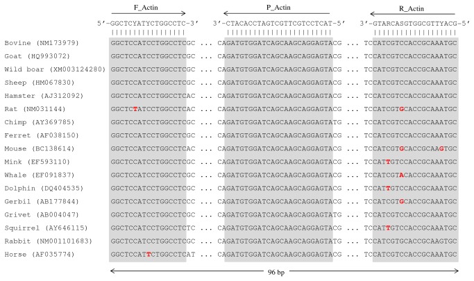
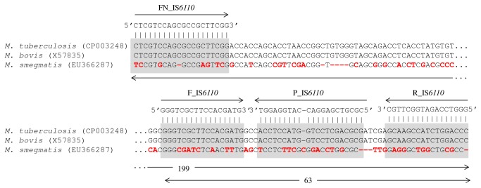
Sequences of IS6110 from MTC members and β-actin gene from a wide range of mammal species were retrieved from NCBI-GenBank. Comparative analysis of these two sets of sequences was achieved through sequence alignment using the CLUSTAL X v2.0 software [29]. Complementary regions for a β-actin gene-targeted mammals-universal TaqMan probe and flanking degenerated primers were found after visual inspection of the respective alignments (Table 3; Figure 1). The amplification of β-actin gene served as control to detect inhibition of the PCR reactions when using DNA extracted from tissues as template. The IS6110-targeted probe (P_IS6110) and respective flanking primers (F_IS6110 and R_IS6110) were retrieved from the study of Restrepo and colleagues [30] (Table 3; Figure 2). An additional IS6110-targeted forward primer (FN_IS6110) for use in a semi-nested PCR was designed (Table 3; Figure 2). Probes and primers specificities were tested in silico using the BLAST tools from NCBI-GenBank. All probes and primers targeting IS6110 and β-actin gene were synthesized by MWG Biotech (Germany).
Table 3. Sequences of primers and probes used in this study.
| Primer/Probe | Sequence (5´-3´) | Complementary target |
|---|---|---|
| F_Actin | GGC TCY ATY CTG GCC TC | β-actin gene of mammals |
| R_Actin | GCA YTT GCG GTG SAC RAT G | |
| P_Actin1 | Cy5.5-TAC TCC TGC TTG CTG ATC CAC ATC-BHQ2 | |
| F_IS6110 | GGG TCG CTT CCA CGA TG | IS6110 element of MTC species |
| FN_IS6110 | CTC GTC CAG CGC CGC TTC GG | |
| R_IS6110 | GGG TCC AGA TGG CTT GC | |
| P_IS6110 2 | FAM-CGC GTC GAG GAC CAT GGA GGT-BHQ1 |
1Probe labeled with Cy5.5 fluorophore and BHQ-2 quencher; 2Probe labeled with carboxyfluorescein (FAM) fluorophore and BHQ-1 quencher
Figure 1. Complementary targets of the mammals β-actin gene targeted probe and flanking primers.
Partial alignment of β-actin gene sequences of several mammal species showing the complementary targets of the P_Actin TaqMan probe and respective flanking degenerated primers (F_Actin and R_Actin) (gray boxes). The GenBank access numbers from which the partial sequences were retrieved are indicated for each species inside parenthesis. Mismatches in relation to consensus sequence are highlighted in red.
Figure 2. Complementary targets of the MTC-specific IS6110-targeted probe and flanking primers.
Partial alignment of the IS6110 sequence of Mycobacterium tuberculosis complex members with other IS6110-like sequence recently found in M. smegmatis (GenBank access numbers are indicated inside parenthesis). The complementary targets of the P_IS6110 TaqMan probe and flanking primers (FN_IS6110, F_IS6110 and R_IS6110) are indicated (gray boxes). Mismatches in sequences are highlighted in red.
Amplification assays
The semi-nested real-time PCR amplification assay using DNA extracted from tissue samples as template consisted of two steps: (i) a first standard PCR using primers FN_IS6110 and R_IS6110; and (ii) a second duplex real-time PCR using the previous amplification product as template and a mixture of IS6110 and β-actin gene targeted TaqMan probes (P_IS6110 and P_Actin, respectively) and the corresponding flanking primers (F_IS6110/R_IS6110 and F_Actin/R_Actin, respectively) (Table 3). IS6110-targeted amplification reactions were previously optimized using DNA extracted from pure cultures as template. The first standard PCR step amplifies an MTC-specific 199 bp fragment of the IS6110 (Figure 2). PCR reactions were carried out in a final volume of 25 μl containing 400 μM of deoxynucleotide triphosphates (Promega), 1 U of Taq DNA polymerase (Promega), 3.5 mM of MgCl2 (Promega), 0.8 μM of each primer (FN_IS6110 and R_IS6110), DNase free water (GIBCO) and 5 μl of extracted DNA template. Amplification was performed in a C1000 thermocycler (Bio-Rad) using the following program: initial denaturation step at 95 °C for 10 min, 45 cycles of 30 sec at 95 °C, 30 sec at 65 °C, 30 sec at 72 °C, and a final extension step of 10 min at 72 °C. The amplified products were stored at 4 °C until electrophoresis analyses in a 2% agarose gel or directly used for the second duplex real-time PCR step. Real-time PCR reactions were carried out in a total volume of 20 µl containing 1× SSO Fast Super Mix (Bio-Rad), 0.4 μM of each primer (F_IS6110, R_IS6110, F_Actin and R_Actin), 0.15 μM of each TaqMan probe (P_IS6110 and P_Actin), DNase free water (GIBCO) and 5 µl of the previous PCR products. The thermal cycling conditions were as follows: 1 cycle at 95 °C for 2 min, followed by 45 cycles at 95 °C for 5 s and 60 °C for 10 s. All samples that probed positive for IS6110 were retested for confirmation. Thermal cycling, fluorescent data collection, and data analysis were performed in a CFX96 (Bio-Rad) detection system real-time PCR instrument, according to the manufacturer’s instructions.
Analytical specificity and sensitivity
To investigate whether the IS6110-targeted real-time PCR assay specifically amplifies DNA from MTC members, MTC and non-MTC mycobacterial isolates were tested, as well as other clinically relevant bacteria (Table 1). To estimate the detection threshold of the assay (analytical sensitivity), a standard curve was constructed using 10-fold serial dilutions of DNA extracted from M. tuberculosis H37Ra (ATCC 25177) as template. Each template was run in triplicate. The end-point corresponded to the dilution at which the assay could not detect the target in at least one of the replicates.
Detection limit of the IS6110-targeted semi-nested real time PCR assay
The detection limit of the semi-nested duplex real time PCR assay was assessed using the serially spiked tissue macerate samples. Each template was tested in triplicate.
Diagnostic specificity and sensitivity
The tissue samples were stratified in six typologies (Table 2): I - lesions compatible with tuberculosis are present and M. bovis was cultured from the samples (n = 54); II- lesions are not present but M. bovis was cultured from samples (n = 3); III- absence of any lesions and mycobacteria were not cultured from samples (n = 46); IV- absence of lesions but non-MTC mycobacteria were cultured from samples (n = 5); V- lesions compatible with tuberculosis are present but mycobacteria were not cultured (n = 12); and VI - lesions are present and non-MTC mycobacteria were cultured from samples (n = 8). The culture of M. bovis from tissue samples was used as gold-standard reference method for the computation of the diagnostic sensitivity and specificity of the semi-nested real-time PCR assay. Overall diagnostic sensitivity, specificity and positive (PPV) and negative (NPV) predictive values were computed using all culture positive (types I + II; n = 57) and negative (types III, IV, V and VI; n = 71) tissue samples (Table 2). Additionally, these parameters were also computed using only the bovine culture positive (n = 26) and negative (n = 43) samples (Table 2). Although sample types V and VI were also culture negative for M. bovis, lesions compatible with the disease were found. For the computation of the kappa coefficient, for measuring the agreement between the gold-standard method of bacteriological culture and the IS6110-targeted semi-nested PCR assay, all tissue samples were used (types I - VI, n = 128). Sensitivity, specificity, PPV, NPV and kappa coefficient, with confidence intervals, were computed using the clinical research calculators of the online VassarStats software (http://vassarstats.net).
Results
Design of probes and primers
A novel set of β-actin gene-targeted TaqMan probe and respective flanking primers was designed (Table 3, Figure 1). In silico analysis using the BLAST suite of NCBI-GenBank confirmed that the complementary regions of this probe, and primers, are widespread amongst most mammal species, including livestock animals. In silico analysis also confirmed that the complementary regions of the IS6110-targeted probes and primers were only present in IS6110 sequences of MTC members (Table 3, Figure 2).
Analytical specificity and sensitivity
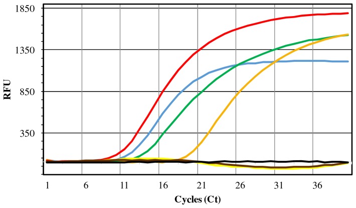
The real-time PCR assay with the IS6110-targeted probe (P_IS6110) and flanking primers (F_IS6110 and R_IS6110) yielded amplification products only when using DNA extracted from MTC members as template (Table 1, Figure 3). No non-specific results were obtained with members of the Mycobacterium avium complex or with strains belonging to other diverse bacterial species. The minimum detection threshold (analytical sensitivity) of this assay was estimated by the construction of a reference curve with serially-diluted suspensions of DNA from M. tuberculosis H37Ra. The analytical sensitivity was estimated in 0.3 fg/µL of M. tuberculosis genomic DNA. A linear relationship between the logarithm of the starting concentration of DNA and the amplification Ct values was obtained, with a -3.222 slope, a Ct = 35 interception in the minimum threshold (0.3 fg/µL) and an R2 = 0.999.
Figure 3. IS6110-targeted real-time PCR amplification curves obtained in specificity tests.
Illustration of the MTC-specific amplification curves obtained in FAM channel during evaluation of the analytical specificity of the IS6110-targeted real-time PCR step. All MTC cultures yielded amplification curves: M. tuberculosis ATCC 25177 (blue line), M. bovis LNIV 13027 (green line), M. bovis ATCC 27291 (orange line) and M. caprae LNIV 17320 (red line). Other bacteria yielded negative results of amplification: M. avium subsp. avium ATCC 25291 (yellow line) and M. avium subsp. paratuberculosis LNIV 39888 (brown line). No amplification was detected in non-template negative control (black line). RFU - Relative Fluorescence Units.
Spiked samples
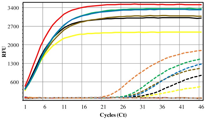
Tissue samples spiked with serially-diluted suspensions of M. tuberculosis cells were used for assessing the detection limit of the β-actin and IS6110-targeted semi-nested duplex real-time PCR assay. The detection threshold was estimated to be one mycobacteria per ml of tissue macerate (Figure 4). The IS6110 amplification curves usually harboured a very low Ct (
Figure 4. Testing of spiked samples with the semi-nested duplex real-time PCR assay.
The analytical sensitivity of the assay was estimated through the use of solutions of DNA extracted from MTC-free tissue samples spiked with serially diluted cellular suspensions of M. tuberculosis ATCC 25177 (ranging from 107 to 10° cells/ml of tissue homogenate). The figure illustrates the IS6110 (solid lines) and mammal β-actin gene (dashed lines) targeted amplification curves obtained in FAM and Cy5.5 channels, respectively, for the several dilutions: 107 (yellow line), 105 (black line), 103 (brown line), 101 (green line) and 10° (blue line) cells. Positive control: M. tuberculosis ATCC 25177 culture (red line); Negative controls: unspiked tissue homogenate (orange line) and non-template control (grey line). RFU - Relative Fluorescence Units.
< 3), meaning that the first standard PCR step yielded abundant amplification products easily detected by the following real time PCR step . The co-amplification of the bovine β-actin gene showed that no apparent inhibition of the real-time PCR step occurred due to the presence of inhibitor components of tissues or excess of bovine DNA, a significant result for the type of specimens used (animal tissues), confirming the efficiency of the extraction and purification procedures used in this work (Figure 4).
Diagnostic specificity and sensitivity
The results of the semi-nested duplex real-time PCR assays using DNA extracted from tissue samples are summarized in Table 2. All but one tissue sample from which M. bovis was cultured (types I and II) yielded positive amplification results, corresponding to a diagnostic sensitivity of 98.2% (CIP95% 89.4-99.9%). Noteworthy, when a subset of these TB culture positive samples was preliminary tested using only the second step of the amplification assay (without the first conventional PCR step), the diagnostic sensitivity was only 40%. Eight DNA-positive tissues were obtained among the 71 M. bovis culture negative samples (types III, IV, V and VI), yielding a diagnostic specificity of 88.7% (CIP95% 78.5-94.7%). Negative controls run with batches of samples did not identify any cross-contaminating DNA. The positive and negative predictive values were estimated in 87.5% (CIP95% 76.3-94.1%) and 98.4% (CIP95% 90.1-99.9%), respectively, for a prevalence of 44.5% M. bovis culture positive samples among all samples analysed. Considering only the bovine samples, the diagnostic sensitivity and specificity of the amplification assay were estimated in 100% (CIP95% 84.0-100%) and 97.7% (CIP95% 86.2-99.9%), respectively, while PPV and NPV were estimated in 96.3% (CIP95% 79.1-99.8%) and 100% (CIP95% 89.6-100%), respectively, for a prevalence of 37.7% M. bovis culture positive samples.
No DNA-positive samples were obtained from samples without TB-compatible lesions and M. bovis isolation (types III and IV) (Table 2). However, eight DNA-positive samples were detected among tissues that were M. bovis culture-negative but that harboured TB-compatible lesions (sample types V and VI) (Table 2).
An observed kappa coefficient was estimated in 0.859 (CIP95% 0.771-0.948) for the overall agreement between the results obtained by the direct application of the IS6110-targeted semi-nested PCR assay to fresh tissue samples and the results obtained from the gold-standard bacteriological culture. The direct detection procedure allowed the detection of MTC infected samples in less than 6 hours while the conventional culture takes about 6 to 12 weeks.
Discussion
The availability of TB confirmatory tests allowing a fast and conclusive detection of tuberculous mycobacteria in suspect animal tissues would be a great advantage in improving the efficiency of TB eradication programs and in decreasing the associated economic burden. Improvements in specificity, sensitivity and detection limit of diagnostic assays are usually introduced by molecular approaches. Nevertheless, only a few diagnostic PCR-based assays have been described for detecting MTC members directly in animal specimens, usually yielding limited sensitivities when compared to the reference bacteriological culture [8-12,15,19]. These moderate sensitivities may well be linked to the increased complexity for disrupting and recovering genomic DNA from the tough mycobacterial cells and to the paucibacillary nature of many animal tissue lesions used for nucleic acids extraction [10]. The presence of amplification inhibitors in crude tissue extracts, namely of large amounts of co-extracted eukaryotic DNA, may represent an additional problem. An option that has been explored to enhance the sensitivity of PCR techniques involves the implementation of more effective DNA extraction and purification methods. Taylor and colleagues were able to increase the M. bovis PCR detection sensitivity in bovine tissue samples with visible lesions, from 70% to 91%, after the inclusion of an additional step of liquid nitrogen freeze-thaw cycles in the DNA extraction procedure [10]. The use of sequence capture or immunomagnetic separation approaches was also forecasted for recovering higher yields of mycobacterial DNA from samples [8,9,12,21]. Nevertheless, these DNA extraction approaches usually involve more experimental steps and expensive equipments and consumables.
In this work, an alternative approach was evaluated for enhancing MTC detection sensitivities directly from fresh animal tissues. An adapted and optimized user-friendly DNA extraction protocol, mainly based in the use of simple commercially obtainable extraction kits, was coupled with an IS6110-targeted semi-nested real-time PCR assay that allows the direct detection of MTC members in animal tissue specimens with very high sensitivity, specificity and positive and negative predictive values, namely in bovine specimens. These two last parameters are dependent on the prevalence of the tested condition in the population under study, as well as on the sensitivity and specificity of the testing assays. The prevalence of MTC (mostly M. bovis) infected samples among all TB-suspect samples submitted to the reference laboratory during the 2002-2010 period (n = 6364) was approximately 40% (reviewed by Cunha and colleagues [27]), which compares well with the prevalence values found in this work and used for the computation of PPV and NPV. Therefore, the PPV and NPV parameters computed may be considered good indicators of the performance of our IS6110-targeted assay for assessing the presence of MTC in specimens collected from animals clinically suspected of having tuberculosis and submitted for confirmatory culture analysis in a major reference laboratory.
Eight DNA-positive amplification results were obtained from tissue samples from which M. bovis could not be cultured (Table 2). However, lesions compatible with TB were observed in these tissues during histological analysis (sample types V and VI) (Table 2). The culture of tissue samples for the isolation of M. bovis and of other MTC members, followed by molecular or biochemical identification procedures, is usually the gold-standard method to validate alternative diagnostic assays. However, it is known that bacteriological culture is slow and laborious and can yield ambiguous or false-negative results, e.g. due to the presence of non-viable mycobacteria, raising concerns about its effectiveness as comparison reference method [19,31]. The results of culture assays may be affected by several factors, such as the harsh processing and decontamination procedures of samples, which can also have a harmful effect on M. bovis viability, as well as the growth media and incubation conditions used and the constrained distribution of mycobacteria in tissues [32]. Therefore, although TB-like lesions identified by histopathology can be induced by other bacteria or mycobacteria, the positive amplification results suggest that MTC members were most probably associated with the observed lesions and that the PCR test is more sensitive than bacteriological culture for detecting these pathogens in animal tissues. Non-MTC mycobacteria such as M. avium and M. scrofulaceum were cultured from five of those eight DNA-positive tissue samples (Table 2). These non-MTC mycobacteria may potentially overgrow any M. bovis isolate present in the sample and mask its presence [19]. Regardless of some discrepancies, the kappa measure of agreement between bacteriological culture and the semi-nested PCR was estimated in 0.859. Although the criteria for judging kappa statistic are not completely objective nor universally accepted, this value may allow us to infer an "almost perfect" agreement between the two MTC detection methods [33].
The enhancement of the MTC detection rates using the semi-nested amplification assay need to be balanced against the associated increased risk of cross contamination of samples. Therefore, we should emphasize the need of working in a veterinary diagnosis laboratory harboring good practice standard conditions for molecular analysis, which include working in separate clean rooms and the use of positive and negative controls.
In a preliminary survey the testing of TB positive and negative tissue samples employed a standard real-time PCR assay (corresponding to the second step of the semi-nested approach). The comparison of this assay with the reference of bacteriological culture showed a diagnostic sensitivity of only 40% (data not shown). The inclusion of the first step of conventional IS6110-targeted PCR amplification, in a semi-nested design, allowed to increase the sensitivity of the assay to near 100%. Previous studies found no significant improvements in the detection of MTC members in animal tissue samples using nested PCR assays, including with real-time PCR formats [10,14,15]. Nevertheless, the performance of PCR detection systems is highly dependent on the efficiency of the primers and probes, even when using the same genomic targets such as the IS6110 element [34]. The MTC-specific IS6110-targeted primers and probe used in this work were shown to be highly efficient for detecting tuberculous mycobacteria in animal tissue samples, although the isolates of the most relevant species, M. bovis, usually contain only one copy of this insertion sequence. It has recently been found that IS6110-like elements may be present in other non-MTC mycobacteria such as M. smegmatis [35]. However, the probe and respective flanking primers used show no relevant complementary regions with these IS6110-like nucleotide sequences (Figure 2).
The protocol for direct detection of MTC from fresh animal tissues, using an optimized DNA extraction and purification procedure coupled with a semi-nested real time PCR assay described in this work, was shown to be highly accurate and much faster than bacteriological culture, reducing the time for confirmatory TB diagnosis from several weeks to few hours, thus also potentially decreasing the arrest period of the suspect animal carcass and herd. The assay is amenable to future automation possibilities regarding both the DNA extraction and amplification steps. It may also allow the detection of MTC members when these pathogens become nonviable and non-cultivable or are overgrown by other less fastidious bacteria or mycobacteria also present in tissue samples. The use of a novel β-actin gene targeted probe, and respective flanking primers, with complementary targets in most mammal species, allowed to assess the presence of amplification inhibitors in the DNA extracts. Although the test is not able to distinguish between different members of the MTC, particularly M. bovis, the identification of any tuberculous mycobacteria infection in domestic or wildlife animals could have public health implications.
Acknowledgments
Celeste Matos, Lurdes Clemente, Madalena Monteiro, Paula Mendonça and Paulo Carvalho (INIAV, I.P.) are acknowledged for excellent technical assistance, providing bacterial strains and performing pathological analysis.
Funding Statement
MVC acknowledges funding from Fundação para a Ciência e Tecnologia (FCT), Portugal ("Ciência 2008'' research contract and PTDC/CVT/117794/2010 research project); JI acknowledges funding from FCT, Portugal ("Ciência 2007'' research contract and PTDC/CVT/111634/2009 research project); PC and ASF are a recipients of PhD grants from FCT (SFRH/BD/64136/2009 and SFRH/BD/62317/2009, respectively). The funders had no role in study design, data collection and analysis, decision to publish, or preparation of the manuscript.
References
- 1. van Ingen J, Rahim Z, Mulder A, Boeree MJ, Simeone R et al. (2012) Characterization of Mycobacterium orygis as M. tuberculosis complex subspecies. Emerg Infect Dis 18: 653–655. doi: 10.3201/eid1804.110888. PubMed: 22469053. [DOI] [PMC free article] [PubMed] [Google Scholar]
- 2. Whelan AO, Coad M, Cockle PJ, Hewinson G, Vordermeier M et al. (2010) Revisiting host preference in the Mycobacterium tuberculosis complex: experimental infection shows M. tuberculosis H37Rv to be avirulent in cattle. PLOS ONE 5: e8527. doi: 10.1371/journal.pone.0008527. PubMed: 20049086. [DOI] [PMC free article] [PubMed] [Google Scholar]
- 3. Kubica T, Rüsch-Gerdes S, Niemann S (2003) Mycobacterium bovis subsp. caprae caused one-third of human M. bovis-associated tuberculosis cases reported in Germany between 1999 and 2001. J Clin Microbiol 41: 3070-3077. doi: 10.1128/JCM.41.7.3070-3077.2003. PubMed: 12843046. [DOI] [PMC free article] [PubMed] [Google Scholar]
- 4. Rodríguez E, Sánchez LP, Pérez S, Herrera L, Jiménez MS et al. (2009) Human tuberculosis due to Mycobacterium bovis and M. caprae in Spain, 2004-2007. Int J Tuberc Lung Dis 13: 1536-1541. PubMed: 19919773. [PubMed] [Google Scholar]
- 5. Torres-Gonzalez P, Soberanis-Ramos O, Martinez-Gamboa A, Chavez-Mazari B, Barrios-Herrera MT et al. (2013) Prevalence of latent and active tuberculosis among dairy farm workers exposed to cattle infected by Mycobacterium bovis. PLoS Negl Trop. Drosophila Inf Service 7: e2177. [DOI] [PMC free article] [PubMed] [Google Scholar]
- 6. Sunder S, Lanotte P, Godreuil S, Martin C, Boschiroli ML et al. (2009) Human-to-human transmission of tuberculosis caused by Mycobacterium bovis in immunocompetent patients. J Clin Microbiol 47: 1249-1251. doi: 10.1128/JCM.02042-08. PubMed: 19171683. [DOI] [PMC free article] [PubMed] [Google Scholar]
- 7. Rua-Domenech R (2006) Human Mycobacterium bovis infection in the United Kingdom: incidence, risks, control measures and review of the zoonotic aspects of bovine tuberculosis. Tuberculosis (Edinburgh) 86: 77-109. doi: 10.1016/j.tube.2005.05.002. PubMed: 16257579. [DOI] [PubMed] [Google Scholar]
- 8. Parra A, García N, García A, Lacombe A, Moreno F et al. (2008) Development of a molecular diagnostic test applied to experimental abattoir surveillance on bovine tuberculosis. Vet Microbiol 127: 315-324. doi: 10.1016/j.vetmic.2007.09.001. PubMed: 17954014. [DOI] [PubMed] [Google Scholar]
- 9. Taylor MJ, Hughes MS, Skuce RA, Neill SD (2001) Detection of Mycobacterium bovis in bovine clinical specimens using real-time fluorescence and fluorescence resonance energy transfer probe rapid-cycle PCR. J Clin Microbiol 39: 1272-1278. doi: 10.1128/JCM.39.4.1272-1278.2001. PubMed: 11283040. [DOI] [PMC free article] [PubMed] [Google Scholar]
- 10. Taylor GM, Worth DR, Palmer S, Jahans K, Hewinson RG (2007) Rapid detection of Mycobacterium bovis DNA in cattle lymph nodes with visible lesions using PCR. BMC. Vet Res 3: 12. [DOI] [PMC free article] [PubMed] [Google Scholar]
- 11. Coetsier C, Vannuffel P, Blondeel N, Denef JF, Cocito C et al. (2000) Duplex PCR for differential identification of Mycobacterium bovis, M. avium, and M. avium subsp. paratuberculosis in formalin- fixed paraffin-embedded tissues from cattle. J Clin Microbiol 38: 3048-3054. PubMed: 10921976. [DOI] [PMC free article] [PubMed] [Google Scholar]
- 12. Roring S, Hughes MS, Skuce RA, Neill SD (2000) Simultaneous detection and strain differentiation of Mycobacterium bovis directly from bovine tissue specimens by spoligotyping. Vet Microbiol 74: 227-236. doi: 10.1016/S0378-1135(00)00189-9. PubMed: 10808091. [DOI] [PubMed] [Google Scholar]
- 13. Miller J, Jenny A, Rhyan J, Saari D, Suarez D (1997) Detection of Mycobacterium bovis in formalin-fixed, paraffin-embedded tissues of cattle and elk by PCR amplification of an IS6110 sequence specific for Mycobacterium tuberculosis complex organisms. J Vet Diagn Invest 9: 244-249. doi: 10.1177/104063879700900304. PubMed: 9249162. [DOI] [PubMed] [Google Scholar]
- 14. Wards BJ, Collins DM, de Lisle GW (1995) Detection of Mycobacterium bovis in tissues by polymerase chain reaction. Vet Microbiol 43: 227-240. doi: 10.1016/0378-1135(94)00096-F. PubMed: 7740761. [DOI] [PubMed] [Google Scholar]
- 15. Thacker TC, Harris B, Palmer MV, Waters WR (2011) Improved specificity for detection of Mycobacterium bovis in fresh tissues using IS6110 real-time PCR. BMC. Vet Res 7: 50. [DOI] [PMC free article] [PubMed] [Google Scholar]
- 16. Pounder JI, Anderson CM, Voelkerding KV, Salfinger M, Dormandy J et al. (2010) Mycobacterium tuberculosis complex differentiation by genomic deletion patterns with multiplex polymerase chain reaction and melting analysis. Diagn Microbiol Infect Dis 67: 101-105. doi: 10.1016/j.diagmicrobio.2009.12.014. PubMed: 20227227. [DOI] [PubMed] [Google Scholar]
- 17. Niemann S, Harmsen D, Rüsch-Gerdes S, Richter E (2000) Differentiation of clinical Mycobacterium tuberculosis complex isolates by gyrB DNA sequence polymorphism analysis. J Clin Microbiol 38: 3231-3234. PubMed: 10970363. [DOI] [PMC free article] [PubMed] [Google Scholar]
- 18. Reddington K, Zumla A, Bates M, van Soolingen D, Niemann S et al. (2012) SeekTB- A two stage multiplex real-time PCR based method for the differentiation of the Mycobacterium tuberculosis complex. J Clin Microbiol 50: 2203-2206. doi: 10.1128/JCM.00718-12. PubMed: 22553241. [DOI] [PMC free article] [PubMed] [Google Scholar]
- 19. Liébana E, Aranaz A, Mateos A, Vilafranca M, Gomez-Mampaso E et al. (1995) Simple and rapid detection of Mycobacterium tuberculosis complex organisms in bovine tissue samples by PCR. J Clin Microbiol 33: 33-36. PubMed: 7699061. [DOI] [PMC free article] [PubMed] [Google Scholar]
- 20. Stewart LD, McNair J, McCallan L, Thompson S, Kulakov LA et al. (2012) Production and evaluation of antibodies and phage display-derived peptide ligands for immunomagnetic separation of Mycobacterium bovis . J Clin Microbiol 50: 1598-1605. doi: 10.1128/JCM.05747-11. PubMed: 22322353. [DOI] [PMC free article] [PubMed] [Google Scholar]
- 21. Stewart LD, McNair J, McCallan L, Gordon A, Grant IR (2013) Improved detection of Mycobacterium bovis infection in bovine lymph node tissue using immunomagnetic separation (IMS)-based methods. PLOS ONE 8: e58374. doi: 10.1371/journal.pone.0058374. PubMed: 23469275. [DOI] [PMC free article] [PubMed] [Google Scholar]
- 22. OIE (2013) Bovine Tuberculosis. In: Manual of Diagnostic Tests and Vaccines for Terrestrial Animals 2013. World Organization for Animal Health. [Google Scholar]
- 23. Cunha MV, Matos F, Canto A, Albuquerque T, Alberto JR et al. (2012) Implications and challenges of tuberculosis in wildlife ungulates in Portugal: a molecular epidemiology perspective. Res Vet Sci 92: 225-235. doi: 10.1016/j.rvsc.2011.03.009. PubMed: 21459394. [DOI] [PubMed] [Google Scholar]
- 24. Duarte EL, Domingos M, Amado A, Botelho A (2008) Spoligotype diversity of Mycobacterium bovis and Mycobacterium caprae animal isolates. Vet Microbiol 130: 415-421. doi: 10.1016/j.vetmic.2008.02.012. PubMed: 18417301. [DOI] [PubMed] [Google Scholar]
- 25. Duarte EL, Domingos M, Amado A, Cunha MV, Botelho A (2010) MIRU-VNTR typing adds discriminatory value to groups of Mycobacterium bovis and Mycobacterium caprae strains defined by spoligotyping. Vet Microbiol 143: 299-306. doi: 10.1016/j.vetmic.2009.11.027. PubMed: 20045269. [DOI] [PubMed] [Google Scholar]
- 26. Costa P, Amaro A, Botelho A, Inácio J, Baptista PV (2010) Gold nanoprobes assay for identification of mycobacteria from the Mycobacterium tuberculosis complex. Clin Microbiol Infect 16: 1464-1469. doi: 10.1111/j.1469-0691.2010.03120.x. PubMed: 19930276. [DOI] [PubMed] [Google Scholar]
- 27. Cunha MV, Monteiro M, Carvalho P, Mendonça P, Albuquerque T et al. (2011) Multihost tuberculosis: insights from the portuguese control program. Vet Med Int vol. 2011: 795165: Article 795165 doi: 10.4061/2011/795165. PubMed: 21776353. [DOI] [PMC free article] [PubMed] [Google Scholar]
- 28. Amaro A, Duarte E, Amado A, Ferronha H, Botelho A (2008) Comparison of three DNA extraction methods for Mycobacterium bovis, Mycobacterium tuberculosis and Mycobacterium avium subsp. avium . Lett Appl Microbiol 47: 8–11. doi: 10.1111/j.1472-765X.2008.02372.x. PubMed: 18498320. [DOI] [PubMed] [Google Scholar]
- 29. Larkin MA, Blackshields G, Brown NP, Chenna R, McGettigan PA et al. (2007) Clustal W and Clustal X version 2.0. Bioinformatics 23: 2947-2948. doi: 10.1093/bioinformatics/btm404. PubMed: 17846036. [DOI] [PubMed] [Google Scholar]
- 30. Restrepo BI, Gomez DI, Shipley GL, McCormick JB, Fisher-Hoch SP (2006) Selective enrichment and detection of mycobacterial DNA in paucibacillary specimens. J Microbiol Methods 67: 220-229. doi: 10.1016/j.mimet.2006.03.016. PubMed: 16678925. [DOI] [PMC free article] [PubMed] [Google Scholar]
- 31. Santos N, Geraldes M, Afonso A, Almeida V, Correia-Neves M (2010) Diagnosis of tuberculosis in the wild boar (Sus scrofa): a comparison of methods applicable to hunter-harvested animals. PLOS ONE. 5: e12663. doi: 10.1371/journal.pone.0012663. PubMed: 20844754. [DOI] [PMC free article] [PubMed] [Google Scholar]
- 32. Corner LA, Gormley E, Pfeiffer DU (2012) Primary isolation of Mycobacterium bovis from bovine tissues: conditions for maximising the number of positive cultures. Vet Microbiol 156: 162-171. doi: 10.1016/j.vetmic.2011.10.016. PubMed: 22074859. [DOI] [PubMed] [Google Scholar]
- 33. Landis JR, Koch GG (1977) The measurement of observer agreement for categorical data. Biometrics 33: 159–174. doi: 10.2307/2529310. PubMed: 843571. [DOI] [PubMed] [Google Scholar]
- 34. Savelkoul PH, Catsburg A, Mulder S, Oostendorp L, Schirm J et al. (2006) Detection of Mycobacterium tuberculosis complex with real time PCR: comparison of different primer-probe sets based on the IS6110 element. J Microbiol Methods 66: 177-180. doi: 10.1016/j.mimet.2005.12.003. PubMed: 16427712. [DOI] [PubMed] [Google Scholar]
- 35. Coros A, DeConno E, Derbyshire KM (2008) IS6110, a Mycobacterium tuberculosis complex-specific insertion sequence, is also present in the genome of Mycobacterium smegmatis, suggestive of lateral gene transfer among mycobacterial species. J Bacteriol 190: 3408-3410. doi: 10.1128/JB.00009-08. PubMed: 18326566. [DOI] [PMC free article] [PubMed] [Google Scholar]


