Figure 1.

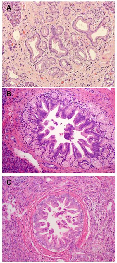
Representative micrographs show pancreatic intraepithelial neoplasia 1 (PanIN1, A), PanIN2 (B) and PanIN3 (C). Hematoxylin & eosin stain, original magnifications: 200×.

Representative micrographs show pancreatic intraepithelial neoplasia 1 (PanIN1, A), PanIN2 (B) and PanIN3 (C). Hematoxylin & eosin stain, original magnifications: 200×.