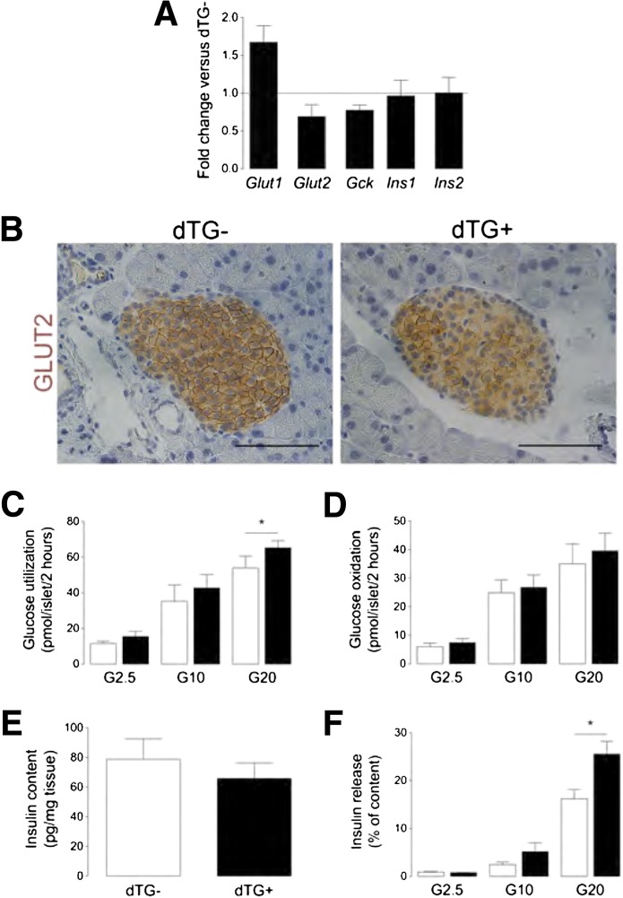
FIG. 4.
Islet hypovascularization and hypoxia do not overtly influence β-cell function. A: Transcript level is increased for Slc2a1 (Glut1) (n = 3), decreased for Slc2a2 (Glut2) (n = 4) and glucokinase (Gck) (n = 4), and unchanged for insulin (Ins1/2) (n = 3) in islets, isolated from dTG+DOX mice. Results are expressed relative to their expression level in dTG-DOX islets, which is set at 1. B: Pancreas sections stained for GLUT2, illustrating decreased membrane expression of GLUT2 in dTG+DOX mice. C and D: In vitro glucose utilization (n = 5) (C) and oxidation (n = 5) (D) are unchanged in islets, isolated from dTG+DOX mice, except for an increase in glucose utilization at 20 mmol/L glucose. E: Pancreas insulin content (n = 5) is unchanged in dTG+DOX mice. F: Insulin release from β-cells, isolated from dTG+DOX mice, is increased at 20 mmol/L glucose (results are expressed as % of total insulin content) (n = 4). All analyses were done after 2 weeks ± DOX. Data were statistically analyzed by one-sample t test (A) or by (un)paired t test (B–F). G2.5, 2.5 mmol/L glucose; G10, 10 mmol/L glucose; G20, 20 mmol/L glucose. *P ≤ 0.05.

