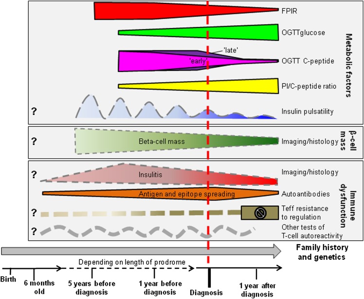
The benefits of residual endogenous insulin secretion in improving metabolic control and reducing complications in patients with type 1 diabetes (T1D) (1) have highlighted the need for early intervention to preserve β-cell mass. However, the time course of β-cell destruction in T1D is still poorly understood (2). Islet autoantibodies indicative of ongoing autoimmunity often appear in early infancy and may be present for many years before diabetes develops (3). Thus, autoantibodies are very useful for identifying individuals at high risk of progression but do not provide precise estimates of time to diabetes onset. Current tests of T-cell activity are poor guides to the rate of progression. Imaging methods for tracking β-cell loss are in development, but are unlikely to be available for several years (4). Metabolic testing is therefore the best approach for monitoring the decline in β-cell capacity (5,6) (Fig. 1).
FIG. 1.
Schematic showing potentially informative tests for tracking the loss of β-cell mass and function. Deterioration of FPIR in the IVGTT and of OGTT responses are currently the best indicators of impending diabetes. Imaging methods for monitoring β-cell mass are still in development. Little is known about the pattern of insulitis and decline in β-cell mass in humans during the prediabetic prodrome. PI/C-peptide, proinsulin-to-C-peptide ratio; Teff, effector T cells.
Several tests have been used to assess β-cell function. These vary in complexity and differ in what they reveal about the residual mass and health of the β-cells (7). Tests of fasting glucose, insulin, or C-peptide can be useful for identifying metabolic decompensation and for estimating insulin sensitivity. Loss of pulsatile insulin secretion (8) and increased proinsulin-to-C-peptide ratio may also indicate declining β-cell function (9). However, stimulation tests, such as the intravenous glucose tolerance test (IVGTT), mixed-meal tolerance test, and oral glucose tolerance test (OGTT), are the methods of choice for monitoring short-term progression (5,10).
The IVGTT measures the insulin or C-peptide response of β-cells to glucose without the influence of incretin hormones released from the gut (7). The early first-phase insulin response (FPIR), defined here as the mean insulin measurement from the samples taken at 1 and 3 min, reflects the rapid release of insulin from the storage granules into the circulation. The IVGTT has been standardized according to glucose load, infusion, and sample collection time to limit variability between tests (11). A reduction in FPIR has long been used as a predictive marker of short-term progression to T1D, and an FPIR below the 1st–10th percentile of healthy control subjects was one inclusion criterion of the parenteral arm of the Diabetes Prevention Trial-Type 1 (DPT-1).
In this issue, Sosenko et al. (12) present new retrospective analysis of IVGTT data from 74 of the oral insulin trial participants from DPT-1 who developed diabetes during follow-up. They observed that the FPIR in these progressors declined slowly from 48 to 18 months prior to diabetes onset, but then fell rapidly during the interval between 18 months and 6 months before diagnosis. This important finding may lead to more precise staging of the prediabetic process in at-risk individuals, thereby facilitating early interventions aimed at preserving β-cell function.
The observation that the FPIR remains relatively stable until 18 months before diabetes onset and then falls rapidly over the following year suggests that the reduction in β-cell mass has reached a tipping point beyond which the remaining β-cells are no longer able to compensate by increases in function. Whether this also reflects a rapid loss of β-cells is not clear, but it is consistent with small early studies that followed IVGTT responses over time in islet cell cytoplasmic antibody (ICA) positive relatives (13,14). Furthermore, recent analysis of OGTT data from progressors from both arms of DPT-1 showed that fasting, 2-h, and integrated glucose levels during OGTTs increased gradually from 30 months to 6 months before diagnosis (15). These increases in glucose levels were accompanied by minimal overall changes in fasting, peak, and integrated C-peptide measures. However, there was a shift of C-peptide responses with a reduction in early (0–30 min) and an increase in late (30–120 min) responses until 6 months before diagnosis. The decline in C-peptide levels then accelerated from 6 months before until 3 months after diabetes onset. Taken together, these studies point to an initial failure of early glucose-stimulated β-cell responses prior to diagnosis; a pattern supported by investigations using the nonglucose secretagogue arginine (14).
The failure of prophylactic parenteral and oral insulin to slow the progression of T1D in at-risk relatives participating in the DPT-1 was a major disappointment, although delayed onset in a subgroup of the oral treatment arm has prompted further trials (16). Recruitment for DPT-1 required screening 103,391 relatives. A key strength of the current study, therefore, is the availability of serial IVGTT measurements on so many well-characterized individuals with normal initial glucose tolerance as they progress to diabetes. Many nonprogressors were also available for comparison. Few, if any, cohorts with equivalent IVGTT follow-up data exist, so it may take time to confirm these findings.
There are a number of limitations inherent to this study. Participants were a very select group of high-risk relatives; progression may be different in those at lower genetic risk or with different risk profiles. First-line screening with ICA as in DPT-1 is now rarely used to select participants for trials, complicating comparison with current cohorts. Only 26 progressors had full FPIR data prior to diagnosis, and therefore smaller changes in FPIR may not have been detected, although the pattern of decline was supported by regression analysis in a further 48 progressors with incomplete data. More frequent IVGTTs during the prediabetic period would have allowed for more precise monitoring of the decline in FPIR. Further, the possible impact of differences in insulin sensitivity on β-cell responses was not investigated (5), even though this may influence β-cell survival (17,18). Use of a more specific insulin assay that did not cross-react with proinsulin may also have improved the discrimination of the test. Finally, some subjects were treated with oral insulin and, although unlikely, this could have altered insulin responses.
Sosenko et al. (12) have highlighted the potential of sequential FPIR measurements to help identify people at particularly high risk of progression, although interindividual variation (14,19) and the relative complexity of the test may limit its application (5). Further work is urgently needed to determine how functional measures of β-cell failure relate to the underlying pathology of β-cell destruction (20).
ACKNOWLEDGMENTS
A.E.L. is funded by a Fulbright/Diabetes UK Moffat Travel Fellowship.
No potential conflicts of interest relevant to this article were reported.
The authors thank Polly Bingley at the University of Bristol, Bristol, U.K., for comments and suggestions.
Footnotes
See accompanying original article, p. 4179.
REFERENCES
- 1.Steffes MW, Sibley S, Jackson M, Thomas W. Beta-cell function and the development of diabetes-related complications in the diabetes control and complications trial. Diabetes Care 2003;26:832–836 [DOI] [PubMed] [Google Scholar]
- 2.von Herrath M, Sanda S, Herold K. Type 1 diabetes as a relapsing-remitting disease? Nat Rev Immunol 2007;7:988–994 [DOI] [PubMed] [Google Scholar]
- 3.Ziegler AG, Rewers M, Simell O, et al. Seroconversion to multiple islet autoantibodies and risk of progression to diabetes in children. JAMA 2013;309:2473–2479 [DOI] [PMC free article] [PubMed] [Google Scholar]
- 4.Di Gialleonardo V, de Vries EF, Di Girolamo M, Quintero AM, Dierckx RA, Signore A. Imaging of β-cell mass and insulitis in insulin-dependent (type 1) diabetes mellitus. Endocr Rev 2012;33:892–919 [DOI] [PubMed] [Google Scholar]
- 5.Xu P, Wu Y, Zhu Y, et al. Diabetes Prevention Trial-Type 1 (DPT-1) Study Group Prognostic performance of metabolic indexes in predicting onset of type 1 diabetes. Diabetes Care 2010;33:2508–2513 [DOI] [PMC free article] [PubMed] [Google Scholar]
- 6.Meier JJ, Menge BA, Breuer TG, et al. Functional assessment of pancreatic beta-cell area in humans. Diabetes 2009;58:1595–1603 [DOI] [PMC free article] [PubMed] [Google Scholar]
- 7.Robertson RP. Estimation of beta-cell mass by metabolic tests: necessary, but how sufficient? Diabetes 2007;56:2420–2424 [DOI] [PubMed] [Google Scholar]
- 8.Bingley PJ, Matthews DR, Williams AJ, Bottazzo GF, Gale EA. Loss of regular oscillatory insulin secretion in islet cell antibody positive non-diabetic subjects. Diabetologia 1992;35:32–38 [DOI] [PubMed] [Google Scholar]
- 9.Truyen I, De Pauw P, Jørgensen PN, et al. Belgian Diabetes Registry Proinsulin levels and the proinsulin:C-peptide ratio complement autoantibody measurement for predicting type 1 diabetes. Diabetologia 2005;48:2322–2329 [DOI] [PubMed] [Google Scholar]
- 10.Greenbaum CJ, Mandrup-Poulsen T, McGee PF, et al. Type 1 Diabetes Trial Net Research Group. European C-Peptide Trial Study Group Mixed-meal tolerance test versus glucagon stimulation test for the assessment of beta-cell function in therapeutic trials in type 1 diabetes. Diabetes Care 2008;31:1966–1971 [DOI] [PMC free article] [PubMed] [Google Scholar]
- 11.Bingley PJ, Colman P, Eisenbarth GS, et al. Standardization of IVGTT to predict IDDM. Diabetes Care 1992;15:1313–1316 [DOI] [PubMed] [Google Scholar]
- 12.Sosenko JM, Skyler JS, Beam CA, et al. Type 1 Diabetes TrialNet and Diabetes Prevention Trial-Type 1 Study Groups Acceleration of the loss of the first-phase insulin response during the progression to type 1 diabetes in Diabetes Prevention Trial–Type 1 participants. Diabetes 2013;62:4179–4183. [DOI] [PMC free article] [PubMed] [Google Scholar]
- 13.Bleich D, Jackson RA, Soeldner JS, Eisenbarth GS. Analysis of metabolic progression to type I diabetes in ICA+ relatives of patients with type I diabetes. Diabetes Care 1990;13:111–118 [DOI] [PubMed] [Google Scholar]
- 14.Chaillous L, Rohmer V, Maugendre D, et al. Differential beta-cell response to glucose, glucagon, and arginine during progression to type I (insulin-dependent) diabetes mellitus. Metabolism 1996;45:306–314 [DOI] [PubMed] [Google Scholar]
- 15.Sosenko JM, Skyler JS, Herold KC, Palmer JP, Type 1 Diabetes TrialNet and Diabetes Prevention Trial-Type 1 Study Groups The metabolic progression to type 1 diabetes as indicated by serial oral glucose tolerance testing in the Diabetes Prevention Trial-Type 1. Diabetes 2012;61:1331–1337 [DOI] [PMC free article] [PubMed] [Google Scholar]
- 16.Skyler JS, Krischer JP, Wolfsdorf J, et al. Effects of oral insulin in relatives of patients with type 1 diabetes: The Diabetes Prevention Trial-Type 1. Diabetes Care 2005;28:1068–1076 [DOI] [PubMed] [Google Scholar]
- 17.Fourlanos S, Narendran P, Byrnes GB, Colman PG, Harrison LC. Insulin resistance is a risk factor for progression to type 1 diabetes. Diabetologia 2004;47:1661–1667 [DOI] [PubMed] [Google Scholar]
- 18.Bingley PJ, Mahon JL, Gale EA, European Nicotinamide Diabetes Intervention Trial Group Insulin resistance and progression to type 1 diabetes in the European Nicotinamide Diabetes Intervention Trial (ENDIT). Diabetes Care 2008;31:146–150 [DOI] [PubMed] [Google Scholar]
- 19.Smith CP, Tarn AC, Thomas JM, et al. Between and within subject variation of the first phase insulin response to intravenous glucose. Diabetologia 1988;31:123–125 [DOI] [PubMed] [Google Scholar]
- 20.Weir GC, Bonner-Weir S. Islet β cell mass in diabetes and how it relates to function, birth, and death. Ann N Y Acad Sci 2013;1281:92–105 [DOI] [PMC free article] [PubMed] [Google Scholar]


