

Abstract
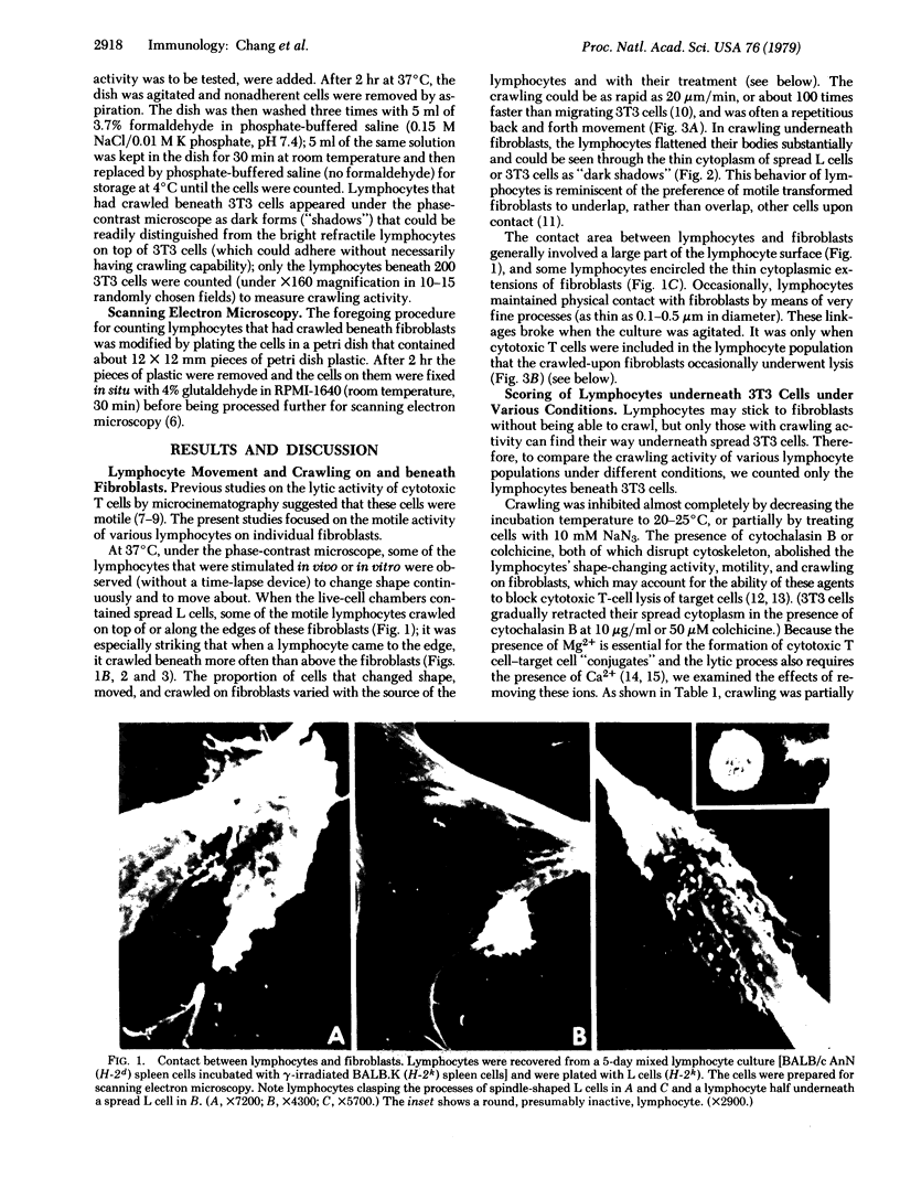
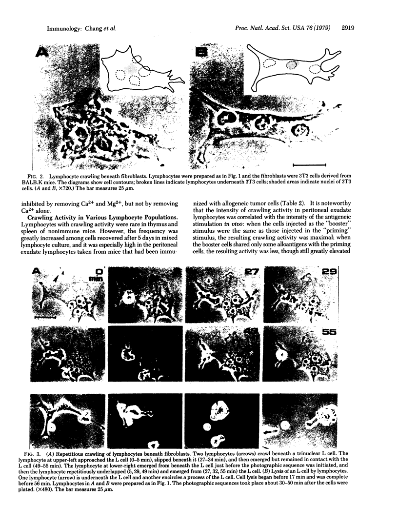
Some lymphocytes become highly motile upon immunological stimulation in vivo or in vitro. When introduced into a culture of 3T3 or L cells and followed by live-cell microscopy, some of these lymphocytes were observed to crawl on top of, along the edges of, and preferentially beneath the attached fibroblasts. The crawling could be as rapid as 20 μm/min, easily detectable without a time-lapse device. The striking ability of crawling lymphocytes to penetrate beneath attached 3T3 cells provided a quantitative means to compare the crawling activity of different lymphocyte populations under various conditions. Crawling was diminished by inhibitors of energy metabolism, by agents that disrupt the cytoskeleton, and by absence of Mg2+ and Ca2+, but not of Ca2+ alone. Crawling lymphocytes were virtually absent in normal thymus and spleen cells. They increased greatly in 5-day mixed lymphocyte cultures and in peritoneal exudate lymphocytes taken after mice had been immunized with allogeneic tumor cells. T cells accounted for most of the crawlers. Of two T-cell leukemias tested, R1+ cells were crawlers whereas EL-4 cells were not. The H-2 haplotype of the 3T3 fibroblasts (i.e., whether syngeneic or allogeneic) had no apparent effect on lymphocyte crawling activity. The crawling may relate to the exploration of cell surface antigens by lymphocytes (immune surveillance), to the mode of action of cytotoxic T cells, to the migration of lymphocytes across blood vessel walls, or to the penetration of lymphocytes into “solid” masses of normal tissue or tumor cells.
Keywords: activated lymphocytes, cell motility, T-cell leukemias, surface antigens
Full text
PDF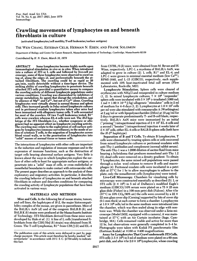
Images in this article
Selected References
These references are in PubMed. This may not be the complete list of references from this article.
- Able M. E., Lee J. C., Rosenau W. Lymphocyte--target cell interaction in vitro. Ultrastructural and cinematographic studies. Am J Pathol. 1970 Sep;60(3):421–434. [PMC free article] [PubMed] [Google Scholar]
- Albrecht-Buehler G. Daughter 3T3 cells. Are they mirror images of each other? J Cell Biol. 1977 Mar;72(3):595–603. doi: 10.1083/jcb.72.3.595. [DOI] [PMC free article] [PubMed] [Google Scholar]
- Berke G., Levey R. H. Cellular immunoabsorbents in transplantation immunity. Specific in vitro deletion and recovery of mouse lymphoid cells sensitized against allogeneic tumors. J Exp Med. 1972 Apr 1;135(4):972–984. doi: 10.1084/jem.135.4.972. [DOI] [PMC free article] [PubMed] [Google Scholar]
- Cantor H., Boyse E. A. Functional subclasses of T-lymphocytes bearing different Ly antigens. I. The generation of functionally distinct T-cell subclasses is a differentiative process independent of antigen. J Exp Med. 1975 Jun 1;141(6):1376–1389. doi: 10.1084/jem.141.6.1376. [DOI] [PMC free article] [PubMed] [Google Scholar]
- Cerottini J. C., Brunner K. T. Reversible inhibition of lymphocyte-mediated cytotoxicity by cytochalasin B. Nat New Biol. 1972 Jun 28;237(78):272–273. doi: 10.1038/newbio237272a0. [DOI] [PubMed] [Google Scholar]
- Doherty P. C., Blanden R. V., Zinkernagel R. M. Specificity of virus-immune effector T cells for H-2K or H-2D compatible interactions: implications for H-antigen diversity. Transplant Rev. 1976;29:89–124. doi: 10.1111/j.1600-065x.1976.tb00198.x. [DOI] [PubMed] [Google Scholar]
- Golstein P., Smith E. T. The lethal hit stage of mouse T and non-T cell-mediated cytolysis: differences in cation requirements and characterization of an analytical "cation pulse" method. Eur J Immunol. 1976 Jan;6(1):31–37. doi: 10.1002/eji.1830060108. [DOI] [PubMed] [Google Scholar]
- Hale A. H., Witte O. N., Baltimore D., Eisen H. N. Vesicular stomatitis virus glycoprotein is necessary for H-2-restricted lysis of infected cells by cytotoxic T lymphocytes. Proc Natl Acad Sci U S A. 1978 Feb;75(2):970–974. doi: 10.1073/pnas.75.2.970. [DOI] [PMC free article] [PubMed] [Google Scholar]
- Harris A. Location of cellular adhesions to solid substrata. Dev Biol. 1973 Nov;35(1):97–114. doi: 10.1016/0012-1606(73)90009-2. [DOI] [PubMed] [Google Scholar]
- Julius M. H., Simpson E., Herzenberg L. A. A rapid method for the isolation of functional thymus-derived murine lymphocytes. Eur J Immunol. 1973 Oct;3(10):645–649. doi: 10.1002/eji.1830031011. [DOI] [PubMed] [Google Scholar]
- Plaut M., Lichtenstein L. M., Henney C. S. Studies on the mechanism of lymphocyte-mediated cytolysis. 3. The role of microfilaments and microtubules. J Immunol. 1973 Mar;110(3):771–780. [PubMed] [Google Scholar]
- Rothstein T. L., Mage M., Jones G., McHugh L. L. Cytotoxic T lymphocyte sequential killing of immobilized allogeneic tumor target cells measured by time-lapse microcinematography. J Immunol. 1978 Nov;121(5):1652–1656. [PubMed] [Google Scholar]
- Sanderson C. J. The mechanism of T cell mediated cytotoxicity. II. Morphological studies of cell death by time-lapse microcinematography. Proc R Soc Lond B Biol Sci. 1976 Jan 20;192(1107):241–255. doi: 10.1098/rspb.1976.0011. [DOI] [PubMed] [Google Scholar]
- Solomon F. Detailed neurite morphologies of sister neurolbastoma cells are related. Cell. 1979 Jan;16(1):165–169. doi: 10.1016/0092-8674(79)90197-1. [DOI] [PubMed] [Google Scholar]
- Sprent J. Role of H-2 gene products in the function of T helper cells from normal and chimeric mice in vivo. Immunol Rev. 1978;42:108–137. doi: 10.1111/j.1600-065x.1978.tb00260.x. [DOI] [PubMed] [Google Scholar]