

Abstract
The cytochrome c maturation system influences the expression of virulence factors in Bacillus anthracis. B. anthracis carries two copies of the ccdA gene, encoding predicted thiol-disulfide oxidoreductases that contribute to cytochrome c maturation, while the closely related organism Bacillus subtilis carries only one copy of ccdA. To investigate the roles of the two ccdA gene copies in B. anthracis, strains were constructed without each ccdA gene, and one strain was constructed without both copies simultaneously. Loss of both ccdA genes results in a reduction of cytochrome c production, an increase in virulence factor expression, and a reduction in sporulation efficiency. Complementation and expression analyses indicate that ccdA2 encodes the primary CcdA in B. anthracis, active in all three pathways. While CcdA1 retains activity in cytochrome c maturation and virulence control, it has completely lost its activity in the sporulation pathway. In support of this finding, expression of ccdA1 is strongly reduced when cells are grown under sporulation-inducing conditions. When the activities of CcdA1 and CcdA2 were analyzed in B. subtilis, neither protein retained activity in cytochrome c maturation, but CcdA2 could still function in sporulation. These observations reveal the complexities of thiol-disulfide oxidoreductase function in pathways relevant to virulence and physiology.
INTRODUCTION
Bacillus anthracis is a Gram-positive, endospore-forming bacterium that is the etiological agent of anthrax. Several virulence factors, including the anthrax toxin and capsule, contribute to virulence in the mammalian host (1). Expression of both the toxin and capsule genes is dependent upon the master virulence regulator AtxA. Loss of atxA strongly decreases transcription of toxin and capsule genes (2, 3) and renders B. anthracis avirulent in animal models of infection (4). The direct interaction between AtxA and the promoters of its target genes has not been demonstrated, and the mechanism by which AtxA regulates its target genes remains unclear. AtxA activity is regulated posttranscriptionally by alternate phosphorylation and dephosphorylation (5), the global regulator CodY (6), and multimerization in response to growth conditions (7). atxA transcription is directly regulated by the transition state regulator AbrB, which connects virulence regulation to the sporulation pathway (8). atxA transcription is also indirectly regulated in response to glucose through the catabolite repressor protein CcpA (9).
We recently reported that loss of the two small c-type cytochromes of B. anthracis, c550 and c551, leads to increased transcription of atxA and increased production of AtxA-regulated virulence factors (10). These observations led us to further investigate the roles of c-type cytochromes and cytochrome c maturation pathways in B. anthracis physiology and virulence. The c-type cytochromes are distinct from other cytochromes due to their covalently attached heme (11). Maturation c-type cytochromes is a complex, multistep process (12). While little is known about cytochrome production and activity in B. anthracis, our understanding is informed by findings in the closely related organism, Bacillus subtilis. B. subtilis carries a system II cytochrome c maturation system that is similar to those found in Gram-positive bacteria, cyanobacteria, the beta-, delta-, and epsilonproteobacteria, and chloroplasts (12, 13). The c-type cytochrome is initially produced as an apoprotein in the cytoplasm, missing its required cofactor, heme. The apocytochrome c is secreted across the membrane by the Sec translocation system. At the membrane, the cytochrome c maturation protein ResA reduces cysteine residues in the heme binding pocket of apocytochrome (14), while ResB and ResC transport and covalently attach heme to the reduced apocytochrome to generate the final, active form of cytochrome c (15, 16).
The thiol-disulfide oxidoreductase protein CcdA is also required for cytochrome c maturation in B. subtilis. CcdA is an integral membrane protein with six transmembrane (TM) domains with conserved cysteine residues in TM1 and TM4 (17, 18). The protein is homologous to a portion of Escherichia coli DsbD (18, 19). CcdA was first identified in B. subtilis during a screen for mutations that result in decreased production of c-type cytochromes (18). Loss of CcdA also results in disruption of late events in spore formation, indicating multiple extracellular functions of CcdA (20, 21). Subsequent work has shown that CcdA accepts electrons from thioredoxin in the cytoplasm and transfers the electrons to ResA at the cell surface (22). Reduced ResA can then reduce a disulfide linkage on apocytochrome c, facilitating covalent attachment of heme and production of functional c-type cytochromes (14, 23). The phenotypes associated with loss of CcdA can be suppressed by mutations in BdbC and BdbD, two other protein thiol-disulfide oxidoreductases that are involved in an oxidative pathway, because the disulfide bond in apocytochrome c that is reduced by ResA is no longer formed (14, 24). In addition to its interactions with ResA, CcdA reduces StoA, which contributes to spore formation and may reduce YneN, which has no known function (12, 25).
In the B. anthracis genome, there are two genes predicted to encode CcdA-like proteins (26), a feature found in members of the pathogenic Bacillus cereus group but not found in many other groups of bacilli (27). It is unclear why B. anthracis carries two copies of ccdA when B. subtilis can carry out both efficient sporulation and c-type cytochrome production with a single copy of ccdA. In this report, we show that the duplicate CcdA proteins have dissimilar activities in the cytochrome c maturation and spore formation pathways and that these differences are reflected in their gene expression profiles. Further, the activities of the two B. anthracis CcdA proteins are altered when expressed in B. subtilis and are distinct from the activity of endogenous B. subtilis CcdA.
MATERIALS AND METHODS
Bacterial strains and growth conditions.
B. anthracis strain 34F2(pXO1+ pXO2−) and its derivatives were routinely grown in LB or brain heart infusion (BHI) broth supplemented with the appropriate antibiotics at the following concentrations: chloramphenicol, 7.5 μg/ml; erythromycin, 5 μg/ml; lincomycin, 25 μg/ml; and kanamycin, 7.5 μg/ml. X-Gal (5-bromo-4-chloro-3-indolyl-β-d-galactopyranoside) (40 μg/ml) was added to LB agar to monitor β-galactosidase activity as necessary. Competent cells of B. anthracis were prepared by following the method of Koehler et al. (28), and electroporation was performed using the Bio-Rad Gene Pulser according to the instructions of the supplier.
B. subtilis strain JH642 and its derivatives were routinely grown in LB or nutrient sporulation medium phosphate (NSMP) (29) supplemented with the appropriate antibiotics at the following concentrations: chloramphenicol, 5 μg/ml; erythromycin, 5 μg/ml; lincomycin, 25 μg/ml; and kanamycin, 5 μg/ml. B. subtilis transformation was performed as previously described (30).
Escherichia coli TG1, C600, and DH5α competent cells were used for the propagation and isolation of all plasmid constructs. E. coli transformation was performed in chemically competent cells as previously described (31). Transformants were selected on LB agar supplemented with ampicillin (100 μg/ml), chloramphenicol (7.5 μg/ml), or kanamycin (30 μg/ml).
Plasmid and strain construction.
Strains and plasmids are listed in Table 1. Oligonucleotide primers are listed in Table S1 in the supplemental material. Markerless gene deletions in B. anthracis and B. subtilis were generated through a modification of the technique of Janes and Stibitz (32), as previously described (10), with the exception that plasmid pSS4332 (33) was used in place of plasmid pBKJ223 (32). The retention of plasmid pXO1 in B. anthracis strains was confirmed by PCR using primers atxAU5′Bam and atxAD3′Pst.
Table 1.
Strains and plasmids used in this study
| Strain or plasmid | Relevant characteristic(s)a | Source |
|---|---|---|
| B. anthracis strains | ||
| 34F2 | pXO1+ pXO2− | Laboratory stock |
| AW-A027 | Markerless deletion of ccdA1 | This study |
| AW-A028 | Markerless deletion of ccdA2 | This study |
| AW-A029 | Markerless deletion of ccdA1 and ccdA2 | This study |
| 34F2ΔresB | Markerless deletion of resB | 10 |
| B. subtilis strains | ||
| JH642 | pheA1 trpC2 | Laboratory stock |
| AW-S002 | Markerless deletion of ccdA | This study |
| Plasmids | ||
| pORI-Cm-I-SceI | pORI-Cm vector with I-SceI recognition site, Cmr | |
| pSS4332 | I-SceI expression plasmid, Kanr | 33 |
| pTCVlac-pagA | pagA-lacZ transcriptional fusion in pTCV-lac, Kanr | 5 |
| pTCVlac-atxA12 | atxA-lacZ transcriptional fusion in pTCV-lac, Kanr | 41 |
| pTCV-lac | Promoterless vector for transcriptional lacZ fusion, Kanr | 35 |
| pAW184 | Regions flanking BAS1647 in pORI-Cm-I-SceI, Cmr | This study |
| pAW186 | Regions flanking BAS3455 in pORI-Cm-I-SceI, Cmr | This study |
| pHP13 | Cloning/shuttle plasmid, Cmr | 34 |
| pAW285 | Xylose-inducible expression plasmid, Cmr | This study |
| pAW304 | ccdA1 expression vector, Cmr | This study |
| pAW305 | ccdA2 expression vector, Cmr | This study |
| pAW306 | B. subtilis ccdA expression vector, Cmr | This study |
| pAW350 | ccdA2-lacZ transcriptional fusion in pTCV-lac, Kanr | This study |
| pAW355 | ccdA1-lacZ transcriptional fusion in pTCV-lac, Kanr | This study |
| pAW360 | Regions flanking B. subtilis ccdA in pORI-Cm-I-SceI, Cmr | This study |
Cmr, chloramphenicol resistance; Kanr, kanamycin resistance.
The expression plasmid pAW285 was created by PCR amplification of JH642 genomic DNA to produce a DNA fragment containing the xylose repressor, xylR, and the XylR-regulated xylA promoter of B. subtilis by using primers xyl5′Eco and xyl3′Bam. The fragment was then cloned into plasmid pHP13 (34) at the EcoRI and BamHI sites. The resulting plasmid allows subcloning of genes downstream of the XylR-repressed xylA promoter, thereby placing their expression under a strong constitutive promoter in B. anthracis that can be further induced by xylose.
β-Galactosidase assays.
B. anthracis strains harboring gene promoter fusions on the replicative vector pTCV-lac (35) were grown at 37°C in LB supplemented with the appropriate antibiotics. β-Galactosidase activity was assayed as described previously, and specific activity was expressed in Miller units (36, 37).
Quantitative RT-PCR.
RNA was extracted from B. anthracis using the UltraClean microbial RNA isolation kit (MoBio, Carlsbad, CA). RNA was treated with Turbo DNase (Life Technologies, Carlsbad, CA) and quantified using an Eppendorf BioSpectrophotometer. Quantitative, real-time reverse transcription (RT)-PCR was performed using the Power SYBR RNA-to-Ct 1-step kit (Life Technologies) on a Prism 7500 Fast real-time PCR system (Life Technologies). Data are presented as threshold cycle (ΔΔCT) values calculated from the results of at least three independent experiments.
TMPD oxidase staining.
The presence of an active c-type cytochrome was verified by a colorimetric assay using N,N,N′,N′-tetramethyl-p-phenylenediamine (TMPD) as an artificial electron donor that can be oxidized by cytochrome caa3. TMPD oxidase staining was performed on B. anthracis or B. subtilis grown on NSMP as previously described (10, 16).
Sporulation analysis.
Sporulation assays were performed using strains grown in NSMP or Schaeffer's sporulation medium (SM) (38), containing chloramphenicol if necessary, at 37°C for 48 h. Sporulation was initially monitored by visualization of cells on a Zeiss Axio Imager microscope and enumeration of spores and vegetative cells from multiple independent fields. Sporulation efficiency was assayed by chloroform treatment and enumeration of spores and vegetative cells as previously described (39). Sporulation efficiency is presented as percentage of spores relative to total viable cells.
Operon and transcription analysis.
Operon organization was determined by conventional RT-PCR by using 34F2 RNA extracted using the UltraClean microbial RNA isolation kit. Following extraction, RNA was treated with Turbo DNase. cDNA was synthesized using SuperScript III reverse transcriptase and random hexamers (Life Technologies). Sets of primers flanking the intergenic regions for each gene pair were used to amplify cDNA. Amplicons were separated by electrophoresis on 1% agarose gels, stained by ethidium bromide, and visualized on a UVP gel documentation system.
For gene expression analysis, the intergenic regions upstream of the first linked gene of each operon were amplified using primers indicated in Table S1 in the supplemental material. The PCR fragments were then cloned into pTCV-lac (35), and sequences were confirmed by DNA sequencing. Transcriptional activities of the promoter fragments were then tested under a variety of conditions using β-galactosidase analysis.
RESULTS
Identification and description of ccdA1 and ccdA2 loci.
Disruption of the cytochrome c maturation pathway in B. anthracis causes increased transcription of virulence genes, the result of the loss of the two small c-type cytochromes c550 and c551 (10). In previous transposon mutagenesis screens for mutants that cause increased transcription of virulence genes, the B. anthracis orthologs of most nonessential genes known to be required for cytochrome c maturation in the model organism B. subtilis were disrupted (10, 16). A gene important to cytochrome c production in B. subtilis is ccdA, which encodes a protein thiol-disulfide oxidoreductase that contributes to covalent attachment of heme to apocytochrome c at the cell surface (18, 21, 24). ccdA is also involved in spore formation in B. subtilis (20). A ccdA disruption was not isolated in a screen for mutants with increased transcription of virulence genes that isolated mutants in most other cytochrome c maturation genes (10). Examination of the B. anthracis genome reveals the presence of two ccdA-like genes. BAS1647 (GenBank accession no. AAT53964.1) encodes a protein with 65% amino acid similarity to B. subtilis ccdA (GenBank accession no. CAB13677.1). BAS3455 (GenBank accession no. AAT55761.1) encodes a protein with 68% amino acid similarity to B. subtilis ccdA. BAS1647 and BAS3455 share 63% amino acid similarity. We will refer to BAS1647 as ccdA1 and BAS3455 as ccdA2. The lack of ccdA disruption mutants in our previous screen may be due to the existence of two complementary copies of ccdA.
The genomic context of both ccdA1 and ccdA2 is different than that of B. subtilis ccdA. B. subtilis ccdA is cotranscribed with two downstream genes, yneI and yneJ, but neither of the downstream genes contribute to sporulation or cytochrome c maturation (20). ccdA1 appears to be part of a large gene cluster containing genes predicted to be involved in multiple cellular processes. Of note is the presence immediately downstream of ccdA1 of an ortholog of yneN, a membrane-bound protein with a thioredoxin-like domain that may interact with ccdA in B. subtilis (25). To determine the structure of a putative ccdA1 operon, linkage analysis was performed by RT-PCR using 34F2 RNA and sets of primers flanking the intergenic regions of each potential gene pair. As shown in Fig. S1 in the supplemental material, amplicons were obtained for each intergenic region between BAS1644 and BAS1649, indicating that transcription of these genes is linked and constitutes an operon. The genes flanking either end of the BAS1644-9 operon are transcribed in the opposite direction and are not part of the operon. These findings are consistent with a recent global transcriptome analysis that identified ccdA1 as part of a six-gene operon (40). Linkage analysis of ccdA2 indicates that it is unlinked to either of the surrounding genes transcribed in the same orientation. The lack of upstream linkage is unsurprising due to the large intergenic region and predicted transcriptional terminator between BAS3456 and ccdA2. A linkage between ccdA2 and BAS3454, a yneJ-like gene, might have been expected given the similarities to the operon organization in B. subtilis (ccdA-yneI-yneJ) (20, 27), but the lack of a yneI-like gene between ccdA2 and the yneJ-like BAS3454 indicates genomic dissimilarities between the two species.
Characterization of ccdA deletion strains.
To investigate the role of ccdA1 and ccdA2 in virulence gene expression, cytochrome c maturation, and sporulation, we created markerless deletion strains in the B. anthracis 34F2 background. Strain AW-A027 is missing ccdA1, strain AW-A028 is missing ccdA2, and strain AW-A029 is missing both ccdA1 and ccdA2. A growth curve in LB of the double-deletion strain is shown in Fig. 1A. Loss of ccdA1 and ccdA2 singly (data not shown) or loss of both in the double deletion did not result in significant growth defects.
Fig 1.
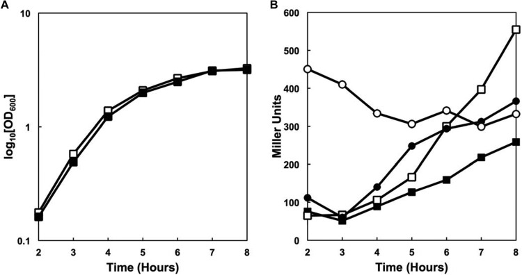
Growth and virulence gene expression in the ccdA1 ccdA2 mutant strain. (A) Cell growth of parental and mutant strains grown in LB at 37°C. ■, 34F2; □, AW-A029 (ccdA1 ccdA2). (B) β-Galactosidase activity in pagA and atxA reporter strains grown in LB supplemented with kanamycin at 37°C. ■, 34F2 pagA-lacZ; ●, 34F2 atxA-lacZ; □, AW-A029 pagA-lacZ; ○, AW-A029 atxA-lacZ.
B. anthracis virulence gene expression was measured by β-galactosidase assays using transcriptional fusions of either the atxA (41) or pagA (5) promoter to an lacZ reporter. As shown in Fig. 1B, loss of both ccdA1 and ccdA2 resulted in increased transcription of atxA during exponential phase and increased transcription of pagA during early stationary phase, similar to the phenotype of a cytochrome c maturation or a cccA cccB knockout mutant, as observed previously (10). As atxA overexpression in exponential phase was shown to be the cause of increased pagA transcription, we focused more carefully on early atxA expression. Figure 2 shows the early atxA expression profile of multiple strains. As expected of the parental 34F2 strain, atxA expression was low at 3 h and remains unchanged at 4 h. In contrast, the resB deletion strain, in which cytochrome c production is completely abolished, showed a more than 5-fold increase in atxA expression at 3 h that decreased significantly at 4 h, consistent with previous findings (10). The atxA transcription profile in the ccdA1 ccdA2 double-deletion strain was similar to that of the resB deletion strain, though the expression level was slightly lower. The ccdA1 deletion strain resembled the parental 34F2 strain with no increased atxA expression. The ccdA2 deletion strain had increased atxA expression relative to that of 34F2 and the ccdA1 deletion strain but lower than that of the double-deletion strain. These results suggested that loss of both ccdA1 and ccdA2 is required for strongly increased virulence gene expression and that there is some degree of complementarity between CcdA1 and CcdA2.
Fig 2.
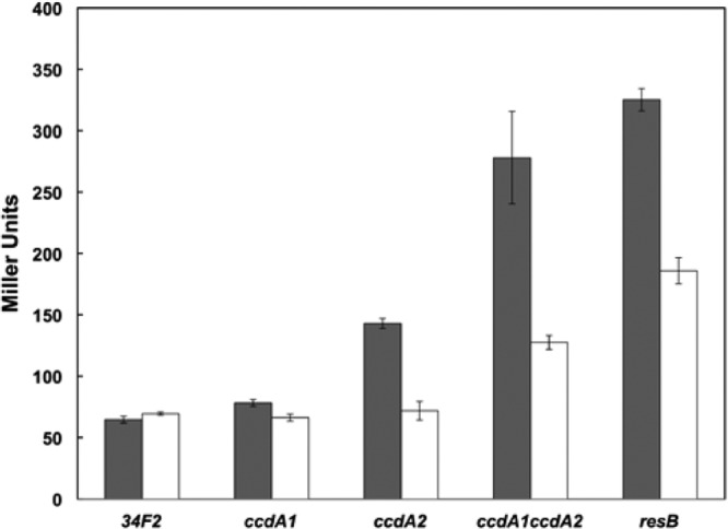
Early atxA expression in ccdA mutant strains. β-Galactosidase activity of parental and mutant strains carrying the atxA-lacZ reporter grown in LB supplemented with kanamycin at 37°C. Filled bars represent atxA expression at 3 h postinoculation, and empty bars represent atxA expression at 4 h postinoculation.
Next, the cytochrome c oxidase activity of the mutants was assayed by TMPD staining. TMPD is an artificial electron donor that interacts specifically with cytochrome c oxidase (cytochrome caa3) and is oxidized to a blue-colored product that stains colonies in the presence of active cytochrome c in the membrane. Parental and mutant strains were grown on NSMP agar plates, stained with TMPD, and scored for the appearance of blue color. As shown in Table 2, the parental 34F2 and the ccdA1 deletion strains were stained using TMPD, indicating active cytochrome c oxidase. In contrast, the ccdA2 and ccdA1 ccdA2 deletion strains were not stained using TMPD, indicating a lack of active cytochrome c oxidase. These findings are consistent with the atxA transcription results, suggesting that loss of ccdA2 results in loss of cytochrome c, while loss of ccdA1 has little effect.
Table 2.
Cytochrome c oxidase activity and sporulation efficiency in strains of B. anthracis
| Plasmid | TMPD staininga |
Sporulation efficiencyb |
||||||
|---|---|---|---|---|---|---|---|---|
| 34F2 | ccdA1 | ccdA2 | ccdA1 ccdA2 | 34F2 | ccdA1 | ccdA2 | ccdA1 ccdA2 | |
| pAW285 (Empty) | + | + | − | − | 53 | 55 | 21 | 23 |
| pAW304 (ccdA1) | + | + | + | + | 52 | 51 | 19 | 20 |
| pAW305 (ccdA2) | + | + | + | + | 50 | 53 | 56 | 53 |
| pAW306 (B. subtilis ccdA) | + | + | + | + | ND | 55 | 57 | 53 |
+, TMPD staining detected; −, no TMPD staining detected.
Sporulation values represent the percentages of spores relative to the total number of viable cells. Results are the averages from three independent assays. Standard errors of the means were less than 10% of the means in all experiments. ND, not determined.
ccdA is also involved in spore formation in B. subtilis in an activity distinct from its role in cytochrome c maturation (20). To test the effect of loss of ccdA1 and ccdA2 in sporulation of B. anthracis, parental and mutant strains were grown on SM. Sporulation efficiency was monitored by both microscopic examination and liquid sporulation assays. As shown in Table 2, the sporulation efficiency of the ccdA1 deletion strain was similar to that of the parental 34F2 strain, while sporulation of the ccdA2 and ccdA1 ccdA2 deletion strains was significantly reduced. Sporulation was also tested using a different sporulation-inducing medium, NSMP, and sporulation efficiency was similar to the results obtained using SM (data not shown). These results suggest that loss of ccdA2 results in decreased sporulation, while loss of ccdA1 has no observable effect on sporulation.
ccdA2 is expressed at higher levels than ccdA1.
Our initial characterization of the ccdA deletion strains suggests that ccdA2 is responsible for all observed phenotypes, while loss of ccdA1 has no measurable effect. This may be due to differential activities of the two protein products, i.e., CcdA1 does not function in cytochrome c maturation and sporulation pathways. Alternatively, the results could be due to different levels of gene expression, i.e., ccdA1 is expressed at much lower levels than ccdA2 such that loss of CcdA1 can easily be compensated for by the presence of abundant CcdA2. We performed quantitative RT-PCR to measure ccdA1 and ccdA2 mRNA levels in the parental 34F2 strain. Assays were performed in triplicate and analyzed by the comparative CT (2−ΔΔCT) method relative to the gyrB control. Under conditions used in the previous phenotypic characterization assays, ccdA2 was expressed at levels 14.2 (±0.91)-fold higher than ccdA1. The higher levels of ccdA2 expression can explain the lack of effect in the ccdA1 single deletion, as the loss of the smaller amount of CcdA1 can easily be compensated by the much more abundant CcdA2, assuming that protein concentrations are consistent with mRNA amounts. This may also explain the increase in atxA expression in the ccdA1 ccdA2 deletion strain relative to that in the ccdA2 deletion strain, as the small amount of CcdA1 production lost in the double mutant further reduces c550 and c551 levels and increases atxA transcription.
ccdA1 expression is reduced in sporulation-inducing medium.
The promoter-containing fragments upstream of the ccdA1 and ccdA2 operons were cloned into the pTCV-lac vector to monitor the temporal pattern of gene expression by β-galactosidase analysis. Growth and the temporal pattern of ccdA1 and ccdA2 expression were examined in rich medium (LB) and in NSMP. As shown in Fig. 3A, strains grew more slowly in NSMP than in LB. Consistent with previous quantitative RT-PCR results, expression of ccdA1 was lower than that of ccdA2 when grown in LB (Fig. 3B). Expression of ccdA2 was not significantly altered when grown in NSMP compared to that in LB. In contrast, expression of ccdA1 was strongly reduced to near background levels when grown in NSMP. Similarly, low ccdA1 expression levels were observed when cells were grown in SM, another commonly used sporulation-inducing medium (data not shown). These data show that expression of ccdA1 is strongly reduced under sporulation-inducing conditions, while expression of ccdA2 is unchanged.
Fig 3.
Gene expression profile of ccdA1 and ccdA2. (A) Cell growth of the parental 34F2 strain carrying ccdA1(pAW355) and ccdA2(pAW350) reporter plasmids grown in the medium indicated supplemented with kanamycin at 37°C. (B) β-Galactosidase activity of the parental 34F2 strain carrying ccdA1(pAW355) and ccdA2(pAW350) reporter plasmids grown in the medium indicated supplemented with kanamycin at 37°C. For both panels: ■, ccdA1-lacZ in LB; ●, ccdA2-lacZ in LB; □, ccdA1-lacZ in NSMP; ○, ccdA2-lacZ in NSMP.
CcdA2 is active in cytochrome c maturation and sporulation, while CcdA1 is active only in cytochrome c maturation.
Our earlier analysis of the ccdA1 and ccdA2 single-deletion strains was hampered by substantial differences in expression. To eliminate the differences in expression and more directly probe the activities of CcdA1 and CcdA2, we took a complementation approach. ccdA1, ccdA2, and B. subtilis ccdA were cloned into the expression plasmid pAW285. This plasmid places the gene of interest under the control of the B. subtilis xylA promoter, which is constitutively active in the absence of added xylose in B. anthracis (data not shown), thereby eliminating differences in transcription from the endogenous promoters.
atxA gene expression was monitored by β-galactosidase analysis in the parental 34F2 strain and the ccdA1 ccdA2 double-deletion strain carrying various plasmids (Fig. 4). The addition of the empty pAW285 or any of the CcdA-expressing plasmids did not significantly alter atxA expression in the 34F2 strain. The ccdA1 ccdA2 strain (AW-A029) carrying empty plasmid overexpressed atxA in a pattern similar to that of our previous data, suggesting the empty plasmid does not affect expression. Addition of plasmids carrying ccdA1, ccdA2, or ccdA of B. subtilis reduced atxA expression levels to that of the parental strain. These data suggest that ccdA1, ccdA2, and B. subtilis ccdA, when expressed at similar levels, can complement the atxA overexpression phenotype associated with loss of CcdA activity.
Fig 4.
Early atxA expression in parental and ccdA1 ccdA2 mutant strains carrying ccdA expression plasmids. β-Galactosidase activity of parental and mutant strains carrying the atxA-lacZ reporter grown in LB supplemented with kanamycin and chloramphenicol at 37°C. Filled bars represent atxA expression at 3 h postinoculation, and empty bars represent atxA expression at 4 h postinoculation.
Cytochrome c maturation in the parental 34F2 and ccdA deletion strains carrying various plasmids was assayed by TMPD oxidase staining (Table 2). The addition of empty or ccdA-expressing plasmids did not alter cytochrome c oxidase activity in the parental 34F2 strain. The ccdA2 and ccdA1 ccdA2 strains transformed with empty plasmid were deficient in TMPD staining, indicating that the empty plasmid does not alter cytochrome c maturation. Consistent with the atxA expression data, the addition of plasmids carrying ccdA1, ccdA2, or ccdA of B. subtilis restored TMPD staining in all deficient strains.
Sporulation efficiency assays were also performed in the complementation strains. As shown in Table 2, the addition of ccdA2 or B. subtilis ccdA returned sporulation to parental strain levels in the ccdA2 and ccdA1 ccdA2 deletion strains. Surprisingly, the addition of ccdA1 was unable to complement the sporulation defect in the ccdA2 and ccdA1 ccdA2 deletion strains. The addition of xylose to the cultures, which increases expression levels almost 5-fold, did not alter sporulation in the presence of the ccdA1 complementation plasmid (data not shown), demonstrating that the inability to complement is not the result of expression levels. While ccdA2 and B. subtilis ccdA can complement all phenotypes associated with loss of CcdA activity, ccdA1 was incapable of complementing sporulation deficiency.
Neither ccdA1 nor ccdA2 can fully complement the loss of ccdA in B. subtilis.
Previous observations indicate that ccdA of B. subtilis provided in trans can fully complement the phenotypes associated with loss of ccdA1 and ccdA2 in B. anthracis. We investigated the converse: whether ccdA1 and ccdA2 can complement the loss of ccdA in B. subtilis. A markerless deletion strain missing ccdA was constructed in the JH642 strain of B. subtilis. This strain, AW-S002, was defective in cytochrome c oxidase activity, as measured by loss of TMPD oxidase activity, and had reduced sporulation efficiency relative to parental JH642. AW-S002 was transformed with the same complementation plasmids previously used in B. anthracis. Unlike in B. anthracis, neither ccdA1 nor ccdA2 could restore TMPD oxidase activity in the ccdA deletion strain (Table 3). In contrast, ccdA of B. subtilis could restore TMPD oxidase activity in the ccdA deletion strain. When sporulation efficiency was measured in the same strains, ccdA2 and B. subtilis ccdA could restore sporulation to parental strain levels, while the addition of ccdA1 did not affect sporulation, observations similar to what was seen in B. anthracis. The same complementation plasmid stocks used for the B. subtilis experiments were again independently transformed into both B. anthracis and B. subtilis, and these strains showed the same phenotypes previously reported, excluding plasmid irregularities, as an explanation of our observations. Neither ccdA1 nor ccdA2 could complement cytochrome c oxidase deficiency, while only ccdA2 could complement sporulation deficiency, indicating that the activities of B. anthracis CcdA1 and CcdA2 are dissimilar when expressed in B. subtilis and B. anthracis.
Table 3.
Cytochrome c oxidase activity and sporulation efficiency in strains of B. subtilis
| Plasmid | TMPD staininga |
Sporulation efficiencyb |
||
|---|---|---|---|---|
| JH642 | ccdA | JH642 | ccdA | |
| pAW285 (Empty) | + | − | 34 | 2 |
| pAW304 (ccdA1) | + | − | 37 | 3 |
| pAW305 (ccdA2) | + | − | 34 | 34 |
| pAW306 (B. subtilis ccdA) | + | + | 35 | 34 |
+, TMPD staining detected; −, no TMPD staining detected.
Sporulation values represent percentages of spores relative to the total number of viable cells. Results are the averages from three independent assays. Standard errors of the means were less than 10% of the means in all experiments.
DISCUSSION
Membrane-bound thiol-disulfide oxidoreductases contribute to several processes important to metabolism and virulence in B. anthracis. There is conservation among components between B. anthracis and the well-characterized model organism B. subtilis. One significant difference lies in the fact that, unlike B. subtilis, which carries one ccdA, B anthracis carries two copies of ccdA. In B. anthracis, CcdA2 functions in cytochrome c maturation, atxA regulation, and sporulation. In contrast, CcdA1 functions in cytochrome c maturation and atxA regulation but has no activity in efficient sporulation. The actual activities of the two proteins are partially masked by the significant differences in expression levels.
The presence of two distinct ccdA genes is a feature found throughout the pathogenic B. cereus group. Comparison of genomic sequences deposited to GenBank reveals a ccdA1 ccdA2 pair in all screened genomes of the pathogenic B. cereus group, including B. anthracis, B. cereus, Bacillus thuringiensis, and Bacillus weihenstephanensis. In contrast, most other nonpathogenic environmental bacilli, including B. subtilis, Bacillus pumilus, and Bacillus halodurans, carry only one ccdA, which is more similar to ccdA1. Interestingly, some other bacilli, including Geobacillus spp. and Bacillus licheniformis, carry multiple ccdA1-like genes. The presence of a distinct ccdA2-like gene appears to be common in the pathogenic bacillus group, suggesting the genetic events that generated two distinct ccdA genes occurred following separation of the B. cereus group from the other bacilli. The potential contribution of ccdA2-like genes to pathogenicity of these organisms may merit further investigation.
The operon organization of ccdA1 and ccdA2 is distinct from that of B. subtilis ccdA. ccdA1 is the fourth gene in a six-gene operon. The purpose of the linkage of these genes is unclear, as most of the genes have no known function, and predictions of activity based on similarity of conserved domains reveals little commonality. A gene of interest in the operon is the fifth gene, BAS1648, just downstream of ccdA1, which is predicted to encode a protein with 59.4% amino acid similarity to B. subtilis YneN. YneN is one of the 8 membrane-bound thiol-disulfide oxidoreductase proteins in B. subtilis, a group which also includes CcdA, ResA, StoA, BdbA, BdbB, BdbC, and BdbD (14, 25, 42, 43). Unlike CcdA, which is a polytopic membrane protein, YneN possesses a single transmembrane segment (24). YneN is unique among this group in that it has no known function in B. subtilis and clearly is not involved in either cytochrome c maturation or sporulation (24, 25). The proximity of the genes suggests that CcdA1 and the YneN-like protein might be a functional pair, with CcdA1 serving as the electron donor to the YneN-like protein, as has been previously suggested (27).
The gene expression profiles of ccdA1 and ccdA2 are consistent with their observed activities. When grown on rich medium, ccdA1 and ccdA2 are expressed robustly. The relevance of significantly higher expression of ccdA2 than ccdA1 in the parental 34F2 strain grown in rich medium is unclear but could perhaps be related to reduced need for ccdA1 given its reduced spectrum of cellular activity. When grown on sporulation-inducing media, ccdA2 expression was unchanged, but ccdA1 expression dropped to near background levels. As CcdA1 is not needed in sporulation, lack of expression under sporulation-inducing conditions could be expected. Experimental induction of artificially high transcription of either ccdA1 or ccdA2 using our complementation plasmids does not alter the phenotypes of the parental strain or the complementation strains without induction, suggesting that increased production of CcdA does not strongly affect B. anthracis physiology.
When the B. anthracis ccdA complementation plasmids were placed in a B. subtilis ccdA deletion strain, a different pattern of activity emerged than that seen in B. anthracis. We found that neither CcdA1 nor CcdA2 was active in cytochrome c maturation, which is odd given the similarity in the target thiol-disulfide oxidoreductase in cytochrome c maturation, ResA, in both organisms. CcdA2 could restore sporulation, while CcdA1 could not, identical to the situation in B. anthracis. This, too, is unexpected in light of our previous findings given the low degree of similarity between the CcdA sporulation target thiol-disulfide oxidoreductase, StoA, and any other predicted thiol-disulfide oxidoreductase in the B. anthracis genome. We cannot rule out the possibility that our in trans complementation approach may not accurately reflect the activities of native CcdA, but the internal consistency of these experiments does highlight functional differences. These observations suggest that specificity of thiol-disulfide oxidoreductases cannot easily be predicted and is dependent upon factors beyond the conservation of the active cysteine residues.
Thiol-disulfide oxidoreductases have been implicated in the pathogenesis of several organisms, including Escherichia coli (44, 45), Vibrio cholerae (46), and Haemophilus influenzae (47), among many others (reviewed in reference 48). The effect of the loss of ccdA1 and ccdA2 on virulence of B. anthracis is unclear. The virulence gene dysregulation upon loss of cytochrome c activity is unique: increased atxA only during exponential phase, which leads to increased toxin expression in stationary phase though a mechanism still under investigation. This effect, however, is seen only under in vitro conditions that do not induce strong toxin production (10). It was recently reported that manipulation of the atxA promoter to overexpress AtxA did not alter virulence in a murine model of infection (49). While this experiment did not directly replicate the unique atxA expression profile found in the loss of ccdA1 and ccdA2, it does raise questions about the relevance of AtxA overexpression. It is also unclear as to whether loss of c-type cytochromes at the cell surface has any effect on virulence of B. anthracis. Beyond the connection between cytochrome c maturation and virulence regulation through AtxA, the association between CcdA and spore formation provides a link to B. anthracis pathogenicity. Spores of B. anthracis are the infectious particle and are capable of persisting outside the host under adverse environmental conditions. Germination of spores to vegetative cells allows production of virulence factors, but the vegetative cells are incapable of initiating infection (1, 50); thus, the process of sporulation is essential to perpetuate the infectious cycle of B. anthracis. Factors that alter or reduce sporulation, such as loss of CcdA1 and CcdA2, disrupt the pathogenic life cycle of B. anthracis and can significantly reduce the infectious success of B. anthracis.
While these observations provide a unique view of the function and specificity of these duplicate ccdA genes, they also highlight the gaps in our knowledge of thiol-disulfide oxidoreductases. Thiol-disulfide oxidoreductases are essential to correct assembly of surface-exposed proteins that are important to many processes in B. subtilis, including sporulation (20, 21), competence (43), cytochrome maturation (18), and lantibiotic production (51). It is certainly possible that thiol-disulfide oxidoreductases could be involved in other as-yet-undefined processes in Gram-positive bacteria. Further, thiol-disulfide oxidoreductases are an interesting drug target, given their roles in the pathogenesis of many organisms and evidence suggesting that inhibitors can block viral infection (52). Additional work is required to define the relationships of CcdA1 and CcdA2 and other thiol-disulfide oxidoreductases to their cognate targets in order to define their activities in bacterial physiology and virulence.
Supplementary Material
Footnotes
Published ahead of print 20 September 2013
Supplemental material for this article may be found at http://dx.doi.org/10.1128/JB.00917-13.
REFERENCES
- 1.Koehler TM. 2009. Bacillus anthracis physiology and genetics. Mol. Aspects Med. 30:386–396 [DOI] [PMC free article] [PubMed] [Google Scholar]
- 2.Uchida I, Hornung JM, Thorne CB, Klimpel KR, Leppla SH. 1993. Cloning and characterization of a gene whose product is a trans-activator of anthrax toxin synthesis. J. Bacteriol. 175:5329–5338 [DOI] [PMC free article] [PubMed] [Google Scholar]
- 3.Uchida I, Makino S, Sekizaki T, Terakado N. 1997. Cross-talk to the genes for Bacillus anthracis capsule synthesis by atxA, the gene encoding the trans-activator of anthrax toxin synthesis. Mol. Microbiol. 23:1229–1240 [DOI] [PubMed] [Google Scholar]
- 4.Dai Z, Sirard JC, Mock M, Koehler TM. 1995. The atxA gene product activates transcription of the anthrax toxin genes and is essential for virulence. Mol. Microbiol. 16:1171–1181 [DOI] [PubMed] [Google Scholar]
- 5.Tsvetanova B, Wilson AC, Bongiorni C, Chiang C, Hoch JA, Perego M. 2007. Opposing effects of histidine phosphorylation regulate the AtxA virulence transcription factor in Bacillus anthracis. Mol. Microbiol. 63:644–655 [DOI] [PubMed] [Google Scholar]
- 6.van Schaik W, Chateau A, Dillies MA, Coppee JY, Sonenshein AL, Fouet A. 2009. The global regulator CodY regulates toxin gene expression in Bacillus anthracis and is required for full virulence. Infect. Immun. 77:4437–4445 [DOI] [PMC free article] [PubMed] [Google Scholar]
- 7.Hammerstrom TG, Roh JH, Nikonowicz EP, Koehler TM. 2011. Bacillus anthracis virulence regulator AtxA: oligomeric state, function and CO2-signalling. Mol. Microbiol. 82:634–647 [DOI] [PMC free article] [PubMed] [Google Scholar]
- 8.Saile E, Koehler TM. 2002. Control of anthrax toxin gene expression by the transition state regulator abrB. J. Bacteriol. 184:370–380 [DOI] [PMC free article] [PubMed] [Google Scholar]
- 9.Chiang C, Bongiorni C, Perego M. 2011. Glucose-dependent activation of Bacillus anthracis toxin gene expression and virulence requires the carbon catabolite protein CcpA. J. Bacteriol. 193:52–62 [DOI] [PMC free article] [PubMed] [Google Scholar]
- 10.Wilson AC, Hoch JA, Perego M. 2009. Two small c-type cytochromes affect virulence gene expression in Bacillus anthracis. Mol. Microbiol. 72:109–123 [DOI] [PMC free article] [PubMed] [Google Scholar]
- 11.Bertini I, Cavallaro G, Rosato A. 2006. Cytochrome c: occurrence and functions. Chem. Rev. 106:90–115 [DOI] [PubMed] [Google Scholar]
- 12.Simon J, Hederstedt L. 2011. Composition and function of cytochrome c biogenesis system II. FEBS J. 278:4179–4188 [DOI] [PubMed] [Google Scholar]
- 13.Kranz R, Lill R, Goldman B, Bonnard G, Merchant S. 1998. Molecular mechanisms of cytochrome c biogenesis: three distinct systems. Mol. Microbiol. 29:383–396 [DOI] [PubMed] [Google Scholar]
- 14.Erlendsson LS, Acheson RM, Hederstedt L, Le Brun NE. 2003. Bacillus subtilis ResA is a thiol-disulfide oxidoreductase involved in cytochrome c synthesis. J. Biol. Chem. 278:17852–17858 [DOI] [PubMed] [Google Scholar]
- 15.Ahuja U, Kjelgaard P, Schulz BL, Thony-Meyer L, Hederstedt L. 2009. Haem-delivery proteins in cytochrome c maturation system II. Mol. Microbiol. 73:1058–1071 [DOI] [PubMed] [Google Scholar]
- 16.Le Brun NE, Bengtsson J, Hederstedt L. 2000. Genes required for cytochrome c synthesis in Bacillus subtilis. Mol. Microbiol. 36:638–650 [DOI] [PubMed] [Google Scholar]
- 17.Page ML, Hamel PP, Gabilly ST, Zegzouti H, Perea JV, Alonso JM, Ecker JR, Theg SM, Christensen SK, Merchant S. 2004. A homolog of prokaryotic thiol disulfide transporter CcdA is required for the assembly of the cytochrome b6f complex in Arabidopsis chloroplasts. J. Biol. Chem. 279:32474–32482 [DOI] [PubMed] [Google Scholar]
- 18.Schiott T, von Wachenfeldt C, Hederstedt L. 1997. Identification and characterization of the ccdA gene, required for cytochrome c synthesis in Bacillus subtilis. J. Bacteriol. 179:1962–1973 [DOI] [PMC free article] [PubMed] [Google Scholar]
- 19.Cho SH, Beckwith J. 2009. Two snapshots of electron transport across the membrane: insights into the structure and function of DsbD. J. Biol. Chem. 284:11416–11424 [DOI] [PMC free article] [PubMed] [Google Scholar]
- 20.Schiott T, Hederstedt L. 2000. Efficient spore synthesis in Bacillus subtilis depends on the CcdA protein. J. Bacteriol. 182:2845–2854 [DOI] [PMC free article] [PubMed] [Google Scholar]
- 21.Schiott T, Throne-Holst M, Hederstedt L. 1997. Bacillus subtilis CcdA-defective mutants are blocked in a late step of cytochrome c biogenesis. J. Bacteriol. 179:4523–4529 [DOI] [PMC free article] [PubMed] [Google Scholar]
- 22.Moller MC, Hederstedt L. 2008. Extracytoplasmic processes impaired by inactivation of trxA (thioredoxin gene) in Bacillus subtilis. J. Bacteriol. 190:4660–4665 [DOI] [PMC free article] [PubMed] [Google Scholar]
- 23.Crow A, Acheson RM, Le Brun NE, Oubrie A. 2004. Structural basis of redox-coupled protein substrate selection by the cytochrome c biosynthesis protein ResA. J. Biol. Chem. 279:23654–23660 [DOI] [PubMed] [Google Scholar]
- 24.Erlendsson LS, Hederstedt L. 2002. Mutations in the thiol-disulfide oxidoreductases BdbC and BdbD can suppress cytochrome c deficiency of CcdA-defective Bacillus subtilis cells. J. Bacteriol. 184:1423–1429 [DOI] [PMC free article] [PubMed] [Google Scholar]
- 25.Erlendsson LS, Moller M, Hederstedt L. 2004. Bacillus subtilis StoA is a thiol-disulfide oxidoreductase important for spore cortex synthesis. J. Bacteriol. 186:6230–6238 [DOI] [PMC free article] [PubMed] [Google Scholar]
- 26.Read TD, Peterson SN, Tourasse N, Baillie LW, Paulsen IT, Nelson KE, Tettelin H, Fouts DE, Eisen JA, Gill SR, Holtzapple EK, Okstad OA, Helgason E, Rilstone J, Wu M, Kolonay JF, Beanan MJ, Dodson RJ, Brinkac LM, Gwinn M, DeBoy RT, Madpu R, Daugherty SC, Durkin AS, Haft DH, Nelson WC, Peterson JD, Pop M, Khouri HM, Radune D, Benton JL, Mahamoud Y, Jiang L, Hance IR, Weidman JF, Berry KJ, Plaut RD, Wolf AM, Watkins KL, Nierman WC, Hazen A, Cline R, Redmond C, Thwaite JE, White O, Salzberg SL, Thomason B, Friedlander AM, Koehler TM, Hanna PC, Kolsto AB, Fraser CM. 2003. The genome sequence of Bacillus anthracis Ames and comparison to closely related bacteria. Nature 423:81–86 [DOI] [PubMed] [Google Scholar]
- 27.Moller M, Hederstedt L. 2006. Role of membrane-bound thiol-disulfide oxidoreductases in endospore-forming bacteria. Antioxid. Redox Signal. 8:823–833 [DOI] [PubMed] [Google Scholar]
- 28.Koehler TM, Dai Z, Kaufman-Yarbray M. 1994. Regulation of the Bacillus anthracis protective antigen gene: CO2 and a trans-acting element activate transcription from one of two promoters. J. Bacteriol. 176:586–595 [DOI] [PMC free article] [PubMed] [Google Scholar]
- 29.Fortnagel P, Freese E. 1968. Analysis of sporulation mutants. II. Mutants blocked in the citric acid cycle. J. Bacteriol. 95:1431–1438 [DOI] [PMC free article] [PubMed] [Google Scholar]
- 30.Wilson AC, Szurmant H. 2011. Transposon-mediated random mutagenesis of Bacillus subtilis. Methods Mol. Biol. 765:359–371 [DOI] [PubMed] [Google Scholar]
- 31.Sambrook J, Russell DW. 2001. Molecular cloning: a laboratory manual, 3rd ed. Cold Spring Harbor Laboratory Press, Cold Spring Harbor, NY [Google Scholar]
- 32.Janes BK, Stibitz S. 2006. Routine markerless gene replacement in Bacillus anthracis. Infect. Immun. 74:1949–1953 [DOI] [PMC free article] [PubMed] [Google Scholar]
- 33.Cybulski RJ, Jr, Sanz P, Alem F, Stibitz S, Bull RL, O'Brien AD. 2009. Four superoxide dismutases contribute to Bacillus anthracis virulence and provide spores with redundant protection from oxidative stress. Infect. Immun. 77:274–285 [DOI] [PMC free article] [PubMed] [Google Scholar]
- 34.Haima P, Bron S, Venema G. 1987. The effect of restriction on shotgun cloning and plasmid stability in Bacillus subtilis Marburg. Mol. Gen. Genet. 209:335–342 [DOI] [PubMed] [Google Scholar]
- 35.Poyart C, Trieu-Cuot P. 1997. A broad-host-range mobilizable shuttle vector for the construction of transcriptional fusions to beta-galactosidase in Gram-positive bacteria. FEMS Microbiol. Lett. 156:193–198 [DOI] [PubMed] [Google Scholar]
- 36.Miller JH. 1972. Experiments in molecular genetics, p 352–355 Cold Spring Harbor Laboratory, Cold Spring Harbor, NY [Google Scholar]
- 37.Wilson AC, Hoch JA, Perego M. 2008. Virulence gene expression is independent of ResDE-regulated respiration control in Bacillus anthracis. J. Bacteriol. 190:5522–5525 [DOI] [PMC free article] [PubMed] [Google Scholar]
- 38.Schaeffer P, Millet J, Aubert JP. 1965. Catabolic repression of bacterial sporulation. Proc. Natl. Acad. Sci. U. S. A. 54:704–711 [DOI] [PMC free article] [PubMed] [Google Scholar]
- 39.Bongiorni C, Stoessel R, Perego M. 2007. Negative regulation of Bacillus anthracis sporulation by the Spo0E family of phosphatases. J. Bacteriol. 189:2637–2645 [DOI] [PMC free article] [PubMed] [Google Scholar]
- 40.Passalacqua KD, Varadarajan A, Ondov BD, Okou DT, Zwick ME, Bergman NH. 2009. Structure and complexity of a bacterial transcriptome. J. Bacteriol. 191:3203–3211 [DOI] [PMC free article] [PubMed] [Google Scholar]
- 41.Bongiorni C, Fukushima T, Wilson AC, Chiang C, Mansilla MC, Hoch JA, Perego M. 2008. Dual promoters control expression of the Bacillus anthracis virulence factor AtxA. J. Bacteriol. 190:6483–6492 [DOI] [PMC free article] [PubMed] [Google Scholar]
- 42.Bolhuis A, Venema G, Quax WJ, Bron S, van Dijl JM. 1999. Functional analysis of paralogous thiol-disulfide oxidoreductases in Bacillus subtilis. J. Biol. Chem. 274:24531–24538 [DOI] [PubMed] [Google Scholar]
- 43.Meima R, Eschevins C, Fillinger S, Bolhuis A, Hamoen LW, Dorenbos R, Quax WJ, van Dijl JM, Provvedi R, Chen I, Dubnau D, Bron S. 2002. The bdbDC operon of Bacillus subtilis encodes thiol-disulfide oxidoreductases required for competence development. J. Biol. Chem. 277:6994–7001 [DOI] [PubMed] [Google Scholar]
- 44.Foreman DT, Martinez Y, Coombs G, Torres A, Kupersztoch YM. 1995. TolC and DsbA are needed for the secretion of STB, a heat-stable enterotoxin of Escherichia coli. Mol. Microbiol. 18:237–245 [DOI] [PubMed] [Google Scholar]
- 45.Zhang HZ, Donnenberg MS. 1996. DsbA is required for stability of the type IV pilin of enteropathogenic Escherichia coli. Mol. Microbiol. 21:787–797 [DOI] [PubMed] [Google Scholar]
- 46.Peek JA, Taylor RK. 1992. Characterization of a periplasmic thiol:disulfide interchange protein required for the functional maturation of secreted virulence factors of Vibrio cholerae. Proc. Natl. Acad. Sci. U. S. A. 89:6210–6214 [DOI] [PMC free article] [PubMed] [Google Scholar]
- 47.Rosadini CV, Wong SM, Akerley BJ. 2008. The periplasmic disulfide oxidoreductase DsbA contributes to Haemophilus influenzae pathogenesis. Infect. Immun. 76:1498–1508 [DOI] [PMC free article] [PubMed] [Google Scholar]
- 48.Heras B, Shouldice SR, Totsika M, Scanlon MJ, Schembri MA, Martin JL. 2009. DSB proteins and bacterial pathogenicity. Nat. Rev. Microbiol. 7:215–225 [DOI] [PubMed] [Google Scholar]
- 49.Dale JL, Raynor MJ, Dwivedi P, Koehler TM. 2012. cis-acting elements that control expression of the master virulence regulatory gene atxA in Bacillus anthracis. J. Bacteriol. 194:4069–4079 [DOI] [PMC free article] [PubMed] [Google Scholar]
- 50.Guidi-Rontani C, Weber-Levy M, Labruyere E, Mock M. 1999. Germination of Bacillus anthracis spores within alveolar macrophages. Mol. Microbiol. 31:9–17 [DOI] [PubMed] [Google Scholar]
- 51.Dorenbos R, Stein T, Kabel J, Bruand C, Bolhuis A, Bron S, Quax WJ, Van Dijl JM. 2002. Thiol-disulfide oxidoreductases are essential for the production of the lantibiotic sublancin 168. J. Biol. Chem. 277:16682–16688 [DOI] [PubMed] [Google Scholar]
- 52.Ryser HJ, Levy EM, Mandel R, DiSciullo GJ. 1994. Inhibition of human immunodeficiency virus infection by agents that interfere with thiol-disulfide interchange upon virus-receptor interaction. Proc. Natl. Acad. Sci. U. S. A. 91:4559–4563 [DOI] [PMC free article] [PubMed] [Google Scholar]
Associated Data
This section collects any data citations, data availability statements, or supplementary materials included in this article.