Abstract
Flavivirus genomes with deletions in the capsid (C) gene are attractive vaccine candidates, as they secrete highly immunogenic subviral particles (SVPs) without generating infectious virus. Here, we report that cytomegalovirus promoter-driven cDNA of West Nile virus Kunjin (KUNV) containing a glycosylation motif in the envelope (E) gene and a combined deletion of alpha helices 1, 2, and 4 in C produces significantly more SVPs than KUNV cDNAs with nonglycosylated E and various other deletions in C.
TEXT
West Nile virus (WNV) is a flavivirus of the Japanese encephalitis (JE) serogroup with a historical distribution in Africa, southeast Europe, central and southern Asia, and Australia (as the predominantly avirulent Kunjin [KUNV] subtype) (1). Viruses of the JE serogroup are maintained in the natural environment through a cycle of avian to ornithophilic Culex mosquito infections (2). In 1999, a virulent strain of WNV was introduced to the United States in New York City, causing a continent-wide epidemic (3, 4). Since this outbreak began, 36,801 febrile cases have been recorded in the United States by the Centers for Disease Control and Prevention (CDC), leading to 1,506 human deaths (information correct as of 11 December 2012 [http://www.cdc.gov/westnile/statsMaps/cumMapsData.html]). WNV spread rapidly across the country, with isolations confirmed in all contiguous states by 2004, and has spread north into Canada and south as far as Argentina (1). There are currently no licensed WNV vaccines for use in humans.
The WNV genome consists of one positive-sense single-stranded RNA molecule 11,022 nucleotides in length encoding one open reading frame (ORF) and flanked by highly structured 5′ and 3′ untranslated regions (UTRs) (5). Specific sequences (e.g., cyclization sequences [CS]) and structural elements (e.g., capsid hairpin [cHP]) within the RNA genome of WNV are absolutely required to facilitate translation and efficient virus replication (reviewed in reference 5). Translation of the ORF produces a single polyprotein greater than 3,000 amino acids (aa) in length which is cleaved co-and posttranslationally to form each of the mature viral proteins. The three structural proteins, capsid protein (C), precursor membrane protein (prM), and envelope glycoprotein (E), together form the WNV virion, and the seven nonstructural proteins (NS1, NS2A, NS2B, NS3, NS4A, NS4B, and NS5) are involved in genome replication, virion assembly, and subversion of host innate immunity (reviewed in reference 5). Proteolytic maturation of the structural proteins is tightly regulated at the C-prM junction. Liberation of mature C by viral NS2B/NS3 (NS2B/3) protease cleavage at the cytosolic face of the endoplasmic reticulum (ER) membrane induces a change at the ER lumenal face of the polyprotein that promotes efficient signalase cleavage of prM. This temporal separation of cleavage events is believed to promote efficient nucleocapsid incorporation into budding virions (6, 7), and as prM is required for correct E folding (8), it may also regulate the secretion of subviral particles. Secretion of virions and subviral particle (SVPs) also appears to be regulated by the presence of Asn-linked glycosylation motifs within the E protein, with particles derived from glycosylated strains transiting the cellular secretory pathways more rapidly and thus producing higher extracellular titers (9–11).
Several strategies are currently being pursued to develop effective vaccines against WNV infection. One of the most promising approaches concerns the introduction of large internal deletions within the C gene of flavivirus genomes to generate replication-competent RNAs that are unable to be packaged into virions while maintaining secretion of immunogenic SVPs (12, 13). Such pseudoinfectious C-deleted vaccines offer the combined benefit of the safety of noninfectious inactivated or subunit vaccines with the robust immune response generated by the replication of live vaccines (12, 13). The C protein of flaviviruses displays an unusual degree of flexibility in regard to both sequence composition and tolerance of internal deletions (14–17). In order to abrogate the packaging functions of the C protein, most groups have had to introduce large internal deletions (12, 13); however, even deletion of up to 30 aa residues in tick-borne encephalitis virus (TBEV) C (hereinafter designated dC28-57, indicating deletion of residues 28 to 57) leads to spontaneous compensatory mutations elsewhere in the capsid (either duplication of residues 24 to 27 and 58 to 78 or a K79I mutation), resulting in generation of infectious virus (15). The only small internal C deletion to effectively ablate the infectious phenotype excised the internal hydrophobic region in alpha helix 2, i.e., dC44-59 in WNV (18). However, Schlick et al. have since demonstrated that deletion of over one-third of the C gene in a sequence entirely spanning alpha helix 2 (dC39-75) can be tolerated in WNV, producing an infectious virus (17). In addition to these concerns, the regions at the start and end of C cannot be deleted, as sequences within the N terminus encoding the cHP/5′CS and within the C terminus corresponding to the NS2B/3 cleavage site must be maintained to ensure RNA replication and proper processing at the C-prM junction. Our study outlines a novel approach for generating C-deleted KUNV genomes in which alpha helices 1, 2, and 4 are removed in two separate segments and the hydrophilic alpha helix 3 (17) is maintained. The conservation of this larger cytosolic moiety (alpha helix 3) led to a significant improvement in SVP secretion compared to that of constructs with deletions of all alpha helices of C and dC44-59. Additional improvements to SVP secretion were also observed upon the incorporation of an Asn-linked glycosylation motif at N154 of the E protein, a feature of many circulating strains of WNV and recent isolates of KUNV, corresponding to an NYS motif at aa 154 to 156 of the E protein (19–21).
Glycosylation of the E protein enhances SVP secretion from constructs with deletions in C.
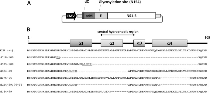
Glycosylation of the E protein has been associated with increased virus secretion in several different flaviviruses (9–11, 22). In order to assess the impact of E glycosylation on SVP secretion from C-deleted WNV cDNAs, we chose to investigate this parameter in the context of large internal deletions similar or analogous to those used in the construction of published vaccine candidates. The dC18-100 deletion has been utilized previously by our group in a KUNV-based single-round infectious particle (SRIP)-producing DNA vaccine (23), and the dC33-100 C deletion is similar to that utilized by the Mason group in their WNV RepliVAX series of vaccines (dC30-101) (24, 25). Plasmids encoding KUNV genomic cDNA under the control of cytomegalovirus promoter and containing these large internal deletions in C (Fig. 1) were constructed from pKUN1 (23, 26). In parallel, analogous plasmids containing the glycosylation motif found within the E protein of other circulating WNV and KUNV strains (19–21) (NYS, generated by the F156S mutation; designated gE) were also constructed. To assist in cloning, peptide sequences encoded by restriction enzyme sites were inserted in the center of each deletion (TS for dC18-100 [SpeI] and AAAKKMH for dC33-100 [NotI and NsiI]) (Fig. 1B). These resulting plasmid DNAs were transfected into cells alongside fully infectious cDNA clones pKUN1 (23, 26) and pKUN1/gE (derivative of pKUN1 with an F156S mutation in E, introducing the NYS glycosylation motif). Culture fluids and cell lysates were harvested at 2 days posttransfection (dpt) and assayed for the presence of any infectious virions (culture fluid) and for the relative abundance of viral C and E proteins (culture fluid and cell lysates) (Fig. 2).
Fig 1.
Schematic representations of the constructs used to analyze the roles of E glycosylation and C deletions in SVP production. (A) Plasmids encoding KUNV cDNA under the control of a cytomegalovirus (CMV) promoter were used to assess the contribution of glycosylation and different C deletions to SVP secretion. (B) Internal deletions in C constructed and analyzed in this study. Translated amino acid sequences of these deletions are aligned with a representation of C secondary structure. Additional residues encoded by SpeI (TS), NotI, and NsiI (AAAKMH) restriction sites inserted to facilitate cloning are underlined. KUN (wt), wild-type KUNV.
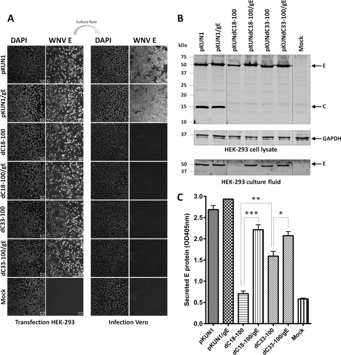
Fig 2.
C-deletion constructs with glycosylated E secrete significantly more SVPs than corresponding nonglycosylated constructs. (A) Cells transfected with each plasmid were stained for KUNV E. Culture fluid derived from this transfection was diluted 1:10 and inoculated onto Vero cells for analysis of infectious particle content via KUNV E staining. Nuclei were counterstained using DAPI. (B) Western blot detection of KUNV E and C protein expression in transfected cell lysate and E secretion into culture fluid. Blots were stained using antibodies specific for WNV C and KUNV E. Detection of GAPDH was utilized as a lysate loading control. (C) Capture ELISA to quantitatively compare E/SVP secretion into culture fluid. As all samples were assayed in parallel, controls are the same for Fig. 2 and 3. Results shown in panels A and B are representative of those of at least three independent experiments, while results shown in panel C are averages from at least three independent experiments. *, significant (P = 0.01 to 0.05); **, very significant (P = 0.001 to 0.01); ***, highly significant (P < 0.001). Mock, mock transfection. OD405nm, optical density at 405 nm.
Immunofluorescence assays were utilized to demonstrate that the constructs were able to express the polyprotein efficiently upon transfection. HEK-293 cells were transfected with equal amounts of plasmid DNAs using Lipofectamine LTX (Life Technologies, Carlsbad, CA). At 1 dpt, cells were fixed with 4% (wt/vol) paraformaldehyde (PFA)–0.1% (vol/vol) Triton X-100 in PBS, and at 2 dpt, culture fluid was harvested from parallel wells. To test for the presence or absence of secreted infectious virus particles, culture fluid was treated with RNase A (20 μg/ml) and RQ1 DNase (4 units/ml) for >1 h on ice, diluted 1:10 with RPMI medium (Life Technologies, Paisley, United Kingdom), and used to infect Vero cells. At 2 days postinfection (dpi), cells were fixed with 4% (wt/vol) PFA–0.1% (vol/vol) Triton X-100 in phosphate-buffered saline (PBS) as described above. Fixed transfected or infected cells were stained with the monoclonal 3.67G antibody (recognizing KUNV E) (19), and nuclei were counterstained with 4′,6′-diamidino-2-phenylindole (DAPI). Each of the C-deleted constructs was shown to replicate efficiently in transfected HEK-293 cells as judged by strong staining with 3.67G (Fig. 2A) and by staining with an antibody recognizing double-stranded RNA (data not shown). As no Vero cells were positive for KUNV E when incubated with culture fluid from cells transfected with C-deleted constructs, we concluded that each of the deletions, dC18-100 and dC33-100, effectively abrogated production of infectious virions (Fig. 2A).
Western blot analysis of lysates and culture fluids from transfected cells was undertaken to discern KUNV protein expression and E secretion from constructs (Fig. 2B). Culture fluid was removed from transfected HEK-293 cells 2 dpt, and cells were lysed with 2× cracking buffer (0.09 M Tris-HCl, 20% [vol/vol] glycerol, 2% [wt/vol] SDS, 0.02% [wt/vol] bromophenol blue) and treated with 5% (vol/vol) β-mercaptoethanol. Culture fluid was similarly lysed via the addition of 8× cracking buffer (0.36 M Tris-HCl, 80% [vol/vol] glycerol, 8% [wt/vol] SDS, 0.08% [wt/vol] bromophenol blue; 1 part buffer to 4 parts culture fluid) and treatment with 5% (vol/vol) β-mercaptoethanol. Cell and culture fluid lysates were denatured at 95°C for 3 min, electrophoresed on 12% polyacrylamide gels, and transferred to nitrocellulose membranes. Western blots were probed with 3.67G anti-KUNV E, a rabbit polyclonal 3E6 anti-WNV C (kindly provided by Tom Hobman), and monoclonal anti-glyceraldehyde-3-phosphate dehydrogenase (anti-GAPDH; Sigma, St. Louis, MO) antibodies. Each of the plasmid constructs demonstrated robust expression of the KUNV polyprotein (as determined by anti-KUNV E staining), while C could only be detected in the lysates of pKUN1- and pKUN1/gE-transfected cells (Fig. 2B). The bands corresponding to E protein for the glycosylation mutants migrated slower than nonglycosylated forms, consistent with previous observations by many groups (9–11, 27). Secreted E protein could also be detected in the culture fluid of transfected cells, with nonglycosylated pKUNdC18-100 appearing to display reduced secretion compared to that of other constructs.
Several groups have demonstrated convincingly that in the presence of prM, E is predominantly secreted in the form of prM-E particles rather than as a soluble protein (8, 10, 28); therefore, the levels of E detected in the supernatant of HEK-293 cells transfected with C-deleted constructs can be equated with the levels of SVPs. The relative amount of secreted E protein (i.e., SVPs) in the culture fluid of transfected HEK-293 cells at 2 dpt (determined as the optimal time in pilot experiments) was analyzed quantitatively using capture enzyme-linked immunosorbent assay (ELISA). Round-bottom 96-well plates were coated overnight at 4°C with monoclonal 3.91D ascetic fluid (anti-KUNV E) (19) in carbonate coating buffer (50 mM Na2CO3, 50 mM NaHCO3; pH 9.6). Plates were subsequently washed with PBST (0.15 M NaCl, 4 mM Na2HPO4, 3.2 mM KH2PO4, 0.05% [vol/vol] Tween 20) and blocked for 1 h at RT with TNETC buffer (10 mM Tris base, 0.2 M NaCl, 1 mM EDTA, 2% [wt/vol] casein, 0.05% [vol/vol] Tween 20). Plates were then washed with PBST and incubated with serial dilutions of culture fluid for 1 h at RT. Following another wash with PBST, plates were incubated with biotinylated monoclonal 4G2 antibody (cross-reactive anti-flavivirus E) (29) for 1 h at RT. Plates were subsequently washed with PBST and incubated for 30 min at RT with streptavidin-horseradish peroxidase (HRP) (Life Technologies, Frederick, MD). After a final wash with PBST, the plates were developed for 1 h at RT with 2,2′-azinobis(3-ethylbenzthiazolinesulfonic acid) (ABTS) substrate buffer (40 mM citric acid, 110 mM Na2HPO4, 10 M ABTS, 0.1% [vol/vol] H2O2), and absorbance at 405 nm was recorded. Data presented in Fig. 2C comprise results from the 1:32 dilution (determined as the optimal dilution for analysis of secreted E in these experiments). Comparing nonglycosylated constructs, there was significantly more E protein (i.e., SVPs) secreted into the culture fluid by pKUNdC33-100 than by pKUNdC18-100 (P = 0.0024). Glycosylation mutants pKUNdC18-100/gE and pKUNdC33-100/gE secreted significantly more E (i.e., SVPs) than their nonglycosylated counterparts pKUNdC18-100 and pKUNdC33-100 (P = 0.0004 and 0.0319, respectively). There was, however, no significant difference in the amount of E (i.e., SVPs) secreted from transfection with pKUNdC18-100/gE and pKUNdC33-100/gE. Collectively, these results demonstrate that glycosylation of the E protein (independent of the internal C-deletion utilized) enhances the secretion of E-containing SVPs.
Glycosylation of the viral envelope in our system has led to an increase in particle secretion, which is in line with other reports (9–11, 22). It should be noted that glycosylation of the E protein has been associated with increased pH stability of virions (27); therefore, it is possible that the increased titers of SVPs noted in this study are due merely to higher stability. However, the published increase in stability was determined solely on the basis of infectivity (27), which may be related to the induction of premature fusogenic conformational rearrangements of E rather than degradation. Even considering this, when the redundant methods of native (ELISA) and reducing (Western blot) conditions were used to analyze the culture fluid, each demonstrated an increase in E content. Thus, bias for a particular conformation of E conferred by experimental methods is unlikely, and the observed phenotype can be assumed to be unrelated to protein rearrangements.
In addition to considering the benefit of glycosylation in regard to SVP production, it is also worth considering the nature of the immune response to be generated. As circulating medically important strains of WNV contain glycosylated E (20, 21), it may be beneficial for the vaccine to elicit immune response against SVPs containing glycosylated E. However, E glycosylation has also been identified as a mechanism of immune evasion in JE virus (JEV), with glycosylated virus failing to generate as strong an immune response as a nonglycosylated variant (30). The true contribution of E glycosylation to vaccine efficacy will only emerge upon vaccine evaluation in laboratory animals by comparing equivalent glycosylated and nonglycosylated constructs.
Smaller internal C deletions that preserve alpha helix 3 promote enhanced SVP secretion.
KUNV genomic cDNA plasmids containing a glycosylation motif in E and different internal C deletions (Fig. 1B) were constructed. Internal C deletions were designed to excise the hydrophobic alpha helix 2 (dC44-59) or to remove alpha helices 1, 2, and the majority of helix 4 (preserving helix 3; dC24-59 and dC76-96 [designated dC24-59;76-96]) in a series of two deletions, each applied singly as controls (dC24-59 and dC76-96). To assist in cloning, additional peptide sequences encoded by restriction sites were inserted in each deletion (TS for dC76-96 [SpeI]; AAAKKMH for dC24-59 and dC44-59 [NotI and NsiI]) (Fig. 1B). HEK-293 cells were transfected with equal amounts of plasmid constructs and cell lysate, and culture fluid was harvested at 2 dpt and assessed for the presence of viral C and E proteins by Western blotting and of secreted infectious virus by infecting Vero cells as described above (Fig. 3). In addition, cells were also analyzed for E expression by immunofluorescence assay (IFA) with 3.67G antibody.
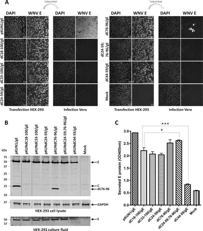
Fig 3.
The construct with a double C deletion, dC24-59;76-96, produces a larger amount of SVPs than other C-deletion constructs. (A) Cells transfected with each plasmid were stained for KUNV E. Culture fluid derived from this transfection was diluted 1:10 and inoculated onto Vero cells for analysis of infectious particle content via KUNV E staining. Nuclei were counterstained using DAPI. (B) Western blot detection of KUNV E and C protein expression in transfected cell lysate and E secretion into culture fluid. Blots were stained using antibodies specific for WNV C and KUNV E. Detection of GAPDH was utilized as a lysate loading control. (C) Capture ELISA to quantitatively compare E/SVP secretion into culture fluid. As all samples were assayed in parallel, controls are the same for Fig. 2 and 3. Results shown in panels A and B are representative of those of at least three independent experiments, while results shown in panel C are averages from at least three independent experiments. *, significant (P = 0.01 to 0.05); **, very significant (P = 0.001 to 0.01); ***, highly significant (P < 0.001).
Each of the C-deleted constructs was able to productively express the KUNV polyprotein in transfected cells as determined by robust detection of E protein (Fig. 3A). Infection of Vero cells with culture fluid from transfected HEK-293 cells demonstrated that the only C-deleted plasmid to retain the ability to produce secreted virus was the construct pKUNdC76-96/gE (Fig. 3A). Western blotting of the transfected HEK-293 cell lysates confirmed robust expression of the E protein from each construct (Fig. 3B). Full-length C protein could only be detected from infectious pKUN1/gE. The only C-deleted construct to express detectable C was pKUNdC76-96/gE; however, this was of a smaller size than C produced by fully infectious pKUN1/gE plasmid, consistent with the size of deletion. Western blot analysis of the culture fluid from transfected cells demonstrated that the construct pKUNdC44-59/gE has a deficiency in the ability to secrete E protein (i.e., SVPs).
E protein capture ELISA was performed with the culture fluid of transfected HEK-293 cells for quantitative assessment of relative SVP secretion as outlined above. Constructs pKUNdC18-100/gE, pKUNdC33-100/gE, pKUNdC24-59/gE, and pKUNdC79-96/gE were not significantly different in their relative levels of E (i.e., SVP) secretion (Fig. 3C). The plasmid pKUNdC44-59/gE was less efficient than pKUNdC18-100/gE (and other C-deleted constructs) in E (i.e., SVP) secretion (P = 0.0004), whereas the dual-deletion construct pKUNdC24-59;76-96/gE secreted significantly more E (i.e., SVPs) than pKUNdC18-100/gE (P = 0.0291) (Fig. 3C). Thus, the noninfectious phenotype and the significantly improved E (and hence SVP) secretion of the pKUNdC24-59;76-96/gE construct make this deletion an optimum candidate for incorporation into the next generation of C-deleted vaccine plasmids.
Although the majority of C-deleted cDNAs investigated in our study proved to be noninfectious, only dC24-59;76-96 demonstrated significantly enhanced SVP secretion compared to our previously published dC18-100 large deletion. Our data demonstrate that while dC44-59 is certainly noninfectious, it appears to have a deficiency in SVP secretion (Fig. 3). Two other small C deletions investigated in this study (dC24-59 and dC76-96) individually are also not optimal for incorporation into the future C-deleted vaccine candidates. Although pKUNdC24-59/gE proved to be noninfectious in this investigation (Fig. 3) and the similar dC26-56 in yellow fever virus (YFV) is also noninfectious (16), studies with TBEV (dC28-57) demonstrated that while the construct was initially unable to secrete virions, passage in mice led to the recovery of viable revertants (15). Deletion of alpha helix 4 (dC76-96 in this study) proved to retain the infectious phenotype (Fig. 3A), as was also observed with dC77-96 for YFV (16). While individually, dC24-59 and dC76-96 deletions are not useful for future vaccine development, combining these two deletions with the addition of a glycosylation motif in E appears to be the most optimal option for future vaccine development.
C-deleted constructs with the highest efficiency of SVP secretion display the lowest degree of hydrophobicity within the remaining residues of C.
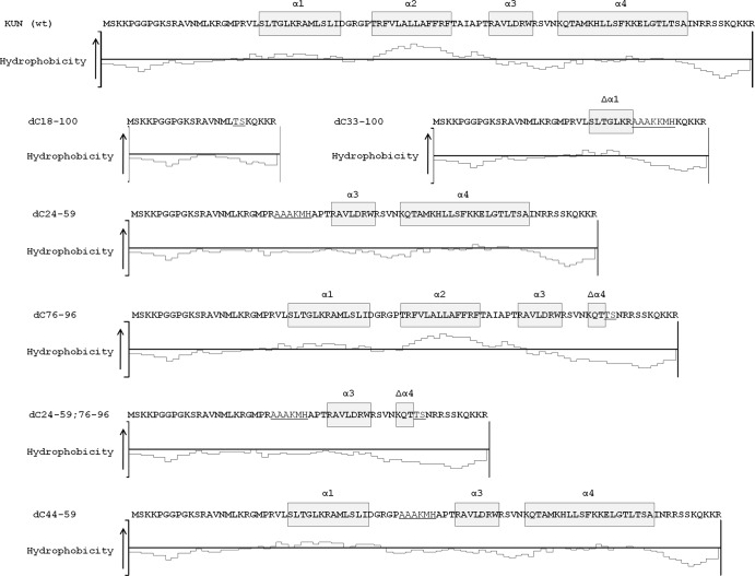
To better understand the relationship between the nature of internal C deletions and the degree of SVP secretion, Kyte-Doolittle hydropathy plots (31) were generated using the program PROTEAN (DNASTAR, Inc.) with a window size of 11 aa (Fig. 4). Wild-type C and dC76-96 are the only forms of C to retain the hydrophobic region spanning alpha helix 2, and it appears significant that these were the only constructs demonstrated to be able to effectively package KUNV genomic RNA into infectious virions (Fig. 3A). Curiously, these were also the only two forms of C that could be detected by the rabbit polyclonal 3E6 antibody via Western blotting (Fig. 3B), potentially indicating the presence of immunodominant epitopes in alpha helix 2. The constructs dC33-100, dC24-59, and dC44-59 display an overall more neutral hydropathy with both hydrophilic and hydrophobic regions. The hydropathy plots of dC18-100 and dC24-59;76-96 demonstrate that these versions of dC have no regions of hydrophobicity; they are entirely hydrophilic or neutral. Thus, it is possible that proteolytic maturation of these internally deleted C proteins by NS2B/3 cleavage from the polyprotein engenders complete dissociation from cellular membranes, so preventing nucleocapsid assembly or potential dC-mediated interference with SVP formation. The increased length of remaining residues of C in the dC24-59;76-96 deletion compared to that of the dC18-100 deletion (58 aa as opposed to 24 aa) may thus assist in viral NS2B/3 protease docking to facilitate C-prM cleavage.
Fig 4.
The double C-deletion dC24-59;76-96 construct preserves a larger section of hydrophilic sequence compared to that for other C-deletion constructs. Kyte-Doolittle plot of the amino acid sequence of KUNV C and dC hydropathy. The plotted values of each residue reflect the average hydrophobicity score of that amino acid and the subsequent 10 residues. Sequences encoded by SpeI (TS), NotI, and NsiI (AAAKMH) restriction sites inserted in the center of C deletions are underlined. The C protein alpha helical secondary structure is represented by boxed residues.
The flavivirus C protein is a logical candidate for the introduction of deletions that will prevent virus spread yet ensure genomic RNA replication. Disruption of the C gene prevents the formation of nucleocapsids yet maintains the ability to generate and secrete immunogenic, noninfectious SVPs, thus making such C-deleted genomes attractive vaccine candidates. This study has demonstrated that the introduction of a F156S mutation in E to force glycosylation significantly improves SVP secretion irrespective of the deletion used in C. Additionally, a combined internal deletion in C (dC24-59;76-96) significantly enhanced the secretion of SVPs from transfected HEK-293 cells. This phenotype correlates with the maintenance of a long stretch of hydrophilic amino acids within the dC24-59;76-96 sequence (Fig. 4). These properties make the dC24-59;76-96 deletion attractive for incorporation into future iterations of C-deleted DNA vaccines against WNV and potentially against other medically important flaviviruses.
ACKNOWLEDGMENTS
The work was funded by the National Health and Medical Research Council (NH&MRC), Australia.
A.A.K. is a research fellow with the NH&MRC.
Footnotes
Published ahead of print 18 September 2013
REFERENCES
- 1.Pesko KN, Ebel GD. 2012. West Nile virus population genetics and evolution. Infect. Genet. Evol. 12:181–190 [DOI] [PMC free article] [PubMed] [Google Scholar]
- 2.Brault AC. 2009. Changing patterns of West Nile virus transmission: altered vector competence and host susceptibility. Vet. Res. 40:43. [DOI] [PMC free article] [PubMed] [Google Scholar]
- 3.Nash D, Mostashari F, Fine A, Miller J, O'Leary D, Murray K, Huang A, Rosenberg A, Greenberg A, Sherman M, Wong S, Layton M. 2001. The outbreak of West Nile virus infection in the New York City area in 1999. N. Engl. J. Med. 344:1807–1814 [DOI] [PubMed] [Google Scholar]
- 4.Lanciotti RS, Roehrig JT, Deubel V, Smith J, Parker M, Steele K, Crise B, Volpe KE, Crabtree MB, Scherret JH, Hall RA, MacKenzie JS, Cropp CB, Panigrahy B, Ostlund E, Schmitt B, Malkinson M, Banet C, Weissman J, Komar N, Savage HM, Stone W, McNamara T, Gubler DJ. 1999. Origin of the West Nile virus responsible for an outbreak of encephalitis in the northeastern United States. Science 286:2333–2337 [DOI] [PubMed] [Google Scholar]
- 5.Roby JA, Funk A, Khromykh AA. 2012. Flavivirus replication and assembly, p 21–49 In Shi PY. (ed), Molecular virology and control of flaviviruses. Caister Academic Press, Wymondham, England [Google Scholar]
- 6.Lobigs M, Lee E. 2004. Inefficient signalase cleavage promotes efficient nucleocapsid incorporation into budding flavivirus membranes. J. Virol. 78:178–186 [DOI] [PMC free article] [PubMed] [Google Scholar]
- 7.Lobigs M, Lee E, Ng ML, Pavy M, Lobigs P. 2010. A flavivirus signal peptide balances the catalytic activity of two proteases and thereby facilitates virus morphogenesis. Virology 401:80–89 [DOI] [PubMed] [Google Scholar]
- 8.Konishi E, Mason PW. 1993. Proper maturation of the Japanese encephalitis virus envelope glycoprotein requires cosynthesis with the premembrane protein. J. Virol. 67:1672–1675 [DOI] [PMC free article] [PubMed] [Google Scholar]
- 9.Lee E, Leang SK, Davidson A, Lobigs M. 2010. Both E protein glycans adversely affect Dengue virus infectivity but are beneficial for virion release. J. Virol. 84:5171–5180 [DOI] [PMC free article] [PubMed] [Google Scholar]
- 10.Hanna SL, Pierson TC, Sanchez MD, Ahmed AA, Murtadha MM, Doms RW. 2005. N-linked glycosylation of West Nile virus envelope proteins influences particle assembly and infectivity. J. Virol. 79:13262–13274 [DOI] [PMC free article] [PubMed] [Google Scholar]
- 11.Li J, Bhuvanakantham R, Howe J, Ng ML. 2006. The glycosylation site in the envelope protein of West Nile virus (Sarafend) plays an important role in replication and maturation processes. J. Gen. Virol. 87:613–622 [DOI] [PubMed] [Google Scholar]
- 12.Mandl CW. 2004. Flavivirus immunization with capsid-deletion mutants: basics, benefits, and barriers. Viral Immunol. 17:461–472 [DOI] [PubMed] [Google Scholar]
- 13.Roby JA, Hall RA, Khromykh AA. 2011. Nucleic acid-based infectious and pseudo-infectious flavivirus vaccines, p 299–320 In Dormitzer PR, Mandl CW, Rapuoli R. (ed), Replicating vaccines: a new generation. Birkhauser Verlag AG, Basel, Switzerland [Google Scholar]
- 14.Kofler RM, Heinz FX, Mandl CW. 2002. Capsid protein C of tick-borne encephalitis virus tolerates large internal deletions and is a favorable target for attenuation of virulence. J. Virol. 76:3534–3543 [DOI] [PMC free article] [PubMed] [Google Scholar]
- 15.Kofler RM, Leitner A, O'Riordain G, Heinz FX, Mandl CW. 2003. Spontaneous mutations restore the viability of tick-borne encephalitis virus mutants with large deletions in protein C. J. Virol. 77:443–451 [DOI] [PMC free article] [PubMed] [Google Scholar]
- 16.Patkar CG, Jones CT, Chang YH, Warrier R, Kuhn RJ. 2007. Functional requirements of the yellow fever virus capsid protein. J. Virol. 81:6471–6481 [DOI] [PMC free article] [PubMed] [Google Scholar]
- 17.Schlick P, Taucher C, Schittl B, Tran JL, Kofler RM, Schueler W, von Gabain A, Meinke A, Mandl CW. 2009. Helices alpha2 and alpha3 of West Nile virus capsid protein are dispensable for assembly of infectious virions. J. Virol. 83:5581–5591 [DOI] [PMC free article] [PubMed] [Google Scholar]
- 18.Seregin A, Nistler R, Borisevich V, Yamshchikov G, Chaporgina E, Kwok CW, Yamshchikov V. 2006. Immunogenicity of West Nile virus infectious DNA and its noninfectious derivatives. Virology 356:115–125 [DOI] [PubMed] [Google Scholar]
- 19.Adams SC, Broom AK, Sammels LM, Hartnett AC, Howard MJ, Coelen RJ, Mackenzie JS, Hall RA. 1995. Glycosylation and antigenic variation among Kunjin virus isolates. Virology 206:49–56 [DOI] [PubMed] [Google Scholar]
- 20.Botha EM, Markotter W, Wolfaardt M, Paweska JT, Swanepoel R, Palacios G, Nel LH, Venter M. 2008. Genetic determinants of virulence in pathogenic lineage 2 West Nile virus strains. Emerg. Infect. Dis. 14:222–230 [DOI] [PMC free article] [PubMed] [Google Scholar]
- 21.Frost MJ, Zhang J, Edmonds JH, Prow NA, Gu XN, Davis R, Hornitzky C, Arzey KE, Finlaison D, Hick P, Read A, Hobson-Peters J, May FJ, Doggett SL, Haniotis J, Russell RC, Hall RA, Khromykh AA, Kirkland PD. 2012. Characterization of virulent West Nile virus Kunjin strain, Australia, 2011. Emerg. Infect. Dis. 18:792–800 [DOI] [PMC free article] [PubMed] [Google Scholar]
- 22.Scherret JH, Mackenzie JS, Khromykh AA, Hall RA. 2001. Biological significance of glycosylation of the envelope protein of Kunjin virus. Ann. N. Y. Acad. Sci. 951:361–363 [DOI] [PubMed] [Google Scholar]
- 23.Chang DC, Liu WJ, Anraku I, Clark DC, Pollitt CC, Suhrbier A, Hall RA, Khromykh AA. 2008. Single-round infectious particles enhance immunogenicity of a DNA vaccine against West Nile virus. Nat. Biotechnol. 26:571–577 [DOI] [PubMed] [Google Scholar]
- 24.Mason PW, Shustov AV, Frolov I. 2006. Production and characterization of vaccines based on flaviviruses defective in replication. Virology 351:432–443 [DOI] [PMC free article] [PubMed] [Google Scholar]
- 25.Widman DG, Ishikawa T, Fayzulin R, Bourne N, Mason PW. 2008. Construction and characterization of a second-generation pseudoinfectious West Nile virus vaccine propagated using a new cultivation system. Vaccine 26:2762–2771 [DOI] [PubMed] [Google Scholar]
- 26.Hall RA, Nisbet DJ, Pham KB, Pyke AT, Smith GA, Khromykh AA. 2003. DNA vaccine coding for the full-length infectious Kunjin virus RNA protects mice against the New York strain of West Nile virus. Proc. Natl. Acad. Sci. U. S. A. 100:10460–10464 [DOI] [PMC free article] [PubMed] [Google Scholar]
- 27.Beasley DWC, Whiteman MC, Zhang SL, Huang CYH, Schneider BS, Smith DR, Gromowski GD, Higgs S, Kinney RM, Barrett ADT. 2005. Envelope protein glycosylation status influences mouse neuroinvasion phenotype of genetic lineage 1 West Nile virus strains. J. Virol. 79:8339–8347 [DOI] [PMC free article] [PubMed] [Google Scholar]
- 28.Schalich J, Allison SL, Stiasny K, Mandl CW, Kunz C, Heinz FX. 1996. Recombinant subviral particles from tick-borne encephalitis virus are fusogenic and provide a model system for studying flavivirus envelope glycoprotein functions. J. Virol. 70:4549–4557 [DOI] [PMC free article] [PubMed] [Google Scholar]
- 29.Gentry MK, Henchal EA, McCown JM, Brandt WE, Dalrymple JM. 1982. Identification of distinct antigenic determinants on Dengue-2 virus using monoclonal-antibodies. Am. J. Trop. Med. Hyg. 31:548–555 [DOI] [PubMed] [Google Scholar]
- 30.Zhang Y, Chen PY, Cao RB, Gu JY. 2011. Mutation of putative N-Linked Glycosylation sites in Japanese encephalitis virus premembrane and envelope proteins enhances humoral immunity in BALB/C mice after DNA vaccination. Virol. J. 8:7. 10.1186/1743-422X-8-7 [DOI] [PMC free article] [PubMed] [Google Scholar]
- 31.Kyte J, Doolittle RF. 1982. A simple method for displaying the hydropathic character of a protein. J. Mol. Biol. 157:105–132 [DOI] [PubMed] [Google Scholar]


