Abstract
Paramyxoviruses, including the human pathogen measles virus (MV) and the avian Newcastle disease virus (NDV), enter host cells through fusion of the viral envelope with the target cell membrane. This fusion is driven by the concerted action of two viral envelope glycoproteins: the receptor binding protein and the fusion protein (F). The MV receptor binding protein (hemagglutinin [H]) attaches to proteinaceous receptors on host cells, while the receptor binding protein of NDV (hemagglutinin-neuraminidase [HN]) interacts with sialic acid-containing receptors. The receptor-bound HN/H triggers F to undergo conformational changes that render it competent to mediate fusion of the viral and cellular membranes. The mechanism of fusion activation has been proposed to be different for sialic acid-binding viruses and proteinaceous receptor-binding viruses. We report that a chimeric protein containing the NDV HN receptor binding region and the MV H stalk domain can activate MV F to fuse, suggesting that the signal to the stalk of a protein-binding receptor binding molecule can be transmitted from a sialic acid binding domain. By engineering the NDV HN globular domain to interact with a proteinaceous receptor, the fusion activation signal was preserved. Our findings are consistent with a unified mechanism of fusion activation, at least for the Paramyxovirinae subfamily, in which the receptor binding domains of the receptor binding proteins are interchangeable and the stalk determines the specificity of F activation.
INTRODUCTION
Paramyxoviruses enter their host cells by merging their envelope with that of the target cell in a fusion process driven by viral glycoproteins: a receptor binding protein and a fusion protein (F) (1–5). Most paramyxoviruses (except members of the Pneumovirinae subfamily) initiate the fusion process when the receptor binding protein engages its host cell receptor and activates the adjacent F protein. The F protein then undergoes a series of conformational changes that permit it to insert into the target membrane and merge the viral and cell membranes (reviewed in references 6 and 7). The interaction between the two envelope glycoproteins and the subsequent events that lead to fusion are critical to the entry process (8–12).
The receptor binding protein (hemagglutinin-neuraminidase [HN]) of Newcastle disease virus (NDV) possesses three functions common to many paramyxovirus receptor binding proteins: receptor binding, receptor destroying (neuraminidase), and F activation. When the NDV HN protein attaches to its sialic acid receptor, it activates the F protein to undergo the series of conformational changes that render it fusion competent (8, 9, 11, 13, 14). The HN is a type II membrane protein, with four major domains: the globular head domain, the stalk domain, a membrane-spanning region, and a cytoplasmic domain. Analyses of the structure of NDV HN (15, 16), along with the HNs of human parainfluenza type 3 (HPIV3) (17) and simian virus 5 (SV5) (parainfluenza virus type 5 [PIV5]) (18), have identified the regions responsible for receptor binding/neuraminidase activity in the globular head domains of these proteins (13, 14, 19, 20). Two sialic acid binding sites in the head of NDV have been identified by structural as well as functional studies, and they are referred to here as site I, for the primary sialic acid binding/neuraminidase site, and site II, for the binding/F-activation site (16, 21–23).
In contrast to the HN molecules, the Nipah virus (NiV) receptor binding protein (G) does not have receptor-cleaving activity. NiV G, therefore, once engaged to its ephrin B2/B3 receptor, remains bound (10, 24, 25). The receptor binding protein (H) of measles virus (MV) also lacks receptor cleavage activity, and similarly to NiV G, H does not disengage from its receptors, i.e., the signaling lymphocyte activation molecule (SLAM or CD150), CD46, or nectin-4 (26–30). For these two proteinaceous receptor-binding viruses (NiV and MV), it has been proposed that the F protein is retained in a metastable state by the receptor binding protein until the receptor is engaged; upon receptor engagement, F is released to undergo the rearrangements required for fusion (8, 10, 27, 31–34).
We recently showed that chimeric proteins consisting of the globular head of the sialic acid-binding NDV HN and the stalk domain of the protein receptor-binding NiV G efficiently promoted fusion (23). To study MV fusion, we utilized specific MV H-NDV HN chimeric proteins to show that a sialic acid-binding globular domain can promote MV fusion activation via the stalk of a chimeric receptor binding protein. The efficiency of F activation by the receptor binding protein relates directly to the strength of interaction between the globular head of the binding protein and its receptor. For the members of the Paramyxovirinae subfamily of the paramyxoviruses—whether sialic acid or protein binding—the receptor binding protein transmits the fusion activation signal through the stalk domain of the binding protein to F.
MATERIALS AND METHODS
Transient expression of NDV HN/F, MV H/F, and chimeric cDNA genes.
Transfections were performed according to the Lipofectamine 2000 manufacturer's protocol (Invitrogen).
Cell cultures.
HEK-293T (human epithelial kidney cells) and Vero cells were grown in Dulbecco's modified Eagle's medium (DMEM) (Gibco) supplemented with 10% fetal bovine serum (FBS) and antibiotics in a humidified incubator supplemented with 5% CO2. Vero-His (African green monkey kidney cells expressing anti-6-His-tag scFv) cells were a generous gift from Stephen J. Russell, Mayo Clinic (35).
Plasmids.
The MV H and F genes from wild-type (wt) MV strain G954 (36) and the chimeric cDNAs were codon optimized and synthesized by Epoch Biolabs and subcloned into the mammalian expression vector pCAGGS between EcoRI and BglII sites. The pCAGGS NDV Australia-Victoria (AV) HN and F constructs were generously provided by Ronald Iorio (University of Massachusetts, Worcester, MA). The SLAMF1-GFP ORF clone was obtained in a transfection-ready plasmid from Origene.
HAD assay.
The hemadsorption (HAD) assay was performed and quantified as previously described (37). Briefly, growth medium from 293T cell monolayers cotransfected with HN and F in 24- or 48-well Biocoat plates (Becton Dickinson Labware) was aspirated, replaced with 150 μl of CO2-independent medium (pH 7.3; Gibco), with or without the indicated concentrations of zanamivir and 1% red blood cells (RBCs) in serum-free, CO2-independent medium, and placed at 4°C for 30 min. The wells were then washed three times with 150 μl cold CO2-independent medium. The bound RBCs were lysed with 200 μl RBC lysis solution (0.145 M NH4Cl and 17 mM Tris-HCl), and absorbance was read at 405 nm, using a Spectramax M5 (Molecular Devices) microplate reader.
Cell surface expression.
Monolayers of 293T cells were transiently transfected with HN or F constructs. Cells were washed twice in phosphate-buffered saline (PBS) and then incubated with a 1:100 dilution of a pool of anti-NDV HN monoclonal antibodies (sc53561, sc53562, and sc53563; Santa Cruz Biotechnology), anti-MV H antibody (38), or anti-His antibody (Genscript) in 3% bovine serum albumin (BSA) and 0.1% sodium azide in PBS for 1 h. Samples were then washed twice with PBS and incubated with a 1:100 dilution of a fluorescein isothiocyanate (FITC)-conjugated anti-mouse IgG(H+L) (BD Pharmingen). To quantify cell surface proteins in each sample, indirect immunofluorescence was measured by fluorescence-activated cell sorting (FACS) (FACSCalibur; Becton, Dickinson).
Partial removal of sialic acid receptors from RBCs.
Partial receptor depletion of RBCs was achieved by treating 2 ml of a 10% RBC solution in serum-free medium for 2 h at 37°C with 0 to 200 mU of Clostridium perfringens neuraminidase (type X; Sigma Scientific, St. Louis, MO) as previously described (39). Neuraminidase was then removed by washing the RBCs 3 times with serum-free medium. Each set of RBCs was then resuspended in serum-free, CO2-independent medium to achieve final 2% RBC stocks.
Assessment of HN receptor binding avidity with receptor-depleted RBCs.
RBCs partially depleted of their surface sialic acid receptors (described above) were used to determine the relative receptor binding avidities of variant HN molecules as previously described (39). In each experiment, aliquots of the same preparation of depleted RBCs (as described above) were used. The RBCs were overlaid at a final concentration of 1% in 150 μl onto 293T cell monolayers in 48-well plates transiently transfected 24 h prior with receptor-binding protein expression vectors as described above. The plates were incubated at 4°C for 30 min to allow RBC binding. The cell monolayers were then washed at 4°C with cold CO2-independent medium to remove unbound RBCs, bound RBCs were lysed with RBC lysis buffer, and absorbance was read at 405 nm on a Spectramax enzyme-linked immunosorbent assay (ELISA) reader. Results are presented as percent retention of RBCs relative to control cells (undepleted RBCs) versus the degree of receptor depletion (expressed as mU of bacterial neuraminidase). For pretreatment of receptor binding protein-expressing cells with neuraminidase, the monolayers were treated with 25 mU of the enzyme per well in 48-well plates (5 × 10⁵ cells) for 3 h at 37°C, transferred to 4°C until they reach that temperature, and then washed.
β-Gal complementation-based fusion assay.
We previously adapted a fusion assay based on alpha complementation of β-galactosidase (β-Gal) (40, 41). In this assay, receptor-bearing cells (Vero-His cells) expressing the omega peptide of β-Gal are mixed with envelope glycoprotein-coexpressing 293T cells that also express the alpha peptide of β-Gal. Cell fusion, which leads to complementation, is stopped by lysing the cells, and after addition of the substrate, fusion is quantified on a Spectramax M5 microplate reader.
RESULTS
Chimeric viral glycoproteins containing the MV H stalk domain and NDV HN globular head (H-HN) are expressed and bind to sialic acid-containing receptors.
To determine whether the globular head of MV H is essential for activation of MV F or if the sialic acid-binding NDV globular domain could activate MV fusion, we designed a series of chimeric proteins with MV H stalk domains of different lengths and with residue changes that abolish the H stalk's ability to activate MV F (42) (Fig. 1A). Three chimeric proteins were used to study fusion signal propagation between the head and stalk regions: (i) H1–130 HN124–571, (ii) H1–160 HN124–571, and (iii) H1–160(C139S-C154W) HN124–571. The stalk domain of H1–160 HN124–571 includes the regions reportedly required for F interaction and F triggering as well as residues C139 and C154, which are required for dimerization (42). The shorter stalk of the chimeric protein H1–130 HN124–571, however, lacks the residues required for F interaction and triggering. Finally, the chimeric protein H1–160(C139S-C154W) HN124–571 contains the domains required for F interaction and triggering, but the cysteine residues C139 and C154 (mentioned above) were mutated in order to prevent dimerization. The cell surface expression of the chimeric proteins was measured by FACS analysis (Fig. 1B) using a pool of anti-NDV AV HN monoclonal antibodies. All of the chimeric proteins had a cell surface expression level of around 50% of NDV AV HN expression.
Fig 1.
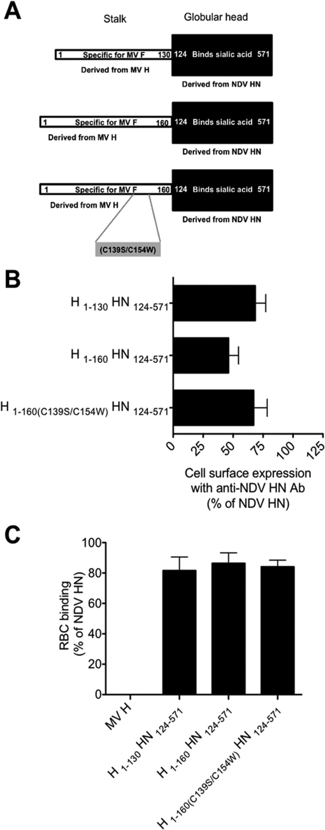
Chimeric proteins containing the MV H stalk domain and the NDV globular head are efficiently expressed and bind sialic acid receptors. (A) Schematic structures of three MV H-NDV HN chimeric proteins. The MV H stalk domains H1–130, H1–160, and H1–160(C139S-C154W) were cloned in frame with NDV AV HN124–571. (B) FACS analysis results for cell surface expression. Cells were transfected with the chimeric glycoproteins H1–130 HN124–571, H1–160 HN124–571, and H1–160(C139S-C154W) HN124–571 and analyzed for cell surface expression. The FACS results were compared to NDV AV HN cell surface expression by using a set of monoclonal antibodies as described in Materials and Methods. (C) Receptor-bearing cell binding of the chimeric proteins.
We assessed receptor binding for each binding protein in an RBC binding assay (41). Cells expressing the indicated proteins were allowed to bind RBCs at 4°C for 30 min (Fig. 1C) and then washed, and the bound RBCs were quantified. The receptor binding efficacies of the expressed chimeric proteins were similar to that of NDV AV HN (Fig. 1C).
Chimeric viral glycoproteins containing the MV H stalk domain and NDV HN globular head (H-HN) promote MV F-mediated fusion.
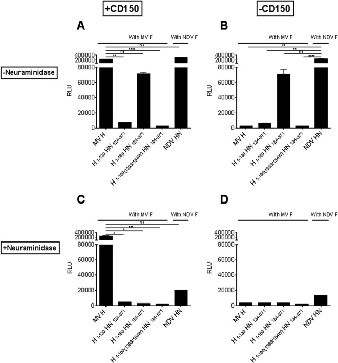
The fusion properties of the chimeric proteins were compared in a β-galactosidase complementation assay. Cells coexpressing the indicated receptor binding proteins with MV F together with the alpha peptide of β-Gal were overlaid with cells expressing the omega peptide and either the MV receptor CD150 (Fig. 2A and C) or empty vector (Fig. 2B and D). The cells were also left untreated (Fig. 2A and B) or treated with neuraminidase (Fig. 2C and D). Upon cell-to-cell fusion, the alpha and omega peptides reconstitute β-galactosidase activity, proportional to the extent of fusion. For the experiment in Fig. 2A, we used cells expressing CD150 to compare the fusion-promoting activity of MV H with that of our chimeric proteins in the presence of MV F. As expected, MV H and F effectively promoted fusion, and only the chimeric protein H1–160 HN124–571 achieved approximately 30% of the MV H/F-mediated fusion. Fusion was abolished in H1–130 HN124–571 and H1–160(C139S-C154W) HN124–571, both of which are chimeric proteins lacking the cysteine residues at positions 139 and 154, which prevents dimerization of MV H (42). These data are consistent with a recent report that short H stems require stabilizing oligomerization tags in order to maintain their F-triggering function (43).
Fig 2.
Chimeric proteins containing the MV H stalk domain and the NDV globular head activate MV F. A cell-to-cell fusion assay was performed with 293T cells bearing the chimeric proteins with target (Vero-His) cells transfected with the MV receptor CD150 (A and C) or empty vector (B and D). Cell-to-cell fusion was measured in the presence (C and D) or absence (A and B) of neuraminidase by a β-Gal complementation assay as described in Materials and Methods. The values are means (with standard deviations [SD]) for results from triplicate samples in a representative experiment repeated at least three times. RLU, relative light units. **, P < 0.01; ***, P < 0.005; ****, P < 0.001 (Kruskal-Wallis test/Dunn's multiple comparison test).
In Fig. 2B, we compared the fusion mediated by MV H and F with that promoted by the chimeric proteins in the presence of MV F but no receptor for MV H. In this case, the MV H- and F-expressing cells did not fuse with the target cells because of the lack of receptors, while the H1–160 HN124–571 chimeric protein achieved approximately the same amount of fusion as that shown in Fig. 2A. Therefore, the NDV globular head activated the MV H stalk domain by interacting with sialic acid receptors independently of CD150. To confirm that the fusion observed in the presence of the chimeric protein was indeed due to the engagement of sialic acid by the NDV HN head, we assessed fusion in the presence of neuraminidase. The chimeric protein H1–160 HN124–571, which activated fusion in the absence of neuraminidase (Fig. 2A and B), did not promote fusion in the presence of neuraminidase, but fusion promotion by MV H was unaltered in the presence of neuraminidase (Fig. 2C and D).
Differences in receptor avidity of the chimeric H proteins do not account for their differences in promoting fusion.
We asked whether the two chimeric proteins that failed to promote fusion [H1–130 HN124–571 and H1–160(C139S-C154W) HN124–571] had altered receptor avidity compared to that of the fusion promotion-competent chimeric protein (H1–160 HN124–571). We previously used a quantitative receptor avidity assay (14, 22, 39, 41) to show that NDV HN has a low avidity for receptors on RBCs (either human or avian) compared to HPIV3 HNs (21). In this assay, receptor-depleted RBCs are bound to HN-expressing cells in a quantitative hemadsorption (HAD) assay where greater receptor depletion required to reduce binding indicates a higher avidity. At 4°C, the neuraminidase of NDV HN is not active and therefore cannot contribute to RBC release. Binding avidity measurements for the three chimeric proteins are shown in Fig. 3. For cells expressing the chimeric protein H1–130 HN124–571, there was around 50% binding at a depletion level of 24 mU. In the case of the H1–160 HN124–571 protein, there was around 50% binding at a depletion level of 10 mU, and for the cells expressing the NDV wt HN and H1–160 (C139S-C154W) HN124–571, the binding was at 50% at a depletion level of 6 mU. Each chimeric protein had a sialic acid-binding avidity that was higher than that of NDV HN. The avidity of H1–130 HN124–571 was the highest, followed by that of H1–160 HN124–571, indicating that the inability of H1–130 HN124–571 to promote fusion was not due to lower avidity. The avidity of H1–160 (C139S-C154W) HN124–571 was lower than those of the other two chimeras but similar to that of NDV HN. These results indicate that the differences in fusion activation between the chimeric proteins cannot be explained by differences in sialic acid receptor binding but rather due to the functionality of the stalk domain.
Fig 3.
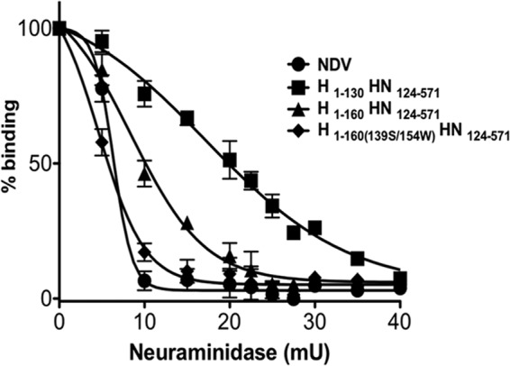
Receptor binding avidities of the chimeric proteins. A panel of RBCs with different degrees of receptor depletion were used to quantify HAD on cell monolayers expressing H1–130 HN124–571, H1–160 HN124–571, H1–160(C139S-C154W) HN124–571, or NDV HN at 4°C. Neuraminidase is not active at 4°C. The extent of binding of each depleted RBC preparation (y axis) is expressed as a percentage of that of the control (i.e., the amount of untreated, nondepleted RBCs bound to cells expressing the corresponding receptor binding protein). The points represent the means for results from triplicate monolayers from 3 representative experiments, with error bars denoting standard deviations.
Activation of MV F by binding of histidine-tagged H1–160 HN124–571 chimeric protein to anti-histidine antibody.
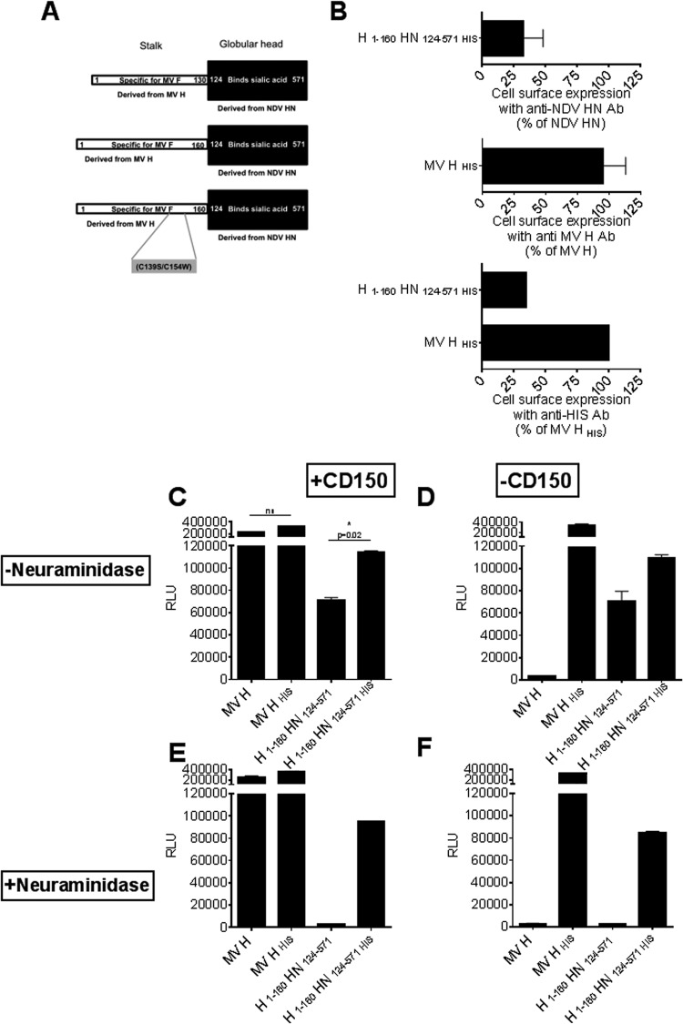
To determine whether the rearrangements in the MV H protein are receptor specific, we tested whether the chimeric protein H1–160 HN124–571 and MV H activate MV F when they engage a nonnatural receptor. We engineered a chimeric protein and MV H with histidine tags at their C termini (Fig. 4A); a histidine tag on MV H has been shown to replace the requirement for a cellular receptor (27, 35). We assessed the expression of these histidine-tagged receptor binding proteins by FACS. While MV H HIS was expressed at a level similar to that of MV H, the expression level of H1–160 HN124–571 HIS was 30% of those of both NDV HN and MV H HIS (Fig. 4B).
Fig 4.
Chimeric proteins containing the MV H stalk domain and the NDV globular head activate MV F. (A) Schematic structures of MV H HIS and H1–160 HN124–571 HIS. (B) FACS analysis results for cell surface expression. Cells were transfected with the MV H HIS and H1–160 HN124–571 HIS proteins and analyzed for cell surface expression by using anti-histidine antibodies. The values are means (with SD) for results from triplicate samples and are representative of an experiment repeated at least four times. (C to F) Cell-to-cell fusion of 293T cells bearing chimeric proteins with anti-histidine antibody-expressing target cells (Vero-His) transfected with the MV receptor CD150 (C and E) or empty vector (D and F). Cell-to-cell fusion was measured in the presence (E and F) or absence (C and D) of neuraminidase by a β-Gal complementation assay as described in Materials and Methods. The values are means (with SD) for results from triplicate samples of a representative experiment repeated at least three times. *, P < 0.05; ns, not significant (Mann-Whitney test).
Fusion promoted by the chimeric proteins was assessed in a β-galactosidase complementation assay, using cells expressing an anti-histidine antibody on their surfaces as the targets (27, 35). Cells coexpressing the indicated receptor binding proteins with MV F together with the alpha peptide were overlaid with Vero cells expressing the anti-histidine antibody and the omega peptide. As shown in Fig. 4C to F, we compared the fusion mediated by MV H HIS/F with fusion mediated by H1–160 HN124–571 HIS/MV F. Engagement of the anti-histidine antibody on the cell surface by the histidine tag on receptor binding proteins led to fusion. Histidine-tagged NDV HN is also fusion promotion competent in the absence of sialic acid receptor and promotes NDV F fusion under these conditions (data not shown). As shown in Fig. 2, the fusion mediated by MV H/F was independent of neuraminidase and depended only on CD150 expression, whereas the fusion promoted by H1–160 HN124–571 was dependent on the presence or absence of neuraminidase but independent of CD150 expression. However, both MV H HIS and H1–160 HN124–571 HIS promoted fusion independently of sialic acid or CD150 receptors. As shown in Fig. 4D, the amounts of fusion promoted by H1–160 HN124–571 HIS and MV H HIS were similar when normalized for the relative expression with anti-His antibody seen in Fig. 4B. Thus, a receptor that bears no resemblance to the natural viral receptors is sufficient for initiating fusion promotion by the receptor binding protein.
MV H can complement the fusion activation function of fusion-impaired chimeric proteins.
An MV H defective in triggering fusion but competent for receptor binding can be complemented by coexpression with an H protein that is fusion triggering competent but receptor blind (44). We asked whether MV H could rescue the fusion of chimeric proteins bearing an MV H stalk that is defective at fusion triggering. We used a chimeric protein carrying the P108S mutation in the MV H stalk domain. Mutation of this particular proline residue, which is highly conserved in the stalk regions of several paramyxovirus receptor binding proteins, impairs fusion promotion for paramyxoviruses, including MV (14, 45, 46). It has been suggested that upon receptor engagement, the globular head domain applies force that results in unwinding of the tightly packed stalk, exposing the central section (containing the conserved proline) that is essential for F triggering (46).
We generated cells that expressed a chimeric protein with the P108S mutation in the stalk [H1–160(P108S) HN124–571 HIS] and assessed fusion with MV F (Fig. 5) by using the β-galactosidase complementation assay with Vero-His target cells. As in Fig. 4, MV H and F mediated fusion only in the presence of CD150. The H1–160(P108S) HN124–571 HIS protein, as expected, did not promote fusion in the presence or absence of CD150 when coexpressed with MV F, but when it was cotransfected with MV H, along with MV F, fusion occurred in the presence of CD150. More importantly, fusion promotion was restored even in the absence of CD150 (Fig. 5B). In this scenario, the NDV HN head of the chimeric protein binds to the anti-His antibody or sialic acid on the Vero-His target cells; however, F activation must occur through the wt MV H stalk, since the chimeric protein's stalk is defective in F activation. Coexpression of the triggering-defective H1–160(P108S) HN124–571 HIS protein, wt MV H, and MV F permitted fusion to occur (Fig. 5), suggesting that the NDV HN globular head domain transmits the fusion activation signal to the stalk of another monomer in an oligomeric complex of MV H and chimeric protein. This result is consistent with an earlier report showing that all heads in the MV H tetramer are not required to engage a receptor in order to promote fusion (44).
Fig 5.
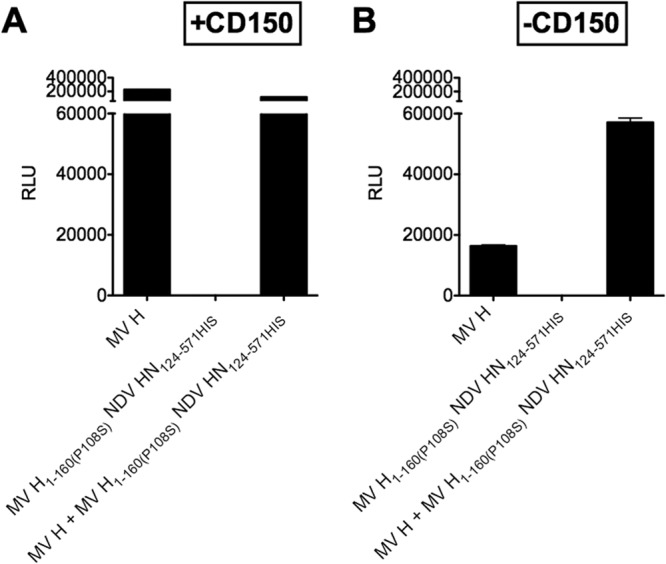
Cis-complementation of the F-triggering activity of fusion-deficient chimeric protein by wt MV H. Cell-to-cell fusion of 293T cells transfected with the indicated receptor binding glycoproteins and MV F with Vero-His cells transfected with the MV receptor CD150 (A) or empty vector (B) was assessed by a β-Gal complementation assay as described in Materials and Methods. Vero-His cells were used as target cells. The values are means (with SD) for results from triplicate samples of a representative experiment repeated at least three times.
DISCUSSION
Paramyxovirus infection relies on specialized fusion machinery that generally consists of a receptor binding protein (either G, H, or HN) and a fusion (F) protein. Upon receptor engagement, the receptor binding proteins activate the fusion cascade by a series of specific movements that begin in the globular head and are transmitted to the stalk domain (11). Analysis of the crystal structure of MV H, either alone or in complex with its receptor, compared to the structures of various HNs and of NiV G, showed major differences with respect to the site of H-receptor interaction (11, 16–18, 27, 30, 34). For MV H, specific head movements have been postulated to be required for transmission of the fusion signal (27, 30).
In this report, chimeric H-HN envelope glycoproteins (15, 16, 42, 46–48) were used to evaluate the interplay between the globular head and the stalk region that leads to fusion protein activation and to determine whether the transmission of the fusion signal from head to stalk is similar whether the virus binds to sialic acid or proteinaceous receptors.
Experimental (21) and crystal structure (16) data for NDV HN identified a second receptor binding site in the globular head of HN. This second binding site is activated by engagement of the primary binding site (22), and we recently showed that activation of NDV HN′s site II is required for its F-triggering activity (23). For MV, however, the dynamics are thought to be different from those of other paramyxoviruses. It was recently postulated that the two dimers of the MV H tetramer shift relative to each other in order to transmit the fusion signal and that rearrangements at the level of the globular head are required for fusion activation (27). Our finding that the NDV HN globular domain coupled with a functional H stalk region can activate MV F-mediated fusion indicates that the MV H globular domain rearrangements are not necessary to transmit the signal to the stalk or that similar rearrangements also take place in the NDV HN globular head. For several paramyxoviruses, we and others have shown that affinity for the receptor is a major determinant of fusion activation (20, 39, 49). The finding that the fusion promoted by the MV-NDV chimeric protein with a histidine tag is similar to that promoted by MV H with a histidine tag supports the notion that receptor affinity is a major determinant for fusion activation. When the receptor is the same—leveling the playing field—fusion is similar regardless of the specific globular head. A model for morbillivirus F activation was recently proposed, in which the stalk domain unfolds to expose a conserved region containing a proline; if unfolding is blocked by introducing disulfide bonds in the stalk domain, F activation does not occur (46). The results presented here suggest that the globular head of NDV can induce such an unfolding in the stalk of MV H. For NiV G, we have proposed that flexibility in the stalk is required for proper fusion activation (50). For MV, too, stalk flexibility is important for fusion activation (46, 48), and a recent report showed that short MV H stems, provided they are properly stabilized, are sufficient for activation of F. In that work, it appeared that either excessive or insufficient tetramer stabilization could deter fusion promotion (43). Future experiments will address the relationship between flexibility of the receptor binding protein stalk and its role in fusion promotion.
The oligomeric state of paramyxovirus receptor binding proteins has been shown to be critical for fusion promotion for several paramyxoviruses (18, 42, 44, 51, 52). We show here that within a tetramer, the receptor-engaged signal can be transmitted from the head of one monomer via the stalk of a different monomer. When chimeric H1–160(P108S) HN124–571 HIS—which is defective in fusion activation—was coexpressed with wt MV H, fusion promotion activity of the oligomeric complex was rescued (Fig. 5). The NDV globular domain transmitted the receptor-engaged fusion signal to F via the functional stalk of wt MV H, indicating that one monomer head can transmit the signal to a different stalk, and also confirming that for MV H, simultaneous engagement of all heads of an oligomer may not be necessary in order for the fusion promotion signal to be transmitted (44).
It has been proposed that for paramyxoviruses that bind a proteinaceous receptor, the receptor binding protein stabilizes the F protein and prevents its natural tendency to form a postfusion structure. This suggests that for these viruses, including measles virus, the role of the receptor binding protein is mainly a repressive one (8, 10, 25, 27, 32, 53–56) and that the classical spring-loaded mechanism applies: the receptor binding protein stabilizes the metastable state of the fusion protein prior to receptor engagement, and upon receptor binding, the fusion protein is released and independently proceeds to fusion (8, 10, 25, 27). While the stalk domain is a major determinant for specific F activation, we propose that the globular head can be interchangeable and that the repressive role, if present, is due to the stalk domain. For HPIV3, HN not only specifically activates fusion after receptor engagement but also prevents premature activation of F before receptor engagement (57), and the stalk domain is responsible for the stabilization effect. The stability of the F protein in the absence of receptor protein may be different for each virus in the family and may be the product of adaptation of the viruses to specific hosts or tissues. In the case of viruses such as HPIV3, the requisite balance of the three activities of HN (binding and cleaving of the receptor and F activation) may necessitate a more readily triggered (i.e., more heat sensitive) F than that of viruses whose receptor binding proteins interact more strongly with their receptor (as is the case for MV and henipaviruses). In the case of a strongly interacting receptor binding protein, receptor engagement is more stable and the F-triggering activity of the receptor binding protein may be protracted; therefore, a more stable F protein may be advantageous. If this hypothesis is correct, we would expect viruses that bind proteinaceous receptors to possess more stable F proteins than those of viruses with sialic acid-binding HNs. A recent report showed that the MV F protein is stable even in the absence of H (58), in agreement with our hypothesis.
Our data suggest that a common mechanism applies to all paramyxoviruses that use a receptor binding protein to activate a fusion protein, including MV. The results are consistent with a unified model for paramyxovirus fusion in which the receptor binding protein is required for F activation, and not simply responsible for releasing F to fuse (52, 59).
ACKNOWLEDGMENTS
We are grateful to Dan and Nancy Paduano for support of innovative research projects, to Ashton Kutcher and Jonathan Ledecky for their support, and to the Friedman Family Foundation for renovation of our laboratories at Weill Cornell Medical College. We thank Jacob Moscona-Skolnik for a critical reading of the manuscript. We thank Stephen J. Russell, Mayo Clinic, for the Vero-His cell line. We acknowledge the flow cytometry support from Sergei Rudchenko in the Flow Cytometry Facility of the Hospital for Special Surgery/Weill Cornell Medical College. We acknowledge the Northeast Center of Excellence for Bio-Defense and Emerging Infectious Disease Research's Proteomics Core for peptide synthesis and purification.
This work was supported by the NIH (NIAID) Northeast Center of Excellence for Biodefense and Emerging Infectious Disease Research (grant U54AI057158 to M.P. and A.M. [the principal investigator for the Center of Excellence grant is W. I. Lipkin] and grant R01AI31971 to A.M.), by NIH grant R21AI100292-01 to M.P., and by a Friedman Research Scholar in Pediatric Infectious Diseases award to M.P.
Footnotes
Published ahead of print 9 October 2013
REFERENCES
- 1.Eckert DM, Kim PS. 2001. Mechanisms of viral membrane fusion and its inhibition. Annu. Rev. Biochem. 70:777–810 [DOI] [PubMed] [Google Scholar]
- 2.White JM, Delos SE, Brecher M, Schornberg K. 2008. Structures and mechanisms of viral membrane fusion proteins: multiple variations on a common theme. Crit. Rev. Biochem. Mol. Biol. 43:189–219 [DOI] [PMC free article] [PubMed] [Google Scholar]
- 3.Harrison SC. 2008. Viral membrane fusion. Nat. Struct. Mol. Biol. 15:690–698 [DOI] [PMC free article] [PubMed] [Google Scholar]
- 4.Weissenhorn W, Hinz A, Gaudin Y. 2007. Virus membrane fusion. FEBS Lett. 581:2150–2155 [DOI] [PMC free article] [PubMed] [Google Scholar]
- 5.Sapir A, Avinoam O, Podbilewicz B, Chernomordik LV. 2008. Viral and developmental cell fusion mechanisms: conservation and divergence. Dev. Cell 14:11–21 [DOI] [PMC free article] [PubMed] [Google Scholar]
- 6.Moscona A. 2005. Entry of parainfluenza virus into cells as a target for interrupting childhood respiratory disease. J. Clin. Invest. 115:1688–1698 [DOI] [PMC free article] [PubMed] [Google Scholar]
- 7.Lamb RA, Paterson RG, Jardetzky TS. 2006. Paramyxovirus membrane fusion: lessons from the F and HN atomic structures. Virology 344:30–37 [DOI] [PMC free article] [PubMed] [Google Scholar]
- 8.Iorio RM, Melanson VR, Mahon PJ. 2009. Glycoprotein interactions in paramyxovirus fusion. Future Virol. 4:335–351 [DOI] [PMC free article] [PubMed] [Google Scholar]
- 9.Dutch RE. 2010. Entry and fusion of emerging paramyxoviruses. PLoS Pathog. 6:e1000881. 10.1371/journal.ppat.1000881 [DOI] [PMC free article] [PubMed] [Google Scholar]
- 10.Lee B, Ataman ZA. 2011. Modes of paramyxovirus fusion: a Henipavirus perspective. Trends Microbiol. 19:389–399 [DOI] [PMC free article] [PubMed] [Google Scholar]
- 11.Plattet P, Plemper RK. 2013. Envelope protein dynamics in paramyxovirus entry. mBio 4:e00413–13. 10.1128/mBio.00413-13 [DOI] [PMC free article] [PubMed] [Google Scholar]
- 12.Bossart KN, Fusco DL, Broder CC. 2013. Paramyxovirus entry. Adv. Exp. Med. Biol. 790:95–127 [DOI] [PMC free article] [PubMed] [Google Scholar]
- 13.Porotto M, Murrell M, Greengard O, Doctor L, Moscona A. 2005. Influence of the human parainfluenza virus 3 attachment protein's neuraminidase activity on its capacity to activate the fusion protein. J. Virol. 79:2383–2392 [DOI] [PMC free article] [PubMed] [Google Scholar]
- 14.Porotto M, Murrell M, Greengard O, Moscona A. 2003. Triggering of human parainfluenza virus 3 fusion protein (F) by the hemagglutinin-neuraminidase (HN): an HN mutation diminishing the rate of F activation and fusion. J. Virol. 77:3647–3654 [DOI] [PMC free article] [PubMed] [Google Scholar]
- 15.Crennell S, Takimoto T, Portner A, Taylor G. 2000. Crystal structure of the multifunctional paramyxovirus hemagglutinin-neuraminidase. Nat. Struct. Biol. 7:1068–1074 [DOI] [PubMed] [Google Scholar]
- 16.Zaitsev V, von Itzstein M, Groves D, Kiefel M, Takimoto T, Portner A, Taylor G. 2004. Second sialic acid binding site in Newcastle disease virus hemagglutinin-neuraminidase: implications for fusion. J. Virol. 78:3733–3741 [DOI] [PMC free article] [PubMed] [Google Scholar]
- 17.Lawrence MC, Borg NA, Streltsov VA, Pilling PA, Epa VC, Varghese JN, McKimm-Breschkin JL, Colman PM. 2004. Structure of the haemagglutinin-neuraminidase from human parainfluenza virus type III. J. Mol. Biol. 335:1343–1357 [DOI] [PubMed] [Google Scholar]
- 18.Yuan P, Thompson TB, Wurzburg BA, Paterson RG, Lamb RA, Jardetzky TS. 2005. Structural studies of the parainfluenza virus 5 hemagglutinin-neuraminidase tetramer in complex with its receptor, sialyllactose. Structure 13:803–815 [DOI] [PubMed] [Google Scholar]
- 19.Russell CJ, Jardetzky TS, Lamb RA. 2001. Membrane fusion machines of paramyxoviruses: capture of intermediates of fusion. EMBO J. 20:4024–4034 [DOI] [PMC free article] [PubMed] [Google Scholar]
- 20.Palermo LM, Porotto M, Greengard O, Moscona A. 2007. Fusion promotion by a paramyxovirus hemagglutinin-neuraminidase protein: pH modulation of receptor avidity of binding sites I and II. J. Virol. 81:9152–9161 [DOI] [PMC free article] [PubMed] [Google Scholar]
- 21.Porotto M, Murrell M, Greengard O, Lawrence M, McKimm-Breschkin J, Moscona A. 2004. Inhibition of parainfluenza type 3 and Newcastle disease virus hemagglutinin-neuraminidase receptor binding: effect of receptor avidity and steric hindrance at the inhibitor binding sites. J. Virol. 78:13911–13919 [DOI] [PMC free article] [PubMed] [Google Scholar]
- 22.Porotto M, Fornabaio M, Greengard O, Murrell MT, Kellogg GE, Moscona A. 2006. Paramyxovirus receptor-binding molecules: engagement of one site on the hemagglutinin-neuraminidase protein modulates activity at the second site. J. Virol. 80:1204–1213 [DOI] [PMC free article] [PubMed] [Google Scholar]
- 23.Porotto M, Salah Z, Devito I, Talekar A, Palmer SG, Xu R, Wilson IA, Moscona A. 2012. The second receptor binding site of the globular head of the Newcastle disease virus (NDV) hemagglutinin-neuraminidase activates the stalk of multiple paramyxovirus receptor binding proteins to trigger fusion. J. Virol. 86:5730–5741 [DOI] [PMC free article] [PubMed] [Google Scholar]
- 24.Vigant F, Lee B. 2011. Hendra and Nipah infection: pathology, models and potential therapies. Infect. Disord. Drug Targets 11:315–336 [DOI] [PMC free article] [PubMed] [Google Scholar]
- 25.Mirza AM, Aguilar HC, Zhu Q, Mahon PJ, Rota PA, Lee B, Iorio RM. 2011. Triggering of the Newcastle disease virus fusion protein by a chimeric attachment protein that binds to Nipah virus receptors. J. Biol. Chem. 286:17851–17860 [DOI] [PMC free article] [PubMed] [Google Scholar]
- 26.Muhlebach MD, Mateo M, Sinn PL, Prufer S, Uhlig KM, Leonard VH, Navaratnarajah CK, Frenzke M, Wong XX, Sawatsky B, Ramachandran S, McCray PB, Cichutek K, von Messling V, Lopez M, Cattaneo R. 2011. Adherens junction protein nectin-4 is the epithelial receptor for measles virus. Nature 480:530–533 [DOI] [PMC free article] [PubMed] [Google Scholar]
- 27.Navaratnarajah CK, Oezguen N, Rupp L, Kay L, Leonard VH, Braun W, Cattaneo R. 2011. The heads of the measles virus attachment protein move to transmit the fusion-triggering signal. Nat. Struct. Mol. Biol. 18:128–134 [DOI] [PMC free article] [PubMed] [Google Scholar]
- 28.Noyce RS, Bondre DG, Ha MN, Lin LT, Sisson G, Tsao MS, Richardson CD. 2011. Tumor cell marker PVRL4 (nectin 4) is an epithelial cell receptor for measles virus. PLoS Pathog. 7:e1002240. 10.1371/journal.ppat.1002240 [DOI] [PMC free article] [PubMed] [Google Scholar]
- 29.Dorig RE, Marcil A, Chopra A, Richardson CD. 1993. The human CD46 molecule is a receptor for measles virus (Edmonston strain). Cell 75:295–305 [DOI] [PubMed] [Google Scholar]
- 30.Hashiguchi T, Ose T, Kubota M, Maita N, Kamishikiryo J, Maenaka K, Yanagi Y. 2011. Structure of the measles virus hemagglutinin bound to its cellular receptor SLAM. Nat. Struct. Mol. Biol. 18:135–141 [DOI] [PubMed] [Google Scholar]
- 31.Plemper RK, Brindley MA, Iorio RM. 2011. Structural and mechanistic studies of measles virus illuminate paramyxovirus entry. PLoS Pathog. 7:e1002058. 10.1371/journal.ppat.1002058 [DOI] [PMC free article] [PubMed] [Google Scholar]
- 32.Iorio RM, Mahon PJ. 2008. Paramyxoviruses: different receptors—different mechanisms of fusion. Trends Microbiol. 16:135–137 [DOI] [PMC free article] [PubMed] [Google Scholar]
- 33.Saphire EO, Oldstone MB. 2011. Measles virus fusion shifts into gear. Nat. Struct. Mol. Biol. 18:115–116 [DOI] [PubMed] [Google Scholar]
- 34.Hashiguchi T, Maenaka K, Yanagi Y. 2011. Measles virus hemagglutinin: structural insights into cell entry and measles vaccine. Front. Microbiol. 2:247. [DOI] [PMC free article] [PubMed] [Google Scholar]
- 35.Nakamura T, Peng KW, Harvey M, Greiner S, Lorimer IA, James CD, Russell SJ. 2005. Rescue and propagation of fully retargeted oncolytic measles viruses. Nat. Biotechnol. 23:209–214 [DOI] [PubMed] [Google Scholar]
- 36.Kouomou DW, Wild TF. 2002. Adaptation of wild-type measles virus to tissue culture. J. Virol. 76:1505–1509 [DOI] [PMC free article] [PubMed] [Google Scholar]
- 37.Porotto M, Greengard O, Poltoratskaia N, Horga M-A, Moscona A. 2001. Human parainfluenza virus type 3 HN-receptor interaction: the effect of 4-GU-DANA on a neuraminidase-deficient variant. J. Virol. 76:7481–7488 [DOI] [PMC free article] [PubMed] [Google Scholar]
- 38.Giraudon P, Wild TF. 1981. Monoclonal antibodies against measles virus. J. Gen. Virol. 54:325–332 [DOI] [PubMed] [Google Scholar]
- 39.Murrell M, Porotto M, Weber T, Greengard O, Moscona A. 2003. Mutations in human parainfluenza virus type 3 hemagglutinin-neuraminidase causing increased receptor binding activity and resistance to the transition state sialic acid analog 4-GU-DANA (zanamivir). J. Virol. 77:309–317 [DOI] [PMC free article] [PubMed] [Google Scholar]
- 40.Moosmann P, Rusconi S. 1996. Alpha complementation of LacZ in mammalian cells. Nucleic Acids Res. 24:1171–1172 [DOI] [PMC free article] [PubMed] [Google Scholar]
- 41.Porotto M, Fornabaio M, Kellogg GE, Moscona A. 2007. A second receptor binding site on human parainfluenza virus type 3 hemagglutinin-neuraminidase contributes to activation of the fusion mechanism. J. Virol. 81:3216–3228 [DOI] [PMC free article] [PubMed] [Google Scholar]
- 42.Plemper RK, Hammond AL, Cattaneo R. 2000. Characterization of a region of the measles virus hemagglutinin sufficient for its dimerization. J. Virol. 74:6485–6493 [DOI] [PMC free article] [PubMed] [Google Scholar]
- 43.Brindley MA, Suter R, Schestak I, Kiss G, Wright ER, Plemper RK. 2013. A stabilized headless measles virus attachment protein stalk efficiently triggers membrane fusion. J. Virol. 87:11693–11703 [DOI] [PMC free article] [PubMed] [Google Scholar]
- 44.Brindley MA, Plemper RK. 2010. Blue native PAGE and biomolecular complementation reveal a tetrameric or higher-order oligomer organization of the physiological measles virus attachment protein H. J. Virol. 84:12174–12184 [DOI] [PMC free article] [PubMed] [Google Scholar]
- 45.Melanson VR, Iorio RM. 2004. Amino acid substitutions in the F-specific domain in the stalk of the Newcastle disease virus HN protein modulate fusion and interfere with its interaction with the F protein. J. Virol. 78:13053–13061 [DOI] [PMC free article] [PubMed] [Google Scholar]
- 46.Ader N, Brindley MA, Avila M, Origgi FC, Langedijk JP, Orvell C, Vandevelde M, Zurbriggen A, Plemper RK, Plattet P. 2012. Structural rearrangements of the central region of the morbillivirus attachment protein stalk domain trigger F protein refolding for membrane fusion. J. Biol. Chem. 287:16324–16334 [DOI] [PMC free article] [PubMed] [Google Scholar]
- 47.Paal T, Brindley MA, St Clair C, Prussia A, Gaus D, Krumm SA, Snyder JP, Plemper RK. 2009. Probing the spatial organization of measles virus fusion complexes. J. Virol. 83:10480–10493 [DOI] [PMC free article] [PubMed] [Google Scholar]
- 48.Apte-Sengupta S, Navaratnarajah CK, Cattaneo R. 2013. Hydrophobic and charged residues in the central segment of the measles virus hemagglutinin stalk mediate transmission of the fusion-triggering signal. J. Virol. 87:10401–10404 [DOI] [PMC free article] [PubMed] [Google Scholar]
- 49.Yuan J, Marsh G, Khetawat D, Broder CC, Wang LF, Shi Z. 2011. Mutations in the G-H loop region of ephrin-B2 can enhance Nipah virus binding and infection. J. Gen. Virol. 92:2142–2152 [DOI] [PMC free article] [PubMed] [Google Scholar]
- 50.Talekar A, Devito I, Salah Z, Palmer SG, Chattopadhyay A, Rose JK, Xu R, Wilson IA, Moscona A, Porotto M. 2013. Identification of a region in the stalk domain of the Nipah virus receptor binding protein that is critical for fusion activation. J. Virol. 87:10980–10996 [DOI] [PMC free article] [PubMed] [Google Scholar]
- 51.Maar D, Harmon B, Chu D, Schulz B, Aguilar HC, Lee B, Negrete OA. 2012. Cysteines in the stalk of the Nipah virus G glycoprotein are located in a distinct subdomain critical for fusion activation. J. Virol. 86:6632–6642 [DOI] [PMC free article] [PubMed] [Google Scholar]
- 52.Porotto M, Palmer SG, Palermo LM, Moscona A. 2012. Mechanism of fusion triggering by human parainfluenza virus type III: communication between viral glycoproteins during entry. J. Biol. Chem. 287:778–793 [DOI] [PMC free article] [PubMed] [Google Scholar]
- 53.Corey EA, Iorio RM. 2007. Mutations in the stalk of the measles virus hemagglutinin protein decrease fusion but do not interfere with virus-specific interaction with the homologous fusion protein. J. Virol. 81:9900–9910 [DOI] [PMC free article] [PubMed] [Google Scholar]
- 54.Aguilar HC, Matreyek KA, Filone CM, Hashimi ST, Levroney EL, Negrete OA, Bertolotti-Ciarlet A, Choi DY, McHardy I, Fulcher JA, Su SV, Wolf MC, Kohatsu L, Baum LG, Lee B. 2006. N-glycans on Nipah virus fusion protein protect against neutralization but reduce membrane fusion and viral entry. J. Virol. 80:4878–4889 [DOI] [PMC free article] [PubMed] [Google Scholar]
- 55.Bishop KA, Hickey AC, Khetawat D, Patch JR, Bossart KN, Zhu Z, Wang LF, Dimitrov DS, Broder CC. 2008. Residues in the stalk domain of the Hendra virus G glycoprotein modulate conformational changes associated with receptor binding. J. Virol. 82:11398–11409 [DOI] [PMC free article] [PubMed] [Google Scholar]
- 56.Plemper RK, Hammond AL, Gerlier D, Fielding AK, Cattaneo R. 2002. Strength of envelope protein interaction modulates cytopathicity of measles virus. J. Virol. 76:5051–5061 [DOI] [PMC free article] [PubMed] [Google Scholar]
- 57.Farzan S, Palermo LM, Yokoyama CC, Orefice G, Fornabaio M, Sarkar A, Kellogg GE, Greengard O, Porotto M, Moscona A. 2011. Premature activation of the paramyxovirus fusion protein before target cell attachment: corruption of the viral fusion machinery. J. Biol. Chem. 286:37945–37954 [DOI] [PMC free article] [PubMed] [Google Scholar]
- 58.Ader N, Brindley M, Avila M, Orvell C, Horvat B, Hiltensperger G, Schneider-Schaulies J, Vandevelde M, Zurbriggen A, Plemper RK, Plattet P. 2013. Mechanism for active membrane fusion triggering by morbillivirus attachment protein. J. Virol. 87:314–326 [DOI] [PMC free article] [PubMed] [Google Scholar]
- 59.Porotto M, Devito I, Palmer SG, Jurgens EM, Yee JL, Yokoyama CC, Pessi A, Moscona A. 2011. Spring-loaded model revisited: paramyxovirus fusion requires engagement of a receptor binding protein beyond initial triggering of the fusion protein. J. Virol. 85:12867–12880 [DOI] [PMC free article] [PubMed] [Google Scholar]


