Abstract
Serological screening and detection of genomic RNA indicates that members of the genus Henipavirus are present not only in Southeast Asia but also in African fruit bats. We demonstrate that the surface glycoproteins F and G of an African henipavirus (M74) induce syncytium formation in a kidney cell line derived from an African fruit bat, Hypsignathus monstrosus. Despite a less broad cell tropism, the M74 glycoproteins show functional similarities to glycoproteins of Nipah virus.
TEXT
Henipaviruses are among the most pathogenic viruses causing disease in humans. Two members of the genus Henipavirus are Hendra virus (HeV) and Nipah virus (NiV), which were first reported in 1994 and 1998, respectively (1). HeV had been isolated from diseased horses in Australia and NiV from pigs in Malaysia (2, 3). Both viruses can cause severe encephalitis in humans with case fatality rates of 40 to 100%. Natural reservoir hosts for henipaviruses are flying foxes of the genus Pteropus (4, 5). The geographic range of pteropid bats comprises Australia and countries of Southeast Asia (6). A related virus, cedar virus (CedPV), causing no clinical disease in animal infections, has been isolated from Pteropus alecto in Australia (7). Evidence for henipaviruses in Africa was suggested when sera from Ghanaian fruit bats were found to contain cross-neutralizing antibodies against NiV and HeV (8). The existence of African henipaviruses was confirmed by the identification of genomic RNA in fecal samples from Eidolon helvum (9). No infectious henipavirus has been isolated so far from African bats.
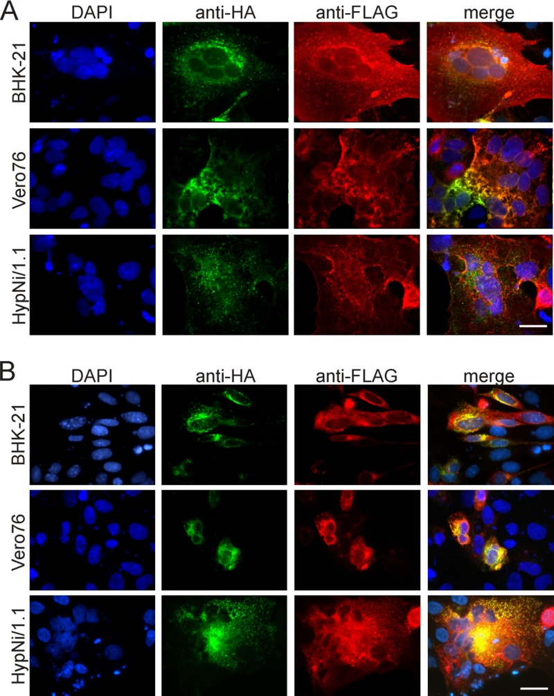
To gain information about the biological activities of the surface glycoproteins of an African henipavirus, the open reading frames of the fusion (F) and attachment (G) proteins of the African bat henipavirus Eid_hel/GH-M74a/GHA/2009 (M74) (10) were inserted into the expression plasmids pCAGGS (M74 F) or pCG1 (M74 G), and expression was compared to that of the corresponding NiV glycoproteins. Amino acid sequence identity between the F and G proteins of M74 and NiV was 55.8 and 26.0%, respectively (10). All F proteins contained a carboxy-terminal hemagglutinin (HA) tag and all G proteins a carboxy-terminal FLAG tag. The F and G proteins of both M74 and NiV were expressed in BHK-21 and Vero76 cells and in HypNi/1.1 cells, kidney cells from Hypsignathus monstrosus (11). The expression efficiencies and the fluorescence intensities of the expressed glycoproteins in permeabilized cells were similar for M74 and NiV (not shown). Major differences were detected when the transfected cells expressing both F and G were monitored at 24 h posttransfection (p.t.) for the presence of syncytia. As shown in Fig. 1A, syncytia were observed for BHK-21, Vero76, and HypNi/1.1 cells expressing both the NiV F (green) and NiV G (red) proteins. In contrast, the glycoproteins of M74 induced syncytium formation only in HypNi/1.1 cells (Fig. 1B), not in BHK-21 or Vero76 cells. When the syncytia of a sample characterized by coexpression of F and G were evaluated, the number of nuclei per syncytium ranged between 4 and 10, with a mean value of 5.0 nuclei/syncytium. This number is much lower than the value of 13.5 that was determined for HypNi/1.1 cells expressing the NiV glycoproteins.
Fig 1.
Syncytium formation of cells coexpressing the glycoproteins F and G of M74 and NiV. The three cell lines indicated were cotransfected for the expression of both the F and G proteins of either NiV (A) or M74 (B). At 24 h p.i., cells were stained for F (green), G (red), or nuclei (blue) and analyzed by fluorescence microscopy (scale bar, 25 μm). DAPI, 4′,6-diamidino-2-phenylindole.
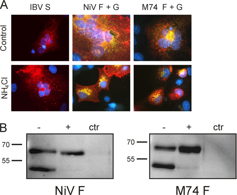
Proteolytic activation of the NiV fusion protein requires recycling from the plasma membrane through an endosomal compartment in an acid-dependent process. Ammonium chloride prevents acidification of endosomal compartments and inhibits syncytium formation induced by NiV F protein (12). In the presence of 25 mM ammonium chloride added 4 h p.t., the F and G proteins of neither NiV nor M74 showed any syncytium formation above background levels (Fig. 2A). As a control, we analyzed the spike (S) glycoprotein of infectious bronchitis virus, an avian coronavirus, which is activated by furin-like proteases (13). Expression of the S protein (14) in HypNi/1.1 cells resulted in syncytium formation which was not inhibited by ammonium chloride (Fig. 2A). Our results show that the proteolytic activation of the fusion activity of M74 F is acid dependent, similar to that of NiV F. The inhibiting effect of ammonium chloride on the proteolytic cleavage of the F proteins was confirmed by Western blotting. In HypNi/1.1 cells expressing the F protein of either NiV or M74, two protein bands are visible, representing the uncleaved precursor F0 and the cleavage product F1 (Fig. 2B). In the samples treated with ammonium chloride, only the uncleaved F0 (upper band) is detectable.
Fig 2.
Effect of ammonium chloride on the function and processing of the surface glycoproteins of M74 and NiV. (A) HypNi/1.1 cells were transfected for expression of M74 F, NiV F, or (as a control) the S protein of IBV and incubated in the absence (control) or presence of ammonium chloride (NH4Cl). At 24 h p.t., the cells were monitored for syncytium formation. The M74 and NiV F and G proteins were stained as described in the legend to Fig. 1. The S protein of IBV was stained by a monoclonal antibody and rhodamine-conjugated secondary antibodies. (B) Transfected cells expressing M74 F, NiV F, or no foreign protein (ctr) were incubated in the presence (+) or absence (−) of NH4Cl and analyzed by Western blotting for the presence of uncleaved precursor F0 (upper band) and cleavage product F1 (lower band). Molecular mass markers (in kilodaltons) are indicated on the left.
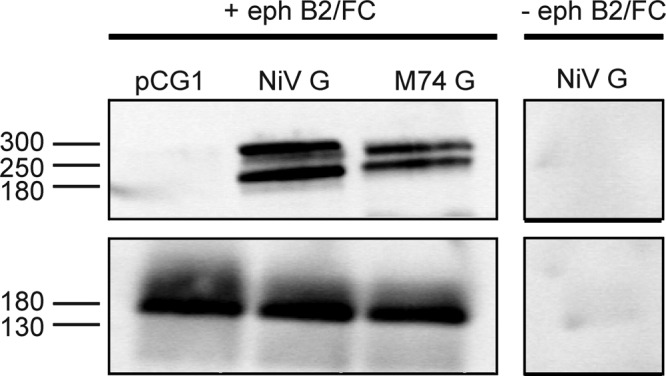
Infection by NiV and HeV is initiated by an attachment process that involves the interaction of the viral G protein with the cell surface receptor, ephrin B2/B3 (15, 16). In a coimmunoprecipitation assay, we analyzed whether ephrin B2 can interact with the G proteins of either NiV or M74. BHK-21 cells were transfected for expression of M74 G or NiV G. At 24 h p.t., cells were lysed and mixed with protein A-Sepharose loaded with soluble mouse ephrin B2/Fc. Precipitates were separated by SDS-polyacrylamide gel electrophoresis and analyzed by Western blotting. G proteins were visualized using mouse anti-FLAG and anti-mouse IgG-horseradish peroxidase (HRP). The presence of the ephrin B2/Fc was confirmed using anti-human IgG-HRP. As shown in Fig. 3, both G proteins were precipitated in an ephrin B2-dependent manner, indicating that the G protein of M74, like the NiV counterpart, interacts with ephrin B2.
Fig 3.

Interaction of the G proteins of M74 and NiV with ephrin B2. The FLAG-tagged G proteins of NiV and M74 were expressed in BHK-21 cells. At 24 h p.t., cells were lysed in NP-40 lysis buffer and mixed with protein A-Sepharose (Sigma-Aldrich) preloaded with soluble Fc-tagged ephrin B2 (+ eph; R&D Systems). − eph, control. After several washings, bound G proteins were released from the beads by boiling in nonreducing sample buffer and analyzed by Western blotting. G proteins were visualized by immunostaining (upper panels) using antibodies against the FLAG tag (Sigma-Aldrich). The presence of ephrin B2 was shown by immunostaining (lower panels) using antibodies against the Fc tag (Dako). Molecular mass markers (in kilodaltons) are indicated on the left.
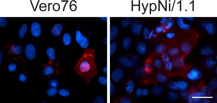
Finally, we analyzed whether differences in surface expression of the viral glycoproteins may explain the observed differences in syncytium formation. As shown in Fig. 4, surface immunofluorescence microscopy revealed the presence of G on the surface of syncytia in HypNi/1.1 cells. Surface expression of G was also found for Vero76 cells expressing both G and F of M74. However, these cells were much lower in number and they were always single cells and not part of a syncytium. Surface expression of F could not be analyzed in the same way, as the HA tag was attached to the cytoplasmic tail. However, as proteolytic cleavage of F (Fig. 2B) was also observed for Vero76 cells (not shown), we conclude that F protein is transported to the surface of these cells. Future experiments are needed to show whether differences in the efficiency of surface transport may explain differences in syncytium formation between NiV and M74.
Fig 4.

Surface expression of M74 G. Cells were transfected for expression of M74 F-HA and G-FLAG. At 24 h p.t., nonpermeabilized cells were stained with anti-FLAG antibody to detect G protein on the surface of transfected cells (scale bar, 25 μm).
Our results show that Asian and African henipaviruses share some characteristics suggesting a similar strategy of virus entry. Above all, we report a cell system that allows the functional characterization of the glycoproteins of African henipaviruses. This cell line may also be helpful in the isolation of infectious henipaviruses from African fruit bats and maybe even from bats of other geographic regions, such as Central America, where henipavirus-like viral RNA sequences have been reported (10).
ACKNOWLEDGMENTS
Part of this work was performed by N.K. and M.H. in partial fulfillment of the requirements for the doctoral degree from University of Veterinary Medicine Hannover and Leibniz University Hannover, respectively.
We are grateful to Roberto Cattaneo for providing expression plasmids.
This work was supported by grants to G.H. from DFG (SFB621 TP B7) and Bundesministerium für Bildung und Forschung (Ecology and Pathogenesis of SARS, an Archetypical Zoonosis, project code 01Kl1005B; and FluResearchNet, project code 01KI1006D). N.K. was funded by a fellowship of the Hannover Graduate School for Veterinary Pathobiology, Neuroinfectiology, and Translational Medicine (HGNI).
Footnotes
Published ahead of print 25 September 2013
REFERENCES
- 1.Wang LF, Yu M, Hansson E, Pritchard LI, Shiell B, Michalski WP, Eaton BT. 2000. The exceptionally large genome of Hendra virus: support for creation of a new genus within the family Paramyxoviridae. J. Virol. 74:9972–9979 [DOI] [PMC free article] [PubMed] [Google Scholar]
- 2.Chua KB, Goh KJ, Wong KT, Kamarulzaman A, Tan PS, Ksiazek TG, Zaki SR, Paul G, Lam SK, Tan CT. 1999. Fatal encephalitis due to Nipah virus among pig-farmers in Malaysia. Lancet 354:1257–1259 [DOI] [PubMed] [Google Scholar]
- 3.Murray K, Selleck P, Hooper P, Hyatt A, Gould A, Gleeson L, Westbury H, Hiley L, Selvey L, Rodwell B, Ketterer P. 1995. A morbillivirus that caused fatal disease in horses and humans. Science 268:94–97 [DOI] [PubMed] [Google Scholar]
- 4.Halpin K, Hyatt AD, Fogarty R, Middleton D, Bingham J, Epstein JH, Rahman SA, Hughes T, Smith C, Field HE, Daszak P. 2011. Pteropid bats are confirmed as the reservoir hosts of henipaviruses: a comprehensive experimental study of virus transmission. Am. J. Trop. Med. Hyg. 85:946–951 [DOI] [PMC free article] [PubMed] [Google Scholar]
- 5.Yob JM, Field H, Rashdi AM, Morrissy C, van der Heide B, Rota P, Abin Adzhar White J, Daniels P, Jamaluddin A, Ksiazek T. 2001. Nipah virus infection in bats (order Chiroptera) in peninsular Malaysia. Emerg. Infect. Dis. 7:439–441 [DOI] [PMC free article] [PubMed] [Google Scholar]
- 6.Hall LS, Richards G. 2000. Flying foxes: fruit and blossom bats of Australia. UNSW Press, Sydney, Australia [Google Scholar]
- 7.Marsh GA, de Jong C, Barr JA, Tachedjian M, Smith C, Middleton D, Yu M, Todd S, Foord AJ, Haring V, Payne J, Robinson R, Broz I, Crameri G, Field HE, Wang LF. 2012. Cedar virus: a novel Henipavirus isolated from Australian bats. PLoS Pathog. 8:e1002836. 10.1371/journal.ppat.1002836 [DOI] [PMC free article] [PubMed] [Google Scholar]
- 8.Hayman DT, Wang LF, Barr J, Baker KS, Suu-Ire R, Broder CC, Cunningham AA, Wood JL. 2011. Antibodies to henipavirus or henipa-like viruses in domestic pigs in Ghana, West Africa. PLoS One 6:e25256. 10.1371/journal.pone.0025256 [DOI] [PMC free article] [PubMed] [Google Scholar]
- 9.Drexler JF, Corman VM, Gloza-Rausch F, Seebens A, Annan A, Ipsen A, Kruppa T, Muller MA, Kalko EK, Adu-Sarkodie Y, Oppong S, Drosten C. 2009. Henipavirus RNA in African bats. PLoS One 4:e6367. 10.1371/journal.pone.0006367 [DOI] [PMC free article] [PubMed] [Google Scholar]
- 10.Drexler JF, Corman VM, Muller MA, Maganga GD, Vallo P, Binger T, Gloza-Rausch F, Rasche A, Yordanov S, Seebens A, Oppong S, Adu Sarkodie Y, Pongombo C, Lukashev AN, Schmidt-Chanasit J, Stocker A, Carneiro AJ, Erbar S, Maisner A, Fronhoffs F, Buettner R, Kalko EK, Kruppa T, Franke CR, Kallies R, Yandoko ER, Herrler G, Reusken C, Hassanin A, Kruger DH, Matthee S, Ulrich RG, Leroy EM, Drosten C. 2012. Bats host major mammalian paramyxoviruses. Nat. Commun. 3:796. [DOI] [PMC free article] [PubMed] [Google Scholar]
- 11.Hoffmann M, Müller MA, Drexler JF, Glende J, Erdt M, Gützkow T, Losemann C, Binger T, Deng H, Schwegmann-Wessels C, Esser KH, Drosten C, Herrler G. 2013. Differential sensitivity of bat cells to infection by enveloped RNA viruses: coronaviruses, paramyxoviruses, filoviruses, and influenza viruses. PLoS One 8:e72942. 10.1371/journal.pone.0072942 [DOI] [PMC free article] [PubMed] [Google Scholar]
- 12.Diederich S, Moll M, Klenk HD, Maisner A. 2005. The Nipah virus fusion protein is cleaved within the endosomal compartment. J. Biol. Chem. 280:29899–29903 [DOI] [PubMed] [Google Scholar]
- 13.Yamada Y, Liu DX. 2009. Proteolytic activation of the spike protein at a novel RRRR/S motif is implicated in furin-dependent entry, syncytium formation, and infectivity of coronavirus infectious bronchitis virus in cultured cells. J. Virol. 83:8744–8758 [DOI] [PMC free article] [PubMed] [Google Scholar]
- 14.Winter C, Schwegmann-Wessels C, Neumann U, Herrler G. 2008. The spike protein of infectious bronchitis virus is retained intracellularly by a tyrosine motif. J. Virol. 82:2765–2771 [DOI] [PMC free article] [PubMed] [Google Scholar]
- 15.Bonaparte MI, Dimitrov AS, Bossart KN, Crameri G, Mungall BA, Bishop KA, Choudhry V, Dimitrov DS, Wang LF, Eaton BT, Broder CC. 2005. Ephrin-B2 ligand is a functional receptor for Hendra virus and Nipah virus. Proc. Natl. Acad. Sci. U. S. A. 102:10652–10657 [DOI] [PMC free article] [PubMed] [Google Scholar]
- 16.Negrete OA, Levroney EL, Aguilar HC, Bertolotti-Ciarlet A, Nazarian R, Tajyar S, Lee B. 2005. EphrinB2 is the entry receptor for Nipah virus, an emergent deadly paramyxovirus. Nature 436:401–405 [DOI] [PubMed] [Google Scholar]


