Abstract
We used a high-density array of real-time PCR assays for commonly reported rodent infectious agents (PRIA) to test naturally infected index mice and sentinel mice exposed by contact and soiled-bedding transfer. PRIA detected 14 pathogens—including viruses, bacteria, fur mites, pinworms, and enteric protozoa—in 97.2% of 28 pooled fecal samples, fur–perianal swabs, and oral swabs from 4 cages containing a total of 10 index mice. Among these pathogens, PRIA (like conventional health monitoring methods) failed to detect Mycoplasma pulmonis, Pasteurella pneumotropica, and Giardia spp. in all of the 9 contact and 9 soiled-bedding sentinels. PRIA demonstrated murine adenovirus and Cryptosporidium and Spironucleus spp. in contact but not soiled-bedding sentinels and detected Helicobacter and pinworms in fewer than half of the soiled-bedding sentinels. Of the 4 species of Helicobacter that species-specific PCR assays identified in index mice, only H. ganmani was found in soiled-bedding and contact sentinels. PRIA detected all of the pathogens in sentinels that were identified by conventional methods. Myobia musculi was detected by PCR in index and sentinel mice but missed by conventional parasitologic examinations. In summary, PRIA reproducibly detected diverse pathogens in heavily pooled specimens collected noninvasively from infected index mice antemortem. The inability of PRIA and conventional health monitoring methods (that is, parasitology, microbiology, and serology) to demonstrate transmission of some pathogens to contact sentinels and the inefficient transmission of others to soiled-bedding sentinels underscores the importance of direct PCR testing to determine the pathogen status of rodents in quarantine and during routine colony surveillance.
Abbreviations: HM, health monitoring; PRIA, PCR array for rodent infectious agents; TNA, total nucleic acid
A requirement for meaningful biomedical research is that the animal models used (typically mice) remain free from infection with specific pathogens, including those that rarely produce disease but still interfere with research by modulating experimental responses and contaminating biologics. To eliminate and then exclude these pathogens, rodents have been rederived or cured of infection and housed behind room- or cage-level barriers, respectively.39 Because no barrier can be guaranteed 100% effective, routine health monitoring (HM) is necessary to verify the SPF status of breeding and research colonies, and imported animals in quarantine. Health monitoring of animals prior to release from quarantine has become especially important given that the genetically engineered mutant mice most frequently exchanged among investigators and institutions have been reported to harbor pathogens frequently.17
Conventional rodent HM programs usually consist of monthly to quarterly serology for prevalent viruses and quarterly to yearly whole-animal assessment, including a comprehensive serologic panel, pathology, parasitology, microbiology, and—since the advent of molecular diagnostics in the 1990s—PCR testing for fastidious microorganisms such as the helicobacters and Mycoplasma pulmonis.34,35 In addition to being determined by test sensitivity and specificity, the correspondence of HM findings to the actual health status of the population being monitored is affected by the degree to which the samples are representative of the population and specimens are suitable for the tests used. For commercial barrier rooms, HM is performed directly on colony animals of both sexes, and multiple age groups. By contrast, rodents from research colonies are rarely made available to be bled or euthanized for conventional HM; consequently, these colonies are monitored indirectly by testing sentinels. Irrespective of the diagnostic approach or test method, detection of a contamination requires that sentinels become infected with the adventitious agent.
Microisolation caging systems have been widely adopted for maintaining and quarantining mice and rats because the cage-level barrier they provide has proven to be very effective at excluding and impeding the spread of adventitious agents. Microisolation cage sentinels may be cohoused with quarantined animals, but for routine surveillance, contact sentinels are impractical because they would have to be moved among colony cages, which would be labor-intensive and undermine the cage-level barrier. Instead, sentinels are kept in separate cages supplied with regular changes of soiled bedding pooled from colony cages. Typically, 1 or 2 sentinel cages are setup for a rack of cages. Reliance on soiled bedding alone to transmit infections to sentinels is problematic because infections with certain respiratory viruses, host-adapted bacteria, and parasites are transmitted inefficiently or not at all via soiled bedding.10,15,24,26,28,38,42 In addition, the ability of microisolation cages to control the spread of infection frequently keeps the percentage of cages with actively infected rodents low. The lower the prevalence of infection, the greater the risk that the pathogen dose in pooled bedding will be insufficient to infect sentinels. The risk may increase as sentinel mice age and become less susceptible to infection, as has been reported for murine parvovirus 1 and mouse rotavirus.3,33
In addition to increasing the risk of missing a contamination, the low prevalence of infection that can occur in microisolation-cage–maintained colonies complicates confirmation of positive findings from sentinels because samples from many cages need to be tested to have an adequate chance of detecting an infection that is limited to a few cages. PCR analysis is frequently the method of choice for this confirmatory testing because it can detect prevalent pathogens in diverse specimens, such as feces and swabs of the skin and oral cavity which can be obtained noninvasively directly from colony animals. Moreover, the high sensitivity that is characteristic of PCR assays permits specimens to be heavily pooled (for example, 10 to 1), thus facilitating broad, representative sampling of cages on a rack. Now that PCR assays have been developed for virtually all of the viruses, bacteria, fungi, and parasites included in rodent SPF exclusion lists, PCR testing of pooled specimens collected directly from colony rodents is increasingly being added to HM programs to augment sentinel testing and enhance detection of pathogens not or inefficiently transmitted in soiled bedding.
One caveat regarding PCR analysis is that it can amplify genomic sequences from nonviable microorganisms . In addition, PCR testing may miss infections with viruses and other agents that are shed transiently, particularly in sentinels that are tested quarterly, although PCR has been shown to detect house hepatitis virus and mouse parvovirus in feces for weeks to months after infected mice are no longer contagious.2,6
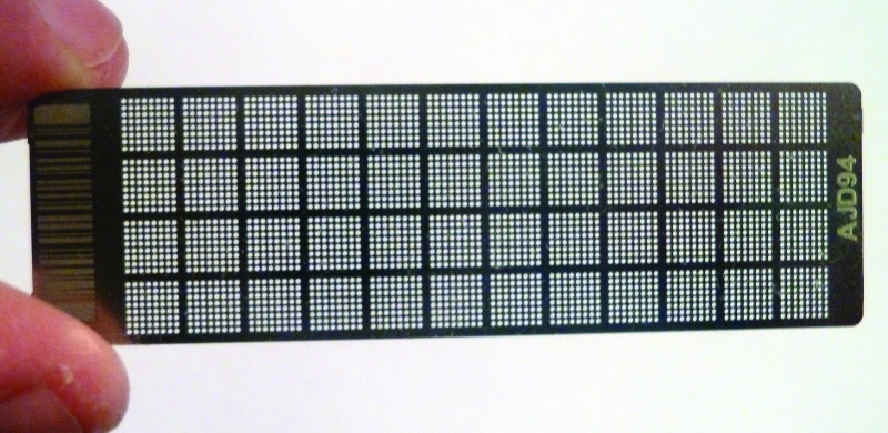
To improve the efficiency and throughput of PCR-based HM, we developed a high-density array of PCR assays for rodent infectious agents (PRIA). For developing PRIA, we chose the OpenArray platform (Applied Biosystems, Life Technologies, Grand Island, NY) because it uses fluorogenic TaqMan PCR reactions, termed ‘real-time’ PCR analysis, because the sequence-specific signal generated by the digestion of a fluorophore-labeled internal probe is measured each amplification cycle. The number of cycles required to reach a threshold signal is inversely related to the copies of microbial DNA added to the reaction.9,13,20,22,46 Particularly for agents such as Helicobacter and Spironucleus spp. that are present in very high copy numbers in specimens from infected animals, estimating copy number is helpful for identifying and discounting low-copy positive results due to cross-contamination from other samples. In addition to being quantitative, the advantages of the TaqMan PCR compared with the standard qualitative gel-based PCR include better specificity due to the internal probe and the elimination of postamplification cross-contamination because reactions do not need to be opened after amplification. By separating individual PCR assays into ‘holes’ (Figure 1), the OpenArray avoids the pitfalls of homogeneous PCR multiplexing, notably competitive inhibition that can cause false-negative results, especially when there are large differences in the genomic copies of multiple agents in a sample.21,32,44
Figure 1.
An OpenArray chip (18 × 0.5 × 63 mm) for PRIA. The chip contains 48 subarrays of 64 reaction holes (volume, 33 µL each), of which 56 are used to perform as many as 28 duplicate individual real-time PCR assays. The 2 chip sets, used to test for the agents listed in Table 1, have a capacity equivalent to that of 56 traditional PCR plates, each comprising 96 wells.
In the current study, we used a PRIA panel comprising PCR assays for commonly excluded or reportable rodent viruses, bacteria, fungi, and parasites (Table 1) to screen naturally infected index mice and sentinel mice exposed by contact and soiled-bedding transfer or by soiled bedding transfer alone. To model direct PCR screening of colony or quarantined mice, PRIA was performed on pools of feces, fur or perianal swabs, and oral swabs repeatedly collected from live index mice during the first 10 d of the study. These PRIA results were compared with those from sentinels tested on study days 28, 56, and 84. In addition, we compared the pathogens detected by PRIA with those identified by using conventional HM.
Table 1.
Pathogens identified in naturally infected index and SPF control mice by conventional (Conv) and PRIA HM at start and end of study
| No. of mice positive for pathogenb | ||||||||
| Index mice | Control mice | |||||||
| Day 0 | Day 84 | Days 0, 28, 56, and 84 | ||||||
| Conventional method | Pathogen | PRIA equivocal rangea | Conv | PRIA | Conv | PRIA | Conv | PRIA |
| Serology | MAdV1&2 | 1–10 | 2 | 1 | 10 | 2 | 0 | 0 |
| MHV | 1–10 | 2 | 1 | 10 | 2 | 0 | 0 | |
| MNV | 10–100 | 0 | 0 | 0 | 0 | 0 | 0 | |
| MRV | 1–10 | 2 | 0 | 9 | 0 | 0 | 0 | |
| MVM&MPV | 1–10 | 2 | 1 | 10 | 5 | 0 | 0 | |
| Reo1&3 | 1–10 | 0 | 0 | 0 | 0 | 0 | 0 | |
| TMEV | 1–10 | 2 | 1 | 10 | 9 | 0 | 0 | |
| CAR bacillus | 10–100 | 0 | 0 | 0 | 0 | 0 | 0 | |
| Clostridium piliforme | 1–10 | 0 | 0 | 0 | 0 | 0 | 0 | |
| M. pulmonis | 1–10 | 0 | 1 | 4 | 6 | 0 | 0 | |
| Microbiology | Bordetella bronchiseptica | 1–10 | 0 | 0 | 0 | 0 | 0 | 0 |
| Campylobacter spp. | 10–100 | 0 | 0 | 0 | 0 | 0 | 0 | |
| Citrobacter rodentium | 1–10 | 0 | 0 | 0 | 0 | 0 | 0 | |
| Corynebacterium bovis | 10–100 | 0 | 0 | 0 | 0 | 0 | 0 | |
| Corynebacterium kutscheri | 1–10 | 0 | 0 | 0 | 0 | 0 | 0 | |
| Klebsiella oxytoca | 1–10 | 0 | 0 | 0 | 0 | 0 | 0 | |
| Klebsiella pneumoniae | 1–10 | 0 | 0 | 0 | 0 | 0 | 0 | |
| P. aeruginosa | 1–10 | 0 | 0 | 10 | 8 | 0 | 0 | |
| P. pneumotropica Heylc | 1–10 | 0 | 0 | 5c | 4 | 0 | 0 | |
| P. pneumotropica Jawetz | 1–10 | 0 | 0 | 5c | 2 | 0 | ||
| Salmonella spp. | 1–10 | 0 | 0 | 0 | 0 | 0 | 0 | |
| Staphylococcus aureus | 1–10 | 0 | 1 | 0 | 0 | 0 | 0 | |
| Streptobacillus moniliformis | 1–10 | 0 | 0 | 0 | 0 | 0 | 0 | |
| Streptococcus pneumonia | 1–10 | 0 | 0 | 0 | 0 | 0 | 0 | |
| β-hemolytic Streptococcus spp.d | 1–10 | 0 | 0 | 0 | 0 | 0 | 0 | |
| Parasitologye | Fur mites | 1–10 | 2 | 1 | 10 | 10 | 0 | 0 |
| Pinworms | 1–10 | 2 | 1 | 8 | 10 | 0 | 0 | |
| Cryptosporidium spp. | 1–10 | NT | 1 | NT | 4 | NT | 0 | |
| Giardia spp. | 1–10 | 2 | 1 | 0 | 4 | 0 | 0 | |
| Spironucleus muris | 100–1000 | 2 | 1 | 3 | 10 | 0 | 0 | |
| PCR | Helicobacter spp. | 100–1000 | 2 | 1 | 10 | 10 | 0 | 0 |
| Pneumocystis murina | 1–10 | 0 | 0 | 0 | 0 | 0 | 0 | |
CAR, cilia-associated respiratory bacillus; MAdV1&2, mouse adenovirus types 1 and 2; MHV, mouse hepatitis virus; MNV, murine norovirus; MPV, mouse parvovirus; MRV, mouse group A rotavirus; MVM, minute virus of mice; NT, not tested; Reo1&3, reovirus types 1 and 3; TMEV, Theiler mouse encephalomyelitis virus.
Samples with copy numbers in the equivocal range were retested by 96-well real-time PCR analysis; when the retest cycle threshold value corresponded to a template copy number within or above the equivocal range, the sample was reclassified as positive.
The same 2 index mice were sampled for conventional and PRIA HM on day 0, but the PRIA specimens were pooled. On day 84, 10 index mice were sampled; 2 control mice each were sampled on days 0, 28, 56, and 84.
Conventional microbiology results for P. pneumotropica were not reported by biotype or confirmed by PCR.
Lancefield groups B, C, and G.
PCR assays for fur mites detected Myobia, Myocoptes, and Radfordia and for pinworms detected Aspiculuris and Syphacia
Materials and Methods
Mice (Mus musculus).
The naturally infected index mice for this study were 6- to 8-wk-old female mice of undetermined genetic background from a pet-rodent population harboring a variety of pathogens. The sentinel and control mice were SPF 3- to 4-wk-old female Crl:CD1 mice (Charles River, Wilmington, MA) reported by the vendor to be free of all agents assayed by PRIA, except for Staphylococcus aureus.
Husbandry.
Index and sentinel mice were housed in sterilized semirigid isolators provided with a 14:10-h light:dark cycle and with 12 changes daily of HEPA-filtered air. Control sentinel mice were held in static filter-top isolator (microisolation) cages. Mice were maintained on γ-irradiated bedding (Aspen Shavings, Northeastern Products, Warrensburg, NY) that was changed at least weekly according to experimental protocols; the mice had ad-libitum access to γ-irradiated feed (Lab Diet 5L79; Purina St Louis, MO) and sterile-filtered water. Exterior surfaces of supply containers were disinfected with chlorine dioxide solution (Clidox S, Pharmacal Research Laboratories, Naugatuck, CT) before being introduced to an isolator or opened in a biologic safety cabinet. All of the animal-use procedures were approved by the local IACUC.
Conventional HM.
The methodologies for conventional (that is, nonPRIA) HM included visual examination for protozoa and parasites, serology for viral and other microbial antibodies, microbiology for bacteria and fungi, and real-time PCR testing for Helicobacter. Mice were transferred to Research Animal Diagnostic Services (Charles River) in static N10 filter-top cages (Ancare, Bellmore, NY), and, on the day of arrival, were euthanized with carbon dioxide and processed in compliance with procedures approved by the local IACUC. After euthanasia, mice were screened for endo- and ectoparasites by macroscopic and microscopic examinations of pelts, perianal tape tests, intestinal contents, and fecal centrifugation concentrates.8,27 Nasal aspirates and gastrointestinal swabs were inoculated onto various nonselective, selective, and enrichment media that were incubated as previously described.30 Preliminary identification of microbial isolates suspected to be primary or opportunistic pathogens was based on colony and cellular morphology; as appropriate, isolates were further characterized by biochemical and immunologic analyses and by species-specific PCR assays.7 Serum samples were screened for microbial antibodies by using a multiplexed fluorometric immunoassay;45 those for which the results were indeterminate, equivocal, or unexpected were retested, typically by using a complementary immunofluorescent assay.18
Collection of specimens for PRIA.
The specimens collected from each mouse, either antemortem or at necropsy, included a fecal pellet, an adhesive swab (Puritan, Guilford, ME) of the fur starting at the head and concluding with the perianal hairs, and a small cotton swab of the oral cavity. Swab tips were excised from the shaft prior to placement in collection tubes. Bronchial wash, nasopharyngeal wash, and lung specimens were added to postmortem collections. Antemortem specimens from index mice were pooled by type and cage. Postmortem specimens were tested by individual mouse and specimen type, with the exception of those collected from index and SPF mice on day 0 of the study, which were pooled by specimen type.
Processing of specimens for PRIA.
Process steps consisted of sample lysate preparation, extraction of total nucleic acid (TNA), reverse transcription of RNA to cDNA, and preamplification. Sample lysates were prepared for TNA extraction as follows. A fecal pool or individual pellet was mixed with approximately 4 times the fecal volume of PBS (pH 7.2; Invitrogen, Carlsbad, CA) in the original collection tube and homogenized by using stainless steel beads in a homgenizer at 20 to 25 Hz for 2 min (TissueLyser, Qiagen, Germantown, MD). The fecal homogenate was clarified by low-speed centrifugation (1,00 × g) . By using the same procedure, approximately 3 mm3 of lung tissue was homogenized in 200 μL of lysis buffer from the TNA isolation kit (MagMAX Total Nucleic Acid Isolation Kit, Life Technologies, Foster City, CA) and clarified. For oral swabs, 500 μL PBS, pH 7.2 was added to each tube that contained 1 or more oral swab tips and vortexed; fur–perianal swab tips were vortexed in 400 to 800 μL of the lysis solution provided in the TNA isolation kit. Particulate concentrates were prepared from the oral swabs as well as the nasopharyngeal and bronchial washes by high-speed centrifugation (21,000 × g) followed by removal of all but approximately 50 μL of supernatant. Sample volumes for TNA isolation were 100 μL clarified fecal homogenate; 275 μL fur–perianal hair lysate; 50 μL per concentrate of oral swab, nasopharyngeal wash, or bronchial wash; and 25 μL lung homogenate.
Prior to TNA isolation, samples prepared from index mouse specimens collected antemortem were combined by adding 275 μL fur–perianal hair lysate and 100 μL clarified fecal lysate to the oral swab concentrate. Other sample lysates were not combined. Each individual or combined sample, including a ‘mock’ sample consisting of lysis buffer only, was spiked with 200 copies of an RNA template transcribed from a plasmid construct containing part of an algal gene. The antemortem samples such as feces and furs have a low nucleic-acid content not easily measured by the standard spectophotometric method; to circumvent this problem, our laboratory depends on the monitoring of a spike RNA added prior to sample extraction. This RNA template is used to evaluate whether the recovery of RNA from TNA isolation and synthesis of cDNA during reverse transcription were adequate. RNA-spiked lysates were homogenized with zirconium beads in a TissueLyser (Qiagen) at 20 to 25 Hz for 15 min before TNA extraction (Kingfisher FLEX 96, ThermoScientific, Waltham, MA) by using a magnetic bead-based kit (MagMAX Total Nucleic Acid Isolation Kit) according to the manufacturer's instructions. The TNA extracted from a sample was eluted in 80 μL of kit elution buffer. A portion (that is, 13 μL) of the TNA eluate was reverse-transcribed by using a random-hexamers–based reverse transcription kit (High Capacity RNA-to-cDNA Kit, Life Technologies, Carlsbad, CA). After reverse transcription, each sample was spiked with 100 copies of a plasmid construct that contained an algal gene sequence that was different from the RNA template used to monitor nucleic acid recovery, to control for sample-mediated inhibition of PCR amplification.31 Preamplification, the final sample-processing step preceding PRIA, was accomplished by using a kit (TaqMan PreAmp Master Mix Kit, Life Technologies) according to the primer concentrations and conditions described in the manufacturer's instructions.
PRIA.
Proprietary PCR primers and MGB TaqMan probes (Life Technologies) for the 32 infectious agents (Table 1) and 2 controls (that is, the RNA-recovery and amplification-inhibition templates), prepared as 20× stocks, were provided to Life Technologies for their manufacture of the PRIA OpenArray chips. Briefly, we used all available sequences in GenBank and other unpublished sequences determined in our laboratory to design PCR assays that detected only the targeted group, genus, species, or strain as defined by the agent category listed in Table 1. All assays fulfilled a set of standard qualification criteria, including a limit of detection of 1 to 10 target copies and sufficient selectivity to amplify only target organisms and exclude heterologous sequences. Each OpenArray chip is divided into 48 subarrays; one sample or control is tested per subarray, which is subdivided into 64 reaction holes (Figure 1). Because tests were performed in duplicate holes, 2 chipsets were needed to accommodate the 38 infectious agent assays. The nucleic-acid recovery and sample-mediated inhibition controls were included in duplicate holes in the subarray configuration of both chipsets. To prepare positive template controls, microbial DNA or cDNA fragments containing the assay target regions were cloned into plasmids. The concentrations of purified plasmids were measured by spectrophotometer (OD260:OD280), and the plasmids were pooled by chipset to 100 and 1000 PTC copies per reaction hole. Chinese hamster ovary DNA, adjusted to 0.41 ng per reaction hole, was used as a negative control template.
After preamplification, samples including the mock sample, 100- and 1000-copy positive template control pools, and the negative template control were each added to master mix (GeneAmp Fast PCR Master Mix, Life) and loaded onto the PRIA arrays by using the AccuFill loader (Life Technologies). OpenArray chips were placed into amplification cassettes, sealed, and then amplified in a thermocycler (OpenArray NT Cycler, Life Technologies) according to the manufacturer's instructions and standard amplification cycling program. The results of the controls for RNA recovery and sample-mediated inhibition were interpreted as acceptable when the their cycle-threshold values were not more than 2.0 above that of the mock sample, which would represent a reduction in template copy number of approximately 0.5 log10. If either of these controls failed, PRIA results were not interpreted, and the sample was retested or reextracted from the original specimen and then retested. The template copies per reaction in a sample were estimated by comparing the average sample and 100-copy positive template control cycle-threshold values; a difference of 3.3 Ct approximately corresponds to a 10-fold difference in copy number.40 Results are presented as the number of PCR template copies per microliter TNA. Negative and positive target copy cutoffs for each assay were established according to the prevalence and persistence of the agent and the copy number range found in field specimens. Sample target copy numbers between the negative and positive cutoffs were classified as equivocal. The equivocal copy number range is shown for each assay in Table 1. When a sample gave an equivocal PRIA result, it was retested by the corresponding 96-well real-time PCR; if the retest cycle threshold value was equivocal or positive, the sample was scored as assay positive.
Additional strain- or species-specific PCR assays were used for select samples and time points to identify pathogens further.
Study design.
On day 0 of the study, 2 naturally infected index and 2 SPF CD1 mice (used as unexposed controls or sentinels) were submitted for conventional and PRIA HM; 4 cages of index mice were placed in an isolator, and fecal, fur swab and oral swab pools for PRIA were prepared from each cage. Three of these cages were used for contact exposure of sentinels by cohousing 3 sentinel with 2 index mice per cage for 11 d; the contact-exposed sentinel mice were then moved to a separate cage (in the same isolator as the index mice) supplied with weekly changes of 50% soiled bedding from the index cage in which they were contact-exposed. The fourth cage, containing 4 index mice, was used as a source of soiled bedding transferred on day 3 and every week thereafter to a second isolator, where it was equally divided among 3 cages of 3 sentinel mice each. The day 3 transfer was included to simulate the build-up of agents in shipping crates. No attempt was made to isolate individual cages within an isolator. Antemortem specimen pools from mice in each of the 4 index cages were collected for PRIA on days 0, 3, 4, 5, 6, 7, 10, and 84. On days 28, 56, and 84, 1 sentinel from each of the 3 contact and 3 bedding-only sentinel cages and 2 control mice were submitted for conventional HM and for PRIA of specimens collected postmortem; the 10 index mice were submitted for conventional HM and PRIA on day 84.
Statistics.
The Fisher Exact test was used to compare the proportion of positive results obtained by PRIA of antemortem specimens collected directly from index mice and postmortem specimens collected from contact and bedding sentinels. The McNemar test was used to compare the proportion of positive results obtained by PRIA and conventional HM. One-way ANOVA followed by posthoc analysis was performed to calculate the statistical significance of differences in PCR target copy numbers for fecal, fur–perianal swab, and oral swab specimens collected antemortem index mouse specimens. The Fisher Exact test was done by using R (http://www.r-project.org/); the McNemar and one-way ANOVA tests were performed in SigmaPlot (Systat Software, San Jose, CA). A P value of less than 0.05 was used to define statistical significance.
Results
Pathogen status of index and control mice.
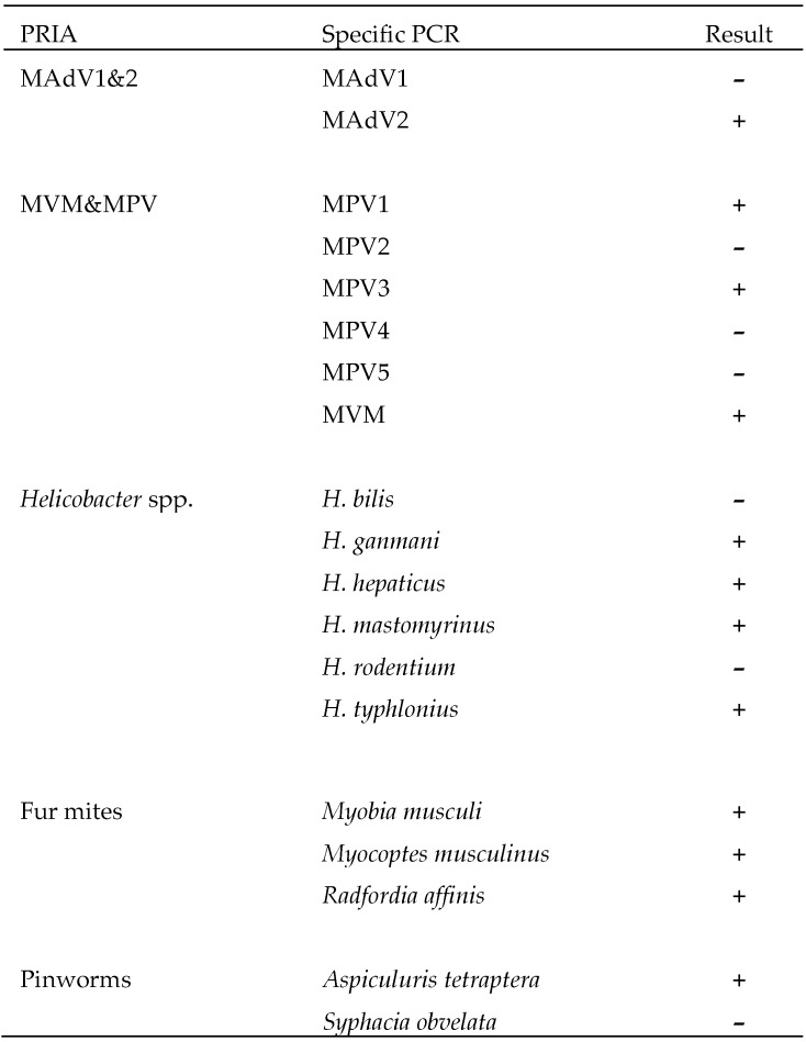
The pathogens detected by PRIA in fecal, fur–perianal and oral swab, lung homogenate and nasopharyngeal and bronchial lavage specimens collected from index mice postmortem on days 0 and 84 are shown in Table 1. They included 4 prevalent viruses, M. pulmonis, P. aeruginosa, P. pneumotropica, S. aureus, Helicobacter spp., fur mites, pinworms and 3 enteric protozoa. To verify these findings and further identify the pathogens, index mouse TNA samples screened by generic PRIA assays for mouse adenovirus, mouse parvoviruses, Helicobacter spp., mites, and pinworms were retested by strain- or species-specific real-time PCR analyses (Figure 2). In summary, these assays detected mouse adenovirus type 2 (but not type 1), mouse parvoviruses types 1 and minute virus of mice (but not type 2, 4, or 5); the helicobacters H. ganmani, H. hepaticus, H. mastomyrinus, and H. typhlonius (but not H. bilis or H. rodentium); the fur mites Myobia musculi, Myocoptes musculinus, and Radfordia affinis; and the pinworm Aspiculuris tetraptera (but not Syphacia obvelata).
Figure 2.
PCR identification of the species or strains of pathogens detected in index mice by PRIA. Myobia musculi was not detected in index mice by direct parasitologic examinations.
Although diagnoses made by PRIA generally were corroborated by conventional HM, some differences were noted. The 2 index mice tested on day 0 were seronegative but PCR-positive for M. pulmonis; of the 10 mice tested when the study concluded on day 84, 4 were seropositive for M. pulmonis as compared with 6 positive by PRIA. Multiplexed fluorometric immunoassays detected serum antibodies to mouse rotavirus, Sendai virus, and pneumonia virus of mice (data not shown), but shedding of these viruses was not detectable by PRIA, which included mouse rotavirus, or 96-well real-time PCR assays for Sendai virus and pneumonia virus of mice. PRIA measured approximately 106 Cryptosporidium template copies per microliter TNA. Although Cryptosporidium was not detected by conventional parasitology, histologic examination of the gastrointestinal tract, considered to be the definitive method for detecting Cryptosporidium,41 was not included; therefore, conventional HM for Cryptosporidium is recorded in Table 1 and 2 as not tested. However, Cryptosporidium infestation was verified by showing that the sequence of a 715-bp segment of an 18S ribosomal RNA gene amplified from index mouse fecal TNA was identical to that of C. parvum (data not shown). Whereas PRIA on day 0 did not detect P. aeruginosa or P. pneumotropica in the postmortem specimen pool, it did in the antemortem specimens collected from the 4 index cages. These results, along with those presented subsequently, show that the pathogen profile of the index mice stayed the same throughout the study. In addition, adventitious infections of unexposed control mice were not detected.
Table 2.
Comparison of PRIA HM of live, naturally infected index mice with conventional and PRIA HM of contact and soiled-bedding sentinels
| No. (%) of positive among no. of mice testeda | ||||||
| Index mice (n = 28) | Contact sentinels (n = 9) | Soiled-bedding sentinels (n = 9) | ||||
| Conventional method | Pathogen | PRIA | Conventional | PRIA | Conventional | PRIA |
| Serology | MAdV1&2b,c | 28 (100%) | 9 (100%) | 9 (100%) | 0 | 0 |
| MHV | 28 (100%) | 9 (100%) | 2 (22%) | 9 (100%) | 3 (33%) | |
| MRV | 0 | 0 | 0 | 0 | 0 | |
| MVM&MPV | 28 (100%) | 9 (100%) | 8 (89%) | 9 (100%) | 9 (100%) | |
| TMEV | 28 (100%) | 9 (100%) | 9 (100%) | 9 (100%) | 6 (67%) | |
| M. pulmonisc,d | 27 (96%) | 0 | 0 | 0 | 0 | |
| Microbiology | P. aeruginosa | 25 (89%) | 3 (33%) | 7 (78%) | 9 (100%) | 8 (89%) |
| P. pneumotropica Heylc,d | 28 (100%) | 0 | 0 | 0 | 0 | |
| P. pneumotropica Jawetzc,d | 25 (89%) | 0 | 0 | 0 | 0 | |
| Parasitology | Fur mites | 28 (100%) | 9 (100%) | 9 (100%) | 3 (33%) | 4 (44%) |
| Pinworms | 28 (100%) | 7 (78%) | 9 (100%) | 7 (78%) | 9 (100%) | |
| Cryptosporidium spp. | 28 (100%) | NT | 8 (89%) | NT | 0 | |
| Giardia spp.c,d | 24 (86%) | 0 | 0 | 0 | 0 | |
| Spironucleus murisb,c | 28 (100%) | 8 (89%) | 9 (100%) | 0 | 0 | |
| PCR | Helicobacter spp.b | 28 (100%) | NA | 9 (100%) | NA | 3 (33%) |
MAdV1&2, mouse adenovirus types 1 and 2; MHV, mouse hepatitis virus; MNV, murine norovirus; MPV, mouse parvovirus; MRV, mouse group A rotavirus; MVM, minute virus of mice; NA, not applicable; NT, not tested; TMEV, Theiler mouse encephalomyelitis virus.
For index mice, the no. of mice tested represents pools by cage of feces, fur–perianal swabs, and oral swabs collected from 4 index mouse cages per time point on days 0, 3, 4, 5, 6, 7, and 10. For sentinel mice, the no. of mice tested represents individual mice; 3 contact and 3 soiled-bedding sentinel mice, each from a different cage, were submitted per time point on days 28, 56, and 84.
The proportion positive was significantly higher (P < 0.05, Fisher exact test) for contact than for soiled-bedding sentinels.
The proportion positive was significantly higher (P < 0.05, Fisher exact test) for index mice than for contact sentinels.
The proportion positive was significantly higher (P < 0.05, Fisher exact test) for index mice than for soiled-bedding sentinels.
Comparison of pathogens detected by PRIA in index and sentinel mice.
Table 2 shows a comparison of pathogens found by PRIA of specimens (including feces, fur–perianal swabs, and oral swabs) collected noninvasively directly from live, naturally infected index mice and those detected indirectly in contact and soiled-bedding sentinels by postmortem conventional and PRIA HM. Agents not detected by PRIA or conventional HM were omitted from Table 2, as was S. aureus because it has been found in the production colony from which the sentinel mice originated. Although not detected by PRIA, mouse rotavirus is listed because the index mice were seropositive for this virus (Table 1).
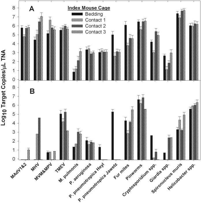
Antemortem sampling of index mice in each of the 4 cages where they were housed was performed at 7 times points between days 0 and 10. Lysates of the different specimen types from a cage were combined prior to TNA extraction (except for the day 0 lysates, which were tested separately to evaluate the effect of specimen type on the number of PCR template copies measured). Therefore, the results shown in Table 2 are the number (and percentage) of samples that were pathogen-positive among the 28 samples tested (that is, 7 time points per cage × 4 cages). For the 14 pathogens listed in Table 2 that were found by PRIA, the average percentage-positive was 97.2% (381 of 392 samples). Moreover, during the first 10 d of the study, the number of PCR template copies per microliter TNA, which ranged from an average from 101 for M. pulmonis to 108 for Spironucleus, did not vary notably between cages of index mice (Figure 3 A). These data, along with those showing pathogens were still detectable by PRIA on day 84 (Table 1, Figure 3 B), demonstrate that the 4 cages of index mice were equivalent sources of infection for the sentinels.
Figure 3.
Number of PRIA template copies per microliter TNA (mean ± 1 SD) by index mouse cage (3 contact and 1 bedding-provider cage) for specimens collected during (A) days 3 to 7 and 10 and (B) day 85 of study. Names of organisms are abbreviated as in Table 1.
Contact and soiled-bedding sentinel groups were exposed to infections of the index mice by contact and then regular transfers of soiled bedding and by soiled-bedding transfer alone, respectively. Each sentinel group consisted of 9 mice equally divided among 3 cages. One third of the sentinel mice (1 mouse per cage) was submitted for postmortem PRIA and conventional HM at each of 3 time points including days 28, 56, and 84. As summarized in Table 2, pathogens that PRIA detected in the index but not sentinel mice were M. pulmonis, P. pneumotropica, and Giardia spp. Those that PRIA determined to be present in contact but not soiled-bedding sentinels were mouse adenovirus, Cryptosporidium spp., and Spironucleus spp. Agents that PRIA detected in both sentinel groups were mouse hepatitis virus, minute virus of mice, mouse parvoviruses, Theiler murine encephalomyelitis virus, P. aeruginosa, fur mites, and pinworms. Transmission of the 4 helicobacter species identified in the index mice (Figure 2) varied substantially. Only H. ganmani could be found in a proportion of soiled-bedding sentinels (that is, 3 of 9) and in all 9 contact sentinels. H. mastomyrinus and H. typhlonius were detected in only 1 and 2 contact sentinels, respectively, whereas H. hepaticus was not present in any sentinel. It is noteworthy that mouse rotavirus, for which index mice were seropositive but PRIA-negative, was not transmitted to sentinels. In addition, pathogens other than those present the index mice were not detected in sentinels (data not shown).
Figure 4 shows the average estimated PCR template copies per microliter TNA at days 28, 56, and 84 for the agents transmitted to the contact and soiled-bedding sentinels. In general, PCR template levels held steady for pathogens that PRIA found in a high percentage of samples. Viral and Cryptosporidium PCR template levels did trend downward over time, with this tendency being most pronounced for MHV, which was undetectable in contact sentinels after day 28. By contrast, fur mite template concentrations increased with time in both sentinel groups.
Figure 4.
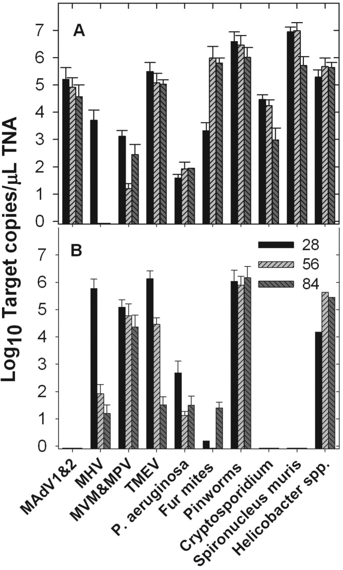
Number of PRIA template copies per microliter TNA (mean ± 1 SD) in specimens collected from (A) contact and (B) soiled-bedding sentinel mice on days 28, 56, and 84 (n = 3 mice for each time point). Names of organisms are abbreviated as in Table 1.
Comparison of pathogens found in sentinels by conventional and PRIA HM.
All 9 pathogens found in sentinels by conventional HM were also detected by PRIA. In contrast, Myobia musculi was shown to be present by PCR assay but was missed by conventional parasitologic examinations. Furthermore, although PRIA detected A. tetraptera DNA in all sentinels, pinworms were not observed in 2 of 3 contact sentinels on day 56 and in 2 of 3 bedding sentinels on day 84. Because the conventional parasitology performed in this study did not include histologic examination of the gastrointestinal tract to detect Cryptosporidium spp, a valid comparison between conventional and PRIA HM for the detection of this agent could not be made. For all pathogens found in sentinels, except for Cyrptosporidium and Helicobacter spp. detected only by PCR, the average percentage of positive samples was 50.0% by microbiology and parasitology compared with 72.2% by corresponding PRIA and 100% by serology compared with 73% by PRIA. Mouse rotavirus, pneumonia virus of mice, and Sendai virus were not detected in contact or bedding sentinels by either PCR analysis or multiplexed fluorometric immunoassays.
Effect of sample type on PCR template copies.
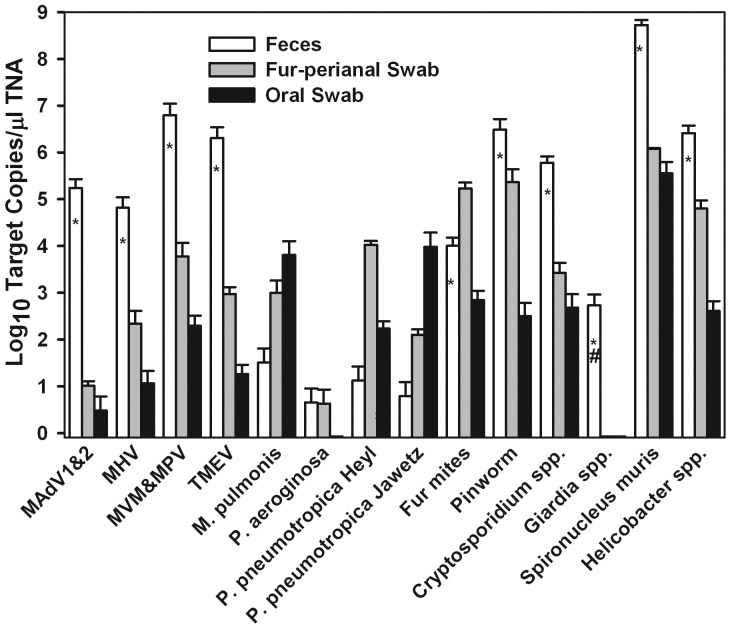
Figure 5 shows the average estimated PCR template copies per microliter TNA by specimen type for the 14 pathogens detected in specimens collected antemortem from index mice on day 0. Fecal specimens contained the highest template concentrations for 9 of these 14 pathogens and were the only specimens in which Giardia spp. was detected. Fur–perianal swab template levels were highest for fur mites and P. pneumotropica Heyl, whereas oral swabs had the highest numbers of genome copies of M. pulmonis and P. pneumotropica Jawetz.
Figure 5.
Number of PRIA template copies per microliter TNA (mean ± 1 SD) by specimen type. Specimens were collected antemortem from 10 index mice on day 0. Target copy levels determined by PRIA were significantly (P < 0.05) higher or lower in pooled feces than in oral (*) or fur–perianal swab (#) specimens. Names of organisms are abbreviated as in Table 1.
Discussion
Genetically engineered mutant mouse models generated and maintained at research facilities worldwide have often been shown to harbor pathogens such as parasites, Helicobacter spp., and murine norovirus.14,30 The growing exchange of these models among investigators and research institutions underscores the importance of HM results that accurately represent the current pathogen status of mouse colonies, particularly before mice are released from quarantine into an SPF facility. In addition to the sensitivity and specificity of a diagnostic technique, its accuracy depends on the suitability of specimens tested and the prevalence of infection. Typically, animals must be euthanized to obtain specimens, which must contain viable intact parasites and microorganisms that are suitable for conventional HM methodologies including direct observation and isolation by culture with subsequent identification. Because a sufficient number of colony mice are seldom available to be euthanized and because mutant models may be immunodeficient and thus not appropriate for serosurveillance, most colony HM has been performed indirectly on sentinel mice exposed to colony infections through contact or routine transfers of soiled bedding. Although the use of contact sentinels is practical and preferable for rodents in quarantine, the exposure of sentinels for routine HM of colonies in popular static and ventilated microisolation cages is predominantly limited to soiled-bedding transfer.
There are a variety of problems with exclusively relying on HM of sentinels to indirectly assess colony pathogen status. First, some important and prevalent pathogens are transmitted inefficiently or not at all through soiled bedding.10,15,24-26,28,38,42 In addition, even for agents transmitted in bedding, there is a risk that the pathogen dose to which sentinels are exposed will be insufficient to infect them when, as can occur in microisolation cages, the prevalence of infection is low and therefore a high proportion of the pooled bedding is from uncontaminated cages. Finally, an adventitious agent may be missed when sentinels are resistant to infection because of their age or strain.
An alternative to indirect sentinel HM is to use PCR analysis to test specimens such as feces and swabs of fur and the upper respiratory tract that can be collected directly from living colony animals by noninvasive means. Although these specimens might be insufficient or unsuitable for conventional HM methodologies, they suffice for PCR testing because detection does not depend on the presence of viable, infectious microorganisms. Moreover, the specificity with which the primers and the internal probes used in real-time assays hybridize to microbial DNA template allows PCR-based methods to detect bacterial pathogens and opportunists that are impractical to isolate from specimens such as feces with complex microflora.
In a previous study, we demonstrated that PCR assessment of fecal specimens collected directly from mice in quarantine diagnosed infections with Helicobacter spp., P. pneumotropica, and murine norovirus that were missed by using or inconsistently found in soiled-bedding sentinels.28 The data presented in the current study extend the comparison of direct and sentinel HM by PCR and conventional methodologies to include contact as well as soiled-bedding sentinels and to the additional viruses and parasites carried by the naturally infected index mice obtained from a pet supplier population. Unique to our study is the description and use of PRIA, an array of real-time PCR assays for rodent infectious agents that we developed to facilitate high-throughput screening for large panels of rodent pathogens. As described in the Introduction, real-time assays are generally more sensitive and specific than are gel-based PCR assays because the generation of a positive signal in real-time assays depends on the hybridization of both an internal probe and the primers to the assay target.
In both our current and previous studies,28 PCR tests detected infections of index mice that were transmitted to sentinels either inconsistently or not at all, thus highlighting the pitfalls of exclusive reliance on sentinel HM. We chose to use index mice that were naturally infected with field strains rather than experimentally infected mice to simplify the experiment and to avoid artifacts that may be associated with experimental infections using potentially attenuated laboratory isolates. However, the variety of concurrent infections harbored by the index mice likely affected their immune systems, susceptibility to infection, duration of the infection, and period of shedding.
To model direct screening of colony or quarantined mice in this study and to evaluate the reproducibility of pathogen detection by PCR of noninvasive specimens, PRIA was performed on TNA samples extracted from pools of feces, fur–perianal swabs, and oral swabs collected from the unanesthetized index mice at 7 time points during the first 10 d of the study; the pathogens detected included mouse adenovirus, mouse hepatitis virus, mouse parvovirus, Theiler murine encephalomyelitis virus, Helicobacter spp., M. pulmonis, P. aeruginosa, the Heyl and Jawetz biotypes of P. pneumotropica, fur mites, pinworms, Cryptosporidium spp., Giardia spp., and Spironucleus muris. The absence of murine norovirus in the index mice was notable given the high prevalence of this agent in laboratory mice. The average percentages of samples that were positive for these 14 pathogens was 97.2%, with 10 pathogens being detected in 100% of samples. Therefore, PRIA results were highly reproducible.
Although the infections of index mice that were detected by using conventional HM on days 0 and 84 (that is, study conclusion) largely corresponded to those detected by PRIA, some differences were noted. Index mice were seropositive but PRIA-negative for mouse rotavirus, pneumonia virus of mice, and Sendai virus, most likely because the mice had recovered from these typically short-lived infections. This explanation is supported by the inability of serology or PCR to demonstrate infection of sentinels by these viruses. Infestations of index mice with the fur mite Myobia musculi, identified by PCR testing, were missed in conventional parasitologic examinations. As noted in the Results, detection of the enteric protozoan Cryptosporidium could not be confirmed by conventional HM because the histologic method best suited to detect this agent was not included in the study. The presence of Cryptosporidium spp., however, was verified by 18S ribosomal RNA gene sequencing. Finally, although PRIA detected A. tetraptera DNA in all sentinels, pinworms were not observed in 2 of the 3 contact sentinels on day 56 and 2 of the 3 soiled-bedding sentinels on day 84. The cyclic nature of the infestations and the difference between contact and soiled-bedding sentinels is likely a result of A. tetraptera’s synchronized life cycle16,29 and earlier infestation of more heavily exposed contact sentinels.
Beginning on day 0 of the study, contact sentinels were housed together with index mice for 11 d and then moved to separate cages supplied weekly with soiled bedding from the index mouse cages. Starting on day 3, bedding sentinels were exposed to infection by weekly transfers of soiled bedding alone. Sentinels were evaluated by conventional and (postmortem) PRIA HM on days 28, 56 and 84. According to the results of this testing, M. pulmonis, P. pneumotropica, and Giardia spp. were not transmitted to sentinels; mouse adenovirus type 2, Cryptosporidium spp., and S. muris spread to contact but not soiled-bedding sentinels, and Helicobacter spp. and fur mites were found in fewer than half of the soiled-bedding sentinels. Of the 4 species of Helicobacter that PCR assays identified in index mice, only H. ganmani was found in soiled-bedding sentinels and consistently in contact sentinels.
The poor or lack of transmission of host-adapted bacteria, including M. pulmonis, P. pneumotropica, and Helicobacter spp., are consistent with previous reports.15,25,42 To our knowledge, the current study is the first to specifically investigate mycoplasmal transmission from naturally infected mice to contact and soiled-bedding sentinels, although transmission among rats by contact has been reported.23 For P. pneumotropica, failure of transmission has been related to the organism's brief survival outside of the host.26,38 The variation in the transmission efficiency of the different Helicobacter species that we observed in the current study has been reported previously.43 The variability appears not to have been caused by different levels of exposure, given that species-specific PCR assays measured similar target copy numbers of H. ganmani in fecal specimens from index mice and that 2 species (H. mastomyrinus and H. typhlonius) that were inefficiently spread to sentinels (data not shown). Our current inability to demonstrate the spread of mouse adenovirus type 2 to soiled-bedding sentinels contrasts with an earlier report.11,12 The lack of transmission of Giardia spp. to contact sentinels was unexpected also, because the authors of a prior investigation had reported that CD1 mice (which we used as sentinels in the current study) were highly susceptible to Giardia infection.5 Although the transmission of Giardia and Spironucleus spp. from experimentally inoculated rodents to contact sentinels has been demonstrated,1,19,36,37 we believe that our current study is the first investigation of the transmission of these agents and Cryptosporidium spp. from naturally infected mice to sentinels.
In summary, the results of our current investigation provide clear evidence that the pathogen status of quarantined or colony rodents is represented more accurately by direct PCR testing of noninvasive specimens collected from the principal animals antemortem than by indirect HM of sentinels, regardless of the diagnostic methodologies used. This difference occurs because PCR assays, especially real-time assays such as those in PRIA, are exquisitely sensitive and specific and, therefore, can detect subinfectious levels of pathogens4 in heavily pooled, highly representative samples. In addition, the time that animals spend in quarantine is reduced from 2 mo for conventional HM of sentinels to just 2 wk for direct PCR HM. Furthermore, direct PCR HM is in harmony with the 3Rs principal, because it reduces and eliminates the need for sentinels in routine colony surveillance and quarantine, respectively. PCR HM also alleviates the animal welfare and logistical issues associated with live animal shipments.
Acknowledgments
We want to recognize members of the health monitoring, bacteriology, and serology departments within the laboratories of Research Animal Diagnostic Services for their cooperation in evaluating the mice used in this study. Special appreciation is extended to Panagiota Momtsios and Delia Muise for their assistance with process, materials, and reagent development. The authors are employees of Charles River Laboratories, which has a direct commercial interest in the subject matter of this manuscript.
References
- 1.Belosevic M, Faubert GM, Maclean JD. 1986. Mouse-to-mouse transmission of infections with Giardia muris. Parasitology 92:595–598 [DOI] [PubMed] [Google Scholar]
- 2.Besselsen DG, Becker MD, Henderson KS, Wagner AM, Banu LA, Shek WR. 2007. Temporal transmission studies of mouse parvovirus 1 in BALB/c and C.B17/Icr–Prkdc(scid) mice. Comp Med 57:66–73 [PubMed] [Google Scholar]
- 3.Besselsen DG, Wagner AM, Loganbill JK. 2000. Effect of mouse strain and age on detection of mouse parvovirus 1 by use of serologic testing and polymerase chain reaction analysis. Comp Med 50:498–502 [PubMed] [Google Scholar]
- 4.Blank WA, Henderson KS, White LA. 2004. Virus PCR assay panels: an alternative to the mouse antibody production test. Lab Anim (NY) 33:26–32 [DOI] [PMC free article] [PubMed] [Google Scholar]
- 5.Brett SJ, Cox FE. 1982. Immunological aspects of Giardia muris and Spironucleus muris infections in inbred and outbred strains of laboratory mice: a comparative study. Parasitology 85:85–99 [DOI] [PubMed] [Google Scholar]
- 6.Compton SR, Ball-Goodrich LJ, Paturzo FX, Macy JD. 2004. Transmission of enterotropic mouse hepatitis virus from immunocompetent and immunodeficient mice. Comp Med 54:29–35 [PubMed] [Google Scholar]
- 7.Dole VS, Banu LA, Fister RD, Nicklas W, Henderson KS. 2010. Assessment of rpoB and 16S rRNA genes as targets for PCR-based identification of Pasteurella pneumotropica. Comp Med 60:427–435 [PMC free article] [PubMed] [Google Scholar]
- 8.Dole VS, Zaias J, Kyricopoulos-Cleasby DM, Banu LA, Waterman LL, Sanders K, Henderson KS. 2011. Comparison of traditional and PCR methods during screening for and confirmation of Aspiculuris tetraptera in a mouse facility. J Am Assoc Lab Anim Sci 50:904–909 [PMC free article] [PubMed] [Google Scholar]
- 9.Gibson UE, Heid CA, Williams PM. 1996. A novel method for real-time quantitative RT–PCR. Genome Res 6:995–1001 [DOI] [PubMed] [Google Scholar]
- 10.Grove KA, Smith PC, Booth CJ, Compton SR. 2012. Age-associated variability in susceptibility of Swiss–Webster mice to MPV and other excluded murine pathogens. J Am Assoc Lab Anim Sci 51:789–796 [PMC free article] [PubMed] [Google Scholar]
- 11.Hashimoto K, Sugiyama T, Sasaki S. 1966. An adenovirus isolated from the feces of mice I. Isolation and identification. Jpn J Microbiol 10:115–125 [DOI] [PubMed] [Google Scholar]
- 12.Hashimoto K, Sugiyama T, Yoshikawa M, Sasaki S. 1970. Intestinal resistance in the experimental enteric infection of mice with a mouse adenovirus. I. Growth of the virus and appearance of a neutralizing substance in the intestinal tract. Jpn J Microbiol 14:381–395 [DOI] [PubMed] [Google Scholar]
- 13.Heid CA, Stevens J, Livak KJ, Williams PM. 1996. Real-time quantitative PCR. Genome Res 6:986–994 [DOI] [PubMed] [Google Scholar]
- 14.Henderson KS. 2008. Murine norovirus, a recently discovered and highly prevalent viral agent of mice. Lab Anim (NY) 37:314–320 [DOI] [PubMed] [Google Scholar]
- 15.Hodzic E, McKisic M, Feng S, Barthold SW. 2001. Evaluation of diagnostic methods for Helicobacter bilis infection in laboratory mice. Comp Med 51:406–412 [PubMed] [Google Scholar]
- 16.Hsieh KY. 1952. The effect of the standard pinworm chemotherapeutic agents on the mouse pinworm Aspiculuris tetraptera. Am J Hyg 56:287–293 [PubMed] [Google Scholar]
- 17.Jacoby RO, Lindsey JR. 1998. Risks of infection among laboratory rats and mice at major biomedical research institutions. ILAR J 39:266–271 [DOI] [PMC free article] [PubMed] [Google Scholar]
- 18.Kendall LV, Steffen EK, Riley LK. 1999. Indirect fluorescent antibody (IFA) assay. Contemp Top Lab Anim Sci 38:68. [PubMed] [Google Scholar]
- 19.Kunstyr I, Schoeneberg U, Friedhoff KT. 1992. Host specificity of Giardia muris isolates from mouse and golden hamster. Parasitol Res 78:621–622 [DOI] [PubMed] [Google Scholar]
- 20.Kutyavin IV, Afonina IA, Mills A, Gorn VV, Lukhtanov EA, Belousov ES, Singer MJ, Walburger DK, Lokhov SG, Gall AA, Dempcy R, Reed MW, Meyer RB, Hedgpeth J. 2000. 3′-minor groove binder-DNA probes increase sequence specificity at PCR extension temperatures. Nucleic Acids Res 28:655–661 [DOI] [PMC free article] [PubMed] [Google Scholar]
- 21.Leslie DE, Azzato F, Ryan N, Fyfe J. 2003. An assessment of the Roche Amplicor Chlamydia trachomatis–Neisseria gonorrhoeae multiplex PCR assay in routine diagnostic use on a variety of specimen types. Commun Dis Intell Q Rep 27:373–379 [PubMed] [Google Scholar]
- 22.Leutenegger CM, Higgins J, Matthews TB, Tarantal AF, Luciw PA, Pedersen NC, North TW. 2001. Real-time TaqMan PCR as a specific and more sensitive alternative to the branched-chain DNA assay for quantitation of simian immunodeficiency virus RNA. AIDS Res Hum Retroviruses 17:243–251 [DOI] [PubMed] [Google Scholar]
- 23.Lindsey JR, Baker HJ, Overcash RG, Cassell GH, Hunt CE. 1971. Murine chronic respiratory disease. Significance as a research complication and experimental production with Mycoplasma pulmonis. Am J Pathol 64:675–708 [PMC free article] [PubMed] [Google Scholar]
- 24.Lindstrom KE, Carbone LG, Kellar DE, Mayorga MS, Wilkerson JD. 2011. Soiled-bedding sentinels for the detection of fur mites in mice. J Am Assoc Lab Anim Sci 50:54–60 [PMC free article] [PubMed] [Google Scholar]
- 25.Livingston RS, Riley LK, Besch-Williford CL, Hook RR, Jr, Franklin CL. 1998. Transmission of Helicobacter hepaticus infection to sentinel mice by contaminated bedding. Lab Anim Sci 48:291–293 [PubMed] [Google Scholar]
- 26.Myers DD, Smith E, Schweitzer I, Stockwell JD, Paigen BJ, Bates R, Palmer J, Smith AL. 2003. Assessing the risk of transmission of three infectious agents among mice housed in a negatively pressurized caging system. Contemp Top Lab Anim Sci 42:16–21 [PubMed] [Google Scholar]
- 27.Parkinson CM, O'Brien A, Albers TM, Simon MA, Clifford CB, Pritchett-Corning KR. 2011. Diagnosis of ecto- and endoparasites in laboratory rats and mice. J Vis Exp 55:e2767. [DOI] [PMC free article] [PubMed] [Google Scholar]
- 28.Perkins CL, Crowley ME, Momtsios P, Henderson KS. 2009. Failure of quarantine bedding sentinels to detect Helicobacter, Pasteurella pneumotropica, and murine norovirus. J Am Assoc Lab Anim Sci 48:537 [Google Scholar]
- 29.Phillipson RF. 1974. Intermittent egg release by Aspiculuris tetraptera in mice. Parasitology 69:207–213 [DOI] [PubMed] [Google Scholar]
- 30.Pritchett-Corning KR, Cosentino J, Clifford CB. 2009. Contemporary prevalence of infectious agents in laboratory mice and rats. Lab Anim 43:165–173 [DOI] [PubMed] [Google Scholar]
- 31.Pritt S, Henderson KS, Shek WR. 2010. Evaluation of available diagnostic methods for Clostridium piliforme in laboratory rabbits (Oryctolagus cuniculus). Lab Anim 44:14–19 [DOI] [PubMed] [Google Scholar]
- 32.Raeymaekers L. 1995. A commentary on the practical applications of competitive PCR. Genome Res 5:91–94 [DOI] [PubMed] [Google Scholar]
- 33.Riepenhoff-Talty M, Lee PC, Carmody PJ, Barrett HJ, Ogra PL. 1982. Age-dependent rotavirus–enterocyte interactions. Proc Soc Exp Biol Med 170:146–154 [DOI] [PubMed] [Google Scholar]
- 34.Riley LK, Franklin CL, Hook RR, Jr, Besch-Williford C. 1996. Identification of murine helicobacters by PCR and restriction enzyme analyses. J Clin Microbiol 34:942–946 [DOI] [PMC free article] [PubMed] [Google Scholar]
- 35.Sanchez S, Tyler K, Rozengurt N, Lida J. 1994. Comparison of a PCR-based diagnostic assay for Mycoplasma pulmonis with traditional detection techniques. Lab Anim 28:249–256 [DOI] [PubMed] [Google Scholar]
- 36.Saxe LJ. 1954. Transfaunation studies on the host specificity of enteric protozoa of rodents. J Protozool 1:220–230 [Google Scholar]
- 37.Schagemann G, Bohnet W, Kunstyr I, Friedhoff KT. 1990. Host specificity of cloned Spironucleus muris in laboratory rodents. Lab Anim 24:234–239 [DOI] [PubMed] [Google Scholar]
- 38.Scharmann W, Heller A. 2001. Survival and transmissibility of Pasteurella pneumotropica. Lab Anim 35:163–166 [DOI] [PubMed] [Google Scholar]
- 39.Shek WR. 2008. Role of housing modalities on management and surveillance strategies for adventitious agents of rodents. ILAR J 49:316–325 [DOI] [PubMed] [Google Scholar]
- 40.Villegas EN, Augustine SA, Villegas LF, Ware MW, See MJ, Lindquist HD, Schaefer FW. 2010. Using quantitative reverse transcriptase PCR and cell-culture plaque assays to determine resistance of Toxoplasma gondii oocysts to chemical sanitizers. J Microbiol Methods 81:219–225 [DOI] [PubMed] [Google Scholar]
- 41.Wasson K. 2007. Protozoa, p 517–549. In: Fox JG, Barthold SW, Davisson MT, Newcomer CE, Quimby FW, Smith AL. The mouse in biomedical research 2nd edition, vol 2. Diseases, Oxford, UK: Elsevier. [Google Scholar]
- 42.Whary MT. 2000. Containment of Helicobacter hepaticus by use of husbandry practices. Comp Med 50:584. [PubMed] [Google Scholar]
- 43.Whary MT, Cline JH, King AE, Hewes KM, Chojnacky D, Salvarrey A, Fox JG. 2000. Monitoring sentinel mice for Helicobacter hepaticus, H rodentium, and H bilis infection by use of polymerase chain reaction analysis and serologic testing. Comp Med 50:436–443 [PubMed] [Google Scholar]
- 44.Whiley DM, Sloots TP. 2005. Comparison of 3 in-house multiplex PCR assays for the detection of Neisseria gonorrhoeae and Chlamydia trachomatis using real-time and conventional detection methodologies. Pathology 37:364–370 [DOI] [PubMed] [Google Scholar]
- 45.Wunderlich ML, Dodge ME, Dhawan RK, Shek WR. 2011. Multiplexed fluorometric immunoassay testing methodology and troubleshooting. J Vis Exp 58:3715. [DOI] [PMC free article] [PubMed] [Google Scholar]
- 46.Yin JL, Shackel NA, Zekry A, McGuinness PH, Richards C, Putten KV, McCaughan GW, Eris JM, Bishop GA. 2001. Real-time reverse transcriptase–polymerase chain reaction (RT-PCR) for measurement of cytokine and growth factor mRNA expression with fluorogenic probes or SYBR Green I. Immunol Cell Biol 79:213–221 [DOI] [PubMed] [Google Scholar]


