

Abstract
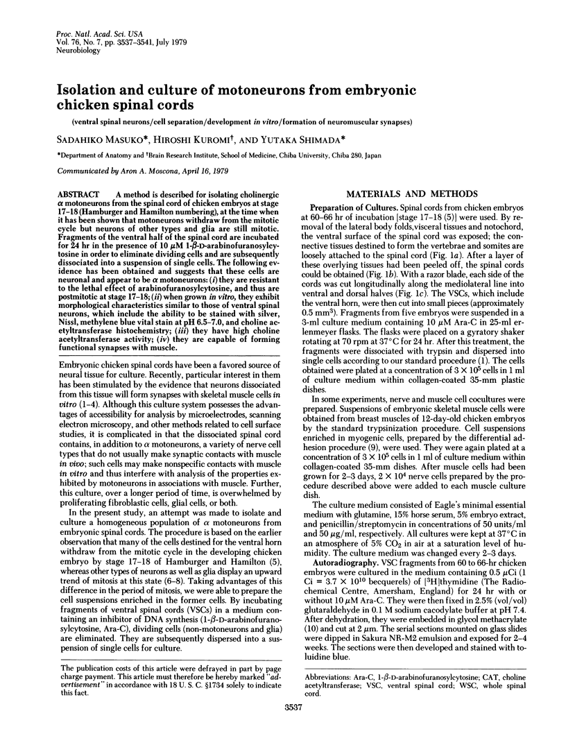
A method is described for isolating cholinergic alpha motoneurons from the spinal cord of chicken embryos at stage 17-18 (Hamburger and Hamilton numbering), at the time when it has been shown that motoneurons withdraw from the mitotic cycle but neurons of other types and glia are still mitotic. Fragments of the ventral half of the spinal cord are incubated for 24 hr in the presence of 10 microM 1-beta-D-arabinofuranosylcytosine in order to eliminate dividing cells and are subsequently dissociated into a suspension of single cells. The following evidence has been obtained and suggests that these cells are neuronal and appear to be alpha motoneurons: (i) they are resistant to the lethal effect of arabinofuranosylcytosine, and thus are postmitotic at stage 17-18; (ii) when grown in vitro, they exhibit morphological characteristics similar to those of ventral spinal neurons, which include the ability to be stained with silver, Nissl, methylene blue vital stain at pH 6.5-7.0, and choline acetyltransferase histochemistry; (iii) they have high choline acetyltransferase activity; (iv) they are capable of forming functional synapses with muscle.
Full text
PDF


Images in this article


Selected References
These references are in PubMed. This may not be the complete list of references from this article.
- Bennett H. S., Wyrick A. D., Lee S. W., McNeil J. H. Science and art in preparing tissues embedded in plastic for light microscopy, with special reference to glycol methacrylate, glass knives and simple stains. Stain Technol. 1976 Mar;51(2):71–97. doi: 10.3109/10520297609116677. [DOI] [PubMed] [Google Scholar]
- Berg D. K., Fischbach G. D. Enrichment of spinal cord cell cultures with motoneurons. J Cell Biol. 1978 Apr;77(1):83–98. doi: 10.1083/jcb.77.1.83. [DOI] [PMC free article] [PubMed] [Google Scholar]
- Burt A. M., Silver A. Histochemistry of choline acetyltransferase: a critical analysis. Brain Res. 1973 Nov 23;62(2):509–516. doi: 10.1016/0006-8993(73)90715-4. [DOI] [PubMed] [Google Scholar]
- FUJITA S. Kinetics of cellular proliferation. Exp Cell Res. 1962 Oct;28:52–60. doi: 10.1016/0014-4827(62)90311-7. [DOI] [PubMed] [Google Scholar]
- Fischbach G. D. Synapse formation between dissociated nerve and muscle cells in low density cell cultures. Dev Biol. 1972 Jun;28(2):407–429. doi: 10.1016/0012-1606(72)90023-1. [DOI] [PubMed] [Google Scholar]
- Fischbach G. D. Synaptic potentials recorded in cell cultures of nerve and muscle. Science. 1970 Sep 25;169(3952):1331–1333. doi: 10.1126/science.169.3952.1331. [DOI] [PubMed] [Google Scholar]
- Fonnum F. A rapid radiochemical method for the determination of choline acetyltransferase. J Neurochem. 1975 Feb;24(2):407–409. doi: 10.1111/j.1471-4159.1975.tb11895.x. [DOI] [PubMed] [Google Scholar]
- Hollyday M., Hamburger V. An autoradiographic study of the formation of the lateral motor column in the chick embryo. Brain Res. 1977 Aug 26;132(2):197–208. doi: 10.1016/0006-8993(77)90416-4. [DOI] [PubMed] [Google Scholar]
- Kano M., Shimada Y. Innervation and acetylcholine sensitivity of skeletal muscle cells differentiated in vitro from chick embryo. J Cell Physiol. 1971 Oct;78(2):233–242. doi: 10.1002/jcp.1040780210. [DOI] [PubMed] [Google Scholar]
- Karon M., Shirakawa S. The locus of action of 1-beta-d-arabinofuranosylcytosine in the cell cycle. Cancer Res. 1969 Mar;29(3):687–696. [PubMed] [Google Scholar]
- Langman J., Guerrant R. L., Freeman B. G. Behavior of neuro-epithelial cells during closure of the neural tube. J Comp Neurol. 1966 Jul;127(3):399–411. doi: 10.1002/cne.901270308. [DOI] [PubMed] [Google Scholar]
- Obata K. Development of neuromuscular transmission in culture with a variety of neurons and in the presence of cholinergic substances and tetrodotoxin. Brain Res. 1977 Jan 1;119(1):141–153. doi: 10.1016/0006-8993(77)90096-8. [DOI] [PubMed] [Google Scholar]
- Richardson K. C. The fine structure of autonomic nerves after vital staining with methylene blue. Anat Rec. 1969 Jul;164(3):359–377. doi: 10.1002/ar.1091640311. [DOI] [PubMed] [Google Scholar]
- SEVIER A. C., MUNGER B. L. TECHNICAL NOTE: A SILVER METHOD FOR PARAFFIN SECTIONS OF NEURAL TISSUE. J Neuropathol Exp Neurol. 1965 Jan;24:130–135. doi: 10.1097/00005072-196501000-00012. [DOI] [PubMed] [Google Scholar]
- Shimada Y., Fischman D. A. Morphological and physiological evidence for the development of functional neuromuscular junctions in vitro. Dev Biol. 1973 Mar;31(1):200–225. doi: 10.1016/0012-1606(73)90332-1. [DOI] [PubMed] [Google Scholar]
- Shimada Y., Fischman D. A., Moscona A. A. Formation of neuromuscular junctions in embryonic cell cultures. Proc Natl Acad Sci U S A. 1969 Mar;62(3):715–721. doi: 10.1073/pnas.62.3.715. [DOI] [PMC free article] [PubMed] [Google Scholar]
- Yaffe D. Retention of differentiation potentialities during prolonged cultivation of myogenic cells. Proc Natl Acad Sci U S A. 1968 Oct;61(2):477–483. doi: 10.1073/pnas.61.2.477. [DOI] [PMC free article] [PubMed] [Google Scholar]