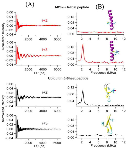
Figure 8.

Three-pulse ESEEM experimental data with a τ=200ns of the i+2 and the i+3 2H-labeled Leu for AchR M2δ helical peptide in lipid bilayer and ubiquitin β-sheet peptide in solution. (A) Time domain, (B) Frequency domain. The inset structural pictures show the location of spin labels and 2H-labeled Leu on AchR M2δ helical peptide and ubiquitin β-sheet peptide. (Adapted from ref. 77 with permission)
