Introduction
A great variety of anomalies of hand extensor musculature have been described over the last century and a half, beginning with Wood (1864) [15–19]. Hand extensor anomalies have been observed in cadaveric cases, during hand surgeries, and through imaging. The continued identification of these anomalies is important in academic studies to examine limb function and characteristics of human development. In addition, reporting limb anomalies is important not only in the clinical diagnosis and treatment of limb dysfunction and injury, but also for awareness in human anatomical and functional variation.
In this case study, we present cadaveric cases of muscular anomalies in the third and fourth extensor compartments of the wrist and anomalous tendons that were observed and identified by students during dissection in a graduate human gross anatomy course. Bilateral anomalies were observed in the extensors of the thumb and index finger in the hands of a 103-year-old female cadaver, and a unilateral anomaly was observed in an 85-year-old female cadaver. Dissection and isolation of the anomalies showed some similarity of the anomalies between the two cadavers, in that the anomalies presented associations between the individual extensors in the third and fourth extensor compartments. Further, the cadavers were received in sequential order through the Body Donation Program at Case Western Reserve University School of Medicine and were investigated for a familial link.
Cadaveric Cases
The cadavers were obtained through the Body Donation Program in the Department of Anatomy at Case Western Reserve University for use in a graduate gross anatomy laboratory course. The cadavers were received in sequential order. Medical, graduate, and undergraduate students participated in the dissections of these cases and the surveys of other cadavers. Once anomalies were identified, the records of these two donations were examined by the program director and determined to have no apparent familial relation.
Case 1
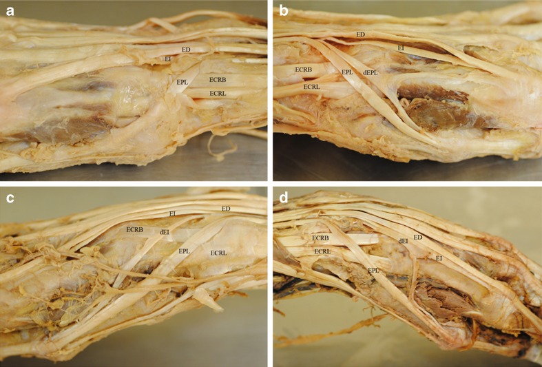
A unilateral anomaly was observed in an 85-year-old female cadaver. Bilateral dissection demonstrated the normal array of tendons of the extensor pollicis longus (EPL), extensor digitorum (ED), and extensor indicis (EI) in the right forearm and hand (Fig. 1a). Dissection of the left forearm and wrist in this cadaver revealed a duplicated tendon and partial muscle belly of the EPL in the third extensor compartment as defined by Rousset et al. (2010) (Fig. 1b) [11]. The partial muscle belly of the duplicated extensor pollicis longus (dEPL) arose from both the normal EPL muscle mass and the interosseous membrane. The tendon of the dEPL bifurcated just superficial to the tendon of the extensor carpi radialis longus, with one tendon branch coursing with the tendon of the normal EI and the other tendon branch coursing with the tendon of the normal EPL. The insertion of the tendon branches coincided with the insertions of the EI and EPL, respectfully. This anomaly is similar to a Type 1e anomaly according to the classification system of Turker et al. (2010), also known as an extensor pollicis et indicis accessories [16]. However, the classification of a dEPL with an anomalous tendon distribution as an extensor pollicis et indicis accessories is insufficient since the extensor pollicis et indicis accessories refers to a tendinous anomaly alone.
Fig. 1.
Case 1 (85-year-old female cadaver): a dorsolateral view of the right forearm and hand showing normal morphology and b dorsolateral view of the left forearm and hand showing a dEPL with a tendon that bifurcates. One tendon branch parallels the tendon of the normal EPL, while the other branch joins the tendon of the EI. Case 2 (103-year-old female cadaver): c dorsolateral view of the right forearm and hand showing a dEI with a tendon that parallels the tendon of the EPL and inserts with the EPL on the distal phalanges of the thumb and d dorsolateral view of the left forearm and hand showing a EI with a bifurcated tendon. One tendon branch courses with the tendon of the normal EI, while the other branch joins the tendon of the EPL. EPL extensor pollicis longus, dEPL duplicated extensor pollicis longus, EI extensor indicis, dEI duplicated extensor indicis, EPB extensor pollicis brevis, ECRB extensor carpi radialis brevis, ECRL extensor carpi radialis longus, ED extensor digitorum
Case 2
Bilateral anomalies were observed in the course of the extensor tendons in a 103-year-old female cadaver. In the right forearm and hand, a duplicated tendon and partial muscle belly of the EI was observed (Fig. 1c). The muscle belly of the duplicated EI arose from the normal EI muscle mass and along the ulna just proximal to the origin of the normal EI. The tendon of the duplicate EI coursed in the fourth extensor compartment, crossed the dorsal thenar surface medial to the tendon of the EPL and inserted with the EPL tendon (Fig. 1c). This anomaly would be classified as Type 1a [16].
In the fourth extensor compartment of the left forearm and hand, a novel anomaly in the extensor musculature was identified. The tendon of a duplicated EI splits into two tendons (Fig. 1d). One branch continued as the tendon of the duplicated EI, while the second less substantial branch formed just deep to the radial artery and coursed across the dorsal surface of the first dorsal interosseous muscle to accompany the tendon of the EPL. The muscle belly of the duplicated EI was similar to that observed in the right upper limb of the same cadaver. The anomaly corresponds, in part, to the Type 2b anomaly [16]; however, the anomaly is a duplicated EI with a branch of the tendon coursing and inserting with the EPL, and has not been described previously.
Discussion
The observation of these anomalies in two cadavers that were received sequentially by the Body Donation Program piqued our interest that the anomalies may represent familial incidence. However, we found no evidence that the individuals were related. Therefore, the presence and similarities in the anomalies between the two cadavers is likely due to chance. The incidence of extensor anomalies in the hand have been reported to be as high as 25 % for the middle finger [8]; however, the incidence of anomalies of the extensor pollicis longus is thought to be much lower [5].
Anomalies of hand and forearm musculature are common and varied. In the case of the EPL, there have been several reports of accessory muscle bellies and supranumerary tendons, as well as the presence of tendons branching from the EI or ED that parallel the course of the EPL tendon [1, 3, 5–7, 9, 12, 14, 16, 19]. However, to our knowledge, this is the first report that describes a bifid tendon of a duplicated EPL with a tendon branch that joins the tendon of the ED or EI. This anomaly, as with other extensor anomalies, may cause hand dysfunction or swelling and pain, such as a third compartment syndrome, and may require significant consideration in hand surgery [2, 10, 13].
The development of these anomalies is likely due to alteration in either the formation of specific muscle masses by myoblasts migrating into the distal limb or by alteration in the muscle patterning dictated by the somatopleure mesoderm [4]. During limb muscle development, coalescing masses of myogenic cells that have moved into the limb from somitic myotomes separate to produce specific muscles in conjunction with epimysium and tendinous development from the surrounding somatopleure mesenchyme. Anomalies may result in aberrant division of the myogenic masses or aberrant deposition of the associated fascial and tendinous components to the skeletal muscle. In the present cases, anomalous division of the extensor digitorum muscle mass into the extensor digitorum, extensor indicis, and extensor pollicis longus muscles may have contributed to the muscle duplications. However, tendon duplications or bifurcations may be a result of aberrant signaling or organization of the mesenchyme rather than the myoblasts.
While muscular anomalies in the upper limb are common, functional differences between individuals with these anomalies and the general population have not been described. This is likely due to the individuals carrying the anomalies being unaware of any difference in limb function from the general population or that the anomaly does not alter function significantly. In most cases, anomalies in muscle or tendon arrangements or attachments do not present a new geometry of muscular function but rather duplicates or enhances the muscle function between two common attachment sites in the limb. For example, a duplicated extensor indicis muscle may increase the force available in extending the index finger; however, the individual carrying the anomaly would have no reference to estimate that their ability to extend this digit is any different from others. Some reports have described third and fourth extensor compartment syndromes in the wrist that contribute to complaints of swelling or pain in the region [6].
A greater concern arises from the impact of these anomalies on surgical procedures in the forearm, wrist, and hand. Surgery to address trauma to the region, to address to manifestations of disease, or to determine reasons for functional impairment may encounter these aberrant tendons during the surgical procedure. If the tendons are cut or damaged, the surgeon will have some difficulty in re-establishing continuity in the tendons, since the functions are unknown at the time. Detection prior to surgery would require imaging with the physician being aware of these types of anomalies. Our observations may help increase the awareness of such anomalies and increase the chances of detection of these anomalies prior to surgery, which may improve surgical outcomes.
Acknowledgments
Conflict of interest
The authors declare that they have no conflict of interest, commercial associations, or intent of financial gain regarding this research.
References
- 1.Abu-Hijleh MF. Extensor pollicis tertius: an additional extensor muscle to the thumb. Plast Reconstr Surg. 1993;92:540–543. doi: 10.1097/00006534-199308000-00023. [DOI] [PubMed] [Google Scholar]
- 2.Beatty JD, Remedios D, McCullough CJ. An accessory extensor tendon of the thumb as a cause of dorsal wrist pain. J Hand Surgery (Br. and Eur.) 2000; 25B:1:110–111 [DOI] [PubMed]
- 3.Chiu DTW. Supernumerary extensor tendon to the thumb: a report on a rare anatomic variation. Plast Reconstr Surg. 1981;68:937–939. doi: 10.1097/00006534-198112000-00017. [DOI] [PubMed] [Google Scholar]
- 4.Christ B, Brand-Saberi B. Limb muscle development. Int J Dev Biol. 2002;46:905–914. [PubMed] [Google Scholar]
- 5.Cohen BE, Haber JL. Supernumerary extensor tendon to the thumb: a case report. Ann Plast Surg. 1996;36:105–107. doi: 10.1097/00000637-199601000-00022. [DOI] [PubMed] [Google Scholar]
- 6.Culver JE. Extensor pollicis and indicis communis tendon: a rare anatomic variation revisited. J Hand Surg (American) 1980;5:548–549. doi: 10.1016/S0363-5023(80)80103-1. [DOI] [PubMed] [Google Scholar]
- 7.De Greef I, De Smet L. Accessory extensor pollicis longus: a case report. Eur J Plast Surg. 2006;28:532–533. doi: 10.1007/s00238-005-0015-0. [DOI] [Google Scholar]
- 8.Klena JC, Riehl JT, Beck JD. Anomalous extensor tendons to the long finger: a cadaveric study of incidence. J Hand Surg (American) 2012;37(5):938–941. doi: 10.1016/j.jhsa.2012.02.014. [DOI] [PubMed] [Google Scholar]
- 9.Komiyama M. Nwe,TM, Toyota N, et al. Variations of the extensor indicis muscle and tendon. J Hand Surgery (Br. and Eur.) 1999;24B:5:575–578 [DOI] [PubMed]
- 10.Masada K, Yasuda M, Takeuchi E, et al. Duplicate extensor tendons of the thumb mimicking rupture of the extensor pollicis longus tendon. Scand J Plast Reconstr Surg Hand Surg. 2003;37(5):318–19. doi: 10.1080/02844310310004668. [DOI] [PubMed] [Google Scholar]
- 11.Rousset P, Vuillemin-Bodaghi V, Laredo J-D, et al. Anatomic variations in the first extensor compartment of the wrist: accuracy of US. Radiology. 2010;257:427–433. doi: 10.1148/radiol.10092265. [DOI] [PubMed] [Google Scholar]
- 12.Sawaizumi T, Nanno M, Ito H. Supernumerary extensor pollicis longus tendon: a case report. J Hand Surg (American) 2003;28A:1014–1017. doi: 10.1016/S0363-5023(03)00382-4. [DOI] [PubMed] [Google Scholar]
- 13.Sookur PA, Am N, Bleakney RR, et al. Accessory muscles: anatomy, symptoms, and radiologic evaluation. RadioGraphics. 2008;28:481–490. doi: 10.1148/rg.282075064. [DOI] [PubMed] [Google Scholar]
- 14.Steichen JB, Petersen DP. Junctura tendium between extensor digitorum communis and extensor pollicis longus. J Hand Surg (American) 1984;9:674–676. doi: 10.1016/S0363-5023(84)80011-8. [DOI] [PubMed] [Google Scholar]
- 15.Tan ST, Smith PJ. Anomalous extensor muscles of the hand: a review. J of Hand Surg. 1999;24:449–455. doi: 10.1053/jhsu.1999.0449. [DOI] [PubMed] [Google Scholar]
- 16.Turker T, Robertson GA, Thirkannad SM. A classification system for anomalies of the extensor pollicis longus. Hand. 2010;5:403–407. doi: 10.1007/s11552-010-9273-9. [DOI] [PMC free article] [PubMed] [Google Scholar]
- 17.Wood J. On some varieties in human myology. Proc R Soc Lond. 1864;13:299–303. [Google Scholar]
- 18.Wood J. Variations in human myology observed during the winter session of 1865-66 at King’s College, London. Proc R Soc Lond. 1867;15:229–244. [Google Scholar]
- 19.Wood J. Variations in human myology observed during the winter session of 1867-68 at King’s College, London. Proc R Soc Lond. 1868;16:483–525. [Google Scholar]


