Abstract
Cell biologists everywhere rejoiced when this year’s Nobel Prize in Physiology or Medicine was awarded to James Rothman, Randy Schekman, and Thomas Südhof for their contributions to uncovering the mechanisms governing vesicular transport. In this article, we highlight their achievements and also pay tribute to the pioneering scientists before them who set the stage for their remarkable discoveries.
In 1974, nearly 40 years ago, the Nobel Prize in Physiology or Medicine was awarded to George E. Palade, Albert Claude, and Christian de Duve for work that effectively established a new field, cell biology. Collectively, the efforts of these three pioneers not only defined the essential features of cells but also how to study them. Correlating morphological observations by electron microscopy with biochemical analysis enabled not only the identification of nearly every major organelle in the eukaryotic cell (although endosomes were missed at that time) but also what their respective functions were. Palade’s efforts demonstrated the now-canonical pathway of protein secretion: synthesis in the endoplasmic reticulum (ER), oligosaccharide processing in the Golgi complex, concentration in secretory granules, and release at the plasma membrane. Palade understood implicitly that the ER, Golgi, secretory granules, and plasma membrane had to be interconnected by a series of vesicular carriers that carried cargo from one station to the next—dissociative transport. He also appreciated that the process had to be regulated if compartment specificity was to be maintained. The need for specificity defined the next major conceptual challenges: how do proteins intended for secretion traverse the compartments of the secretory pathway, how are transport vesicles formed, how do vesicles recognize their appropriate destinations, how does fusion occur after the appropriate destination is reached, and, finally, how are the components from the originating compartment returned or recycled to their sites of origin after fusion with the destination compartment? Palade may have framed these problems, but it was left to the next generation of cell biologists to solve them.
This year’s Nobel Prize in Physiology or Medicine awarded to James Rothman, Randy Schekman, and Thomas Südof recognizes a truly remarkable body of work that provides superb conceptual clarity and mechanistic insight into virtually all of the issues defined by Palade and colleagues. To a large extent, the award also provides a satisfying degree of recognition to the large community of scientists who established the field of “molecular” cell biology. But it was the intellectual leadership, passion, and courage provided by this year’s awardees (Figs. 1 and 2) that played a major role in driving the spectacular advances of the past three decades. Particularly in the case of Rothman and Schekman, the scientific dynamic they helped to generate gave the field focus and excitement, from which came great things. The elegance of their experiments together with the exceptionally clear and simple logic that they presented in their papers moved the field ahead quickly and drew many new converts into membrane trafficking.
Figure 1.

Randy Schekman and James Rothman (center) with many of their former trainees at the American Society for Biochemistry and Molecular Biology meeting on “Biochemistry of Membrane Traffic: Secretory and Endocytic Pathways,” October 2010.
PHOTOGRAPH COURTESY OF THE AMERICAN SOCIETY FOR BIOCHEMISTRY AND MOLECULAR BIOLOGY
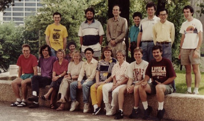
Figure 2.
Thomas Südhof (top row, center) and his laboratory circa 1993.
PHOTOGRAPH COURTESY OF THOMAS SÜDHOF
The first foray into a mechanistic, molecular approach to the cell biological problems defined by Palade was really due to the work of Günter Blobel and his colleagues Peter Walter and Bernhard Dobberstein working at The Rockefeller University. These investigators devised a complex but elegant approach enabling the cell-free reconstitution of the first step of secretion, namely the insertion of newly synthesized proteins into and across the ER membrane. Combined with conventional cold-room biochemistry, Blobel and others were able to provide a detailed understanding of the biochemistry of protein translocation. Blobel was duly awarded the Nobel Prize in Physiology or Medicine for his work in 1999. Influenced by Blobel and also Arthur Kornberg, then chair of the Biochemistry Department at Stanford, Jim Rothman (who was a young faculty member at Stanford in the early 1980s) initiated his courageous effort aimed to reconstitute subsequent steps, namely the transport of secretory and membrane protein cargo to and through the Golgi complex. As is often the case with innovative work that pushes the limits of knowledge, Rothman’s interpretations were on occasion controversial, but there was absolutely no controversy regarding the importance of the various components he and his team identified. These components included soluble factors needed for vesicle formation in the Golgi as well as for vesicle fusion, most notably the COPI coat protein complex, NSF (NEM-sensitive factor) and SNAP (soluble NSF attachment protein). With Richard Scheller, Rothman recognized that the synaptic vesicle–associated proteins cloned and purified by Scheller represented both the docking sites for NSF and SNAP and a key component of the mechanism whereby vesicles recognized and even fused with each other. Indeed, the SNAREs (as these proteins are now called) clearly comprise the core fusion machinery that underlies virtually all membrane fusion events in the cell. SNAREs form a family of proteins that are organelle specific, helping to ensure the specificity of membrane traffic as well as the biochemical and functional identity of individual membrane compartments.
If Rothman’s work began as a quintessential biochemical approach, Randy Schekman’s started at the other end of the spectrum: genetics. Again with great courage, Schekman decided to use the yeast Saccharomyces cerevisiae as a genetically tractable eukaryote to dissect the steps and various components associated with the secretory pathway. At the time, few thought that yeast cells were capable of higher-order processes such as secretion or that their activities had anything to do with mechanisms in animal cells. Yet Schekman and his then graduate student Peter Novick designed a deceptively simple screen to identify secretory (or “sec”) mutants. Their approach was to look for cells that could not secrete by reasoning that continued synthesis of secretory cargo would render the mutant cells more dense. The approach worked, and literally dozens of mutants were discovered, a large number of which could be shown to generate intriguing phenotypes and to control key steps in the secretory, or sometimes even the endocytic, pathway. Although the original sec screens done by Schekman and colleagues did not immediately turn up the SNARE proteins, they did reveal the presence of small Ras-related monomeric GTPases of the Rab family that helped enforce the specificity of vesicle interactions. They also uncovered cytoplasmic coat proteins (COPII) and complex cytosolic “tethers” that serve to gather vesicles at their targets before the final fusion step. When an increasing number of sec mutants began to overlap with components identified by Rothman’s independent biochemical purifications of components required for fusion or vesicle budding, it was clear that both groups (and indeed the field) were on the right track and the transport machinery was universal. Through whatever controversies bubbled up over the years, this basic fact remained unchallenged. Schekman too moved toward the same type of functional biochemical analysis championed by Rothman, and the circle was completed.
Focused on one of the key problems in neurobiology, Thomas Südhof’s efforts may appear less general but are no less important. The synapse represented a special case in the area of membrane traffic since the realization that neuronal transmission reflected the release of quanta of neurotransmitters due to the action potential–triggered fusion of synaptic vesicles with the presynaptic plasma membrane. The work of Cesare Montecucco and colleagues on bacterial toxins provided an important insight, namely that synaptic vesicle release can be blocked by certain bacterial toxins (e.g., botulinum toxin) that act as specific SNARE proteases. Scheller and Rothman had shown that the SNAREs comprised the basic unit of the fusion machinery, but this insight alone did not explain how secretion in the synapse was coupled so tightly to electrical activity. Thomas Südhof’s remarkable body of work, although not growing out of the molecular cell biology community as much as the neuroscience community, provided the conceptual answer: the synaptotagmins. These proteins were found to associate with SNAREs and serve as Ca2+-sensing triggers that temporally linked synaptic vesicle transmission to individual neuronal impulses. In addition, Südhof and colleagues discovered Munc18 in the mouse, which corresponds to the yeast Sec1 protein, and demonstrated that it interacts with the SNARE complex, revealing that Munc18 as well as other members of the Sec1/Munc18-like protein family function as part of the vesicle fusion machinery.
Collectively, these are remarkable achievements that provide conceptual and mechanistic understanding of basic cellular processes at the most fundamental level. It is certainly the case that others, for example Scheller and Novick mentioned here, might just as easily have been included in this award. Regrettably only three are permitted, and there can be no doubt but that the three selected are entirely deserving given not only the nature of their findings but also the scientific leadership they contributed in a myriad of intangible ways to the incredible progress we have witnessed in the post-Palade era of cell biology.
We congratulate our colleagues and friends Jim, Randy, and Thomas for this well-deserved honor. Mazal tov!
Selected publications of interest
- Balch W.E., Dunphy W.G., Braell W.A., Rothman J.E. 1984. Reconstitution of the transport of protein between successive compartments of the Golgi measured by the coupled incorporation of N-acetylglucosamine. Cell. 39:405–416 10.1016/0092-8674(84)90019-9 [DOI] [PubMed] [Google Scholar]
- Hata Y., Slaughter C.A., Südhof T.C. 1993. Synaptic vesicle fusion complex contains unc-18 homologue bound to syntaxin. Nature. 366:347–351 10.1038/366347a0 [DOI] [PubMed] [Google Scholar]
- Kaiser C.A., Schekman R. 1990. Distinct sets of SEC genes govern transport vesicle formation and fusion early in the secretory pathway. Cell. 61:723–733 10.1016/0092-8674(90)90483-U [DOI] [PubMed] [Google Scholar]
- Novick P., Schekman R. 1979. Secretion and cell-surface growth are blocked in a temperature-sensitive mutant of Saccharomyces cerevisiae. Proc. Natl. Acad. Sci. USA. 76:1858–1862 10.1073/pnas.76.4.1858 [DOI] [PMC free article] [PubMed] [Google Scholar]
- Perin M.S., Fried V.A., Mignery G.A., Jahn R., Südhof T.C. 1990. Phospholipid binding by a synaptic vesicle protein homologous to the regulatory region of protein kinase C. Nature. 345:260–263 10.1038/345260a0 [DOI] [PubMed] [Google Scholar]
- Söllner T., Bennett M.K., Whiteheart S.W., Scheller R.H., Rothman J.E. 1993a. A protein assembly-disassembly pathway in vitro that may correspond to sequential steps of synaptic vesicle docking, activation, and fusion. Cell. 75:409–418 10.1016/0092-8674(93)90376-2 [DOI] [PubMed] [Google Scholar]
- Söllner T., Whiteheart S.W., Brunner M., Erdjument-Bromage H., Geromanos S., Tempst P., Rothman J.E. 1993b. SNAP receptors implicated in vesicle targeting and fusion. Nature. 362:318–324 10.1038/362318a0 [DOI] [PubMed] [Google Scholar]


