Abstract
The protein MENIN is the product of the MEN1 gene. Altered MENIN expression is one of the few events that are clearly associated with foregut neuroendocrine tumours (NETs), classical oncogenes or tumor suppressors being not involved. One of the current challenges is to understand how alteration of MENIN expression contributes to the development of these tumours. We hypothesised that MENIN might regulate factors maintaining endocrine-differentiated functions. We chose the insulinoma model, a paradigmatic example of well-differentiated pancreatic NETs, to study whether MENIN interferes with expression of MAFA, a master glucose-dependent transcription factor in differentiated β cells. Immunohistochemical analysis of a series of human insulinomas revealed a correlated decrease of both MENIN and MAFA. Decreased MAFA expression resulting from targeted Men1 ablation was also consistently observed in mouse insulinomas. In vitro analyses using insulinoma cell lines showed that MENIN regulated MAFA protein and mRNA levels, and bound to MafA promoter sequences. MENIN knockdown concomitantly decreased mRNA expression of both MafA and β-cell differentiation markers (Ins1/2, Gck, Slc2a2 and Pdx-1), and in parallel, increased the proliferation rate of tumours as measured by bromodeoxyuridine incorporation. Interestingly, MAFA knockdown alone also increased proliferation rate but did not affect the expression of candidate proliferation genes regulated by MENIN. Finally, MENIN variants with missense mutations detected in patients with MEN1 lost the wild-type MENIN properties to regulate MAFA. Together, our findings unveil a previously unsuspected MENIN/MAFA connection regarding control of the β-cell differentiation/proliferation balance, which could contribute to tumorigenesis.
Keywords: MENIN, MEN1, insulinoma, MAFA, endocrine tumour, differentiation
Introduction
A rise in the incidence and prevalence of neuroendocrine tumours (NETs) has been observed in recent decades, but there has been no improvement in patient survival, mainly owing to a lack of clarity regarding the underlying molecular mechanisms (Modlin et al. 2008; Rindi and Wiedenmann 2012). The MEN1 tumour suppressor gene is one of the few genes for which there is clear evidence of involvement in the pathogenesis of foregut NETs. Alterations of MEN1 are present in patients with multiple endocrine neoplasia type I (MEN1) syndrome (Agarwal et al. 1997; Wautot et al. 2002) characterised by an increased incidence of endocrine tumours in several target tissues (mainly the foregut, the pituitary, parathyroid and thymus glands), as well as in a significant proportion of sporadic pancreatic neuroendocrine tumours (PETs) including insulinomas (Corbo et al. 2010; Jiao et al. 2011).
Insulinomas are tumours characterised by the maintenance of a high level of differentiation (including the capacity to secrete amounts of insulin sufficient to induce functional symptoms), a low proliferation rate and a usually benign behaviour; nevertheless, some cases behave as malignant tumours with metastatic dissemination (de Herder et al. 2006; Metz and Jensen 2008; Shin et al. 2010). In mouse models, targeted Men1 ablation in pancreatic β cells is consistently associated with insulinoma development (Bertolino et al. 2003; Crabtree et al. 2003). A current major challenge in insulinoma and NETs research is to understand how the loss of appropriate expression of the Men1 gene product, MENIN, contributes to the development of these endocrine tumours (Gracanin et al. 2009). MENIN is a protein that is ubiquitously expressed even in non-endocrine tissues. It is found mainly in the nucleus and can function either as a tumour suppressor in several types of endocrine cells or as a tumour promoter in other tissue types (Gracanin et al. 2009). MENIN is translated from several transcripts of about 2.8 kb in size and is initiated from a large number of transcription start sites (Fromaget et al. 2003) Multiple MENIN-interacting proteins have been described, suggesting that the protein has a wide spectrum of activities (Yang and Hua 2007). Regulation of endocrine-specific determinants remains to be clarified.
Accumulating evidence suggests that MENIN mediates chromatin interactions, especially between transcription factors (TFs) and their targets and is involved, either directly or indirectly, in the fine tuning of the balance between proliferation/apoptosis and differentiation (Yang and Hua 2007). It has been shown in several cellular contexts that decreased MENIN expression is associated with increased proliferation and modified apoptotic sensitivity (Bazzi et al. 2008; Hussein et al. 2007; Schnepp et al. 2004; Wu and Hua 2011); however, the consequences of MENIN alterations on endocrine cell differentiation, the mechanisms of such alterations and their contributions to NET tumorigenesis have not yet been explored. Insulinoma offers an attractive model to study these interactions. In contrast to other NETs there are several animal and cellular models of insulinoma currently available that provide opportunities for exploring the role of MENIN in the control of differentiation processes. In addition, the mechanisms of β-cell differentiation and of the TFs involved are intensively studied. MENIN has been implicated in the control of adaptive β-cell proliferation, both in normal physiological contexts such as during pregnancy, and in pathological conditions such as type 1 diabetes, but the underlying mechanisms remain unknown (Karnik et al. 2007; Yang et al. 2010). The molecular mechanisms responsible for MENIN loss of function and the resulting irreversible proliferation/survival leading to tumour development also remain to be elucidated.
MAFA (v-MAF musculoaponeurotic fibrosarcoma oncogene homolog A), is a protein belonging to the large-MAF TF family, whose members have been shown to function as oncogenes or tumour suppressors in different tissues but not in β-cells (Pouponnot et al. 2006). In addition, MAFA is a master glucose-regulated TF that contributes to the maintenance of β-cell differentiation and controls, either directly or indirectly, the expression of target genes implicated in β-cell functions, including Ins-1/2 (encoding insulin), Slc2a2 (encoding the glucose transporter GLUT2), Gck (encoding glucokinase) and Pdx-1 (Aramata et al. 2007; Yang and Cvekl 2007). The level of MAFA in β cells is tightly controlled mainly by glucose, the major growth and differentiation regulator of β cells. Short-term glucose stimulation (up to 24 h) induces a transient increase in MAFA expression and exerts coordinate stimulatory effects both on hormone synthesis and cell proliferation (Liu et al. 2009), whereas prolonged glucose exposure (>24 h) decreases MAFA expression and is deleterious to β-cell function and survival (Martens and Pipeleers 2009). MAFA is thus considered a barometer of β-cell health (Hang and Stein 2011).
In this study, we investigated whether MENIN can regulate the expression of MAFA, a major TF that maintains β-cell differentiation (Aramata et al. 2007). We hypothesised that decreased MENIN expression or impaired MENIN function might result in altered MAFA expression and function, along with disruption of the differentiation/proliferation balance. In this study, we used both in vivo and in vitro insulinoma models to determine whether MENIN could regulate MAFA expression. We also evaluated the functional consequences of MENIN mutations present in patients with MEN1 after MAFA regulation. Our data show for the first time, that MENIN interferes with the β-cell-specific MAFA pathway, thus opening a new field of investigation into the molecular mechanisms involved in pancreatic endocrine tumorigenesis.
Materials and methods
Human tissues
A series of samples from human insulin-expressing PETs were obtained from a tissue biobank (Tumorothèque des Hospices Civils de Lyon, supported by the Institut National du Cancer and the French Ministry of Health), which adheres to French ethical rules and regulations. The tissue in this bank is either from patients who have given their informed consent for their tissue to be used or is from samples that qualify for research use according to French regulations. The research project was approved by the biobank steering committee.
All tissue samples (n = 15) used in this study were obtained from surgical resections. The patients from whom the tissues were obtained (2 males and 13 females) had a mean age of 47.5 years (range 25 to 79 years). Of these 15 tumours, 10 were functional at the time of removal and could be termed insulinomas, whereas the remaining 5 were non-functional tumours, despite insulin expression being detected in most or all tumour cells by immunohistochemistry (IHC). According to the 2010 World Health Organization (WHO) classification, the 10 functional tumours were classified as neuroendocrine neoplasms G1, and the other 5 were neuroendocrine neoplasms G2. One tumour was associated with MEN1 syndrome, while the others were sporadic. Three patients had distant metastases.
Murine tissues
Pancreatic islets and insulinomas were isolated from 12-month-old Men1F/F-RipCre− and Men1F/F-RipCre+ mice, respectively, by hand-picking the tissues from dissociated pancreases on dark-field dishes under a dissecting microscope, as previously described (Fontaniere, et al. 2006).
IHC analysis
For the IHC study, formalin-fixed, paraffin wax-embedded tissue was cut into 4-μm-thick sections. Whole sections, containing both tumour and adjacent peritumoral tissues, were used. Tissue sections were incubated with antibodies to MENIN (Epitomics, Burlingame, CA, USA) and to MAFA (Bethyl Laboratory Inc., Montgomery, TX, USA) at appropriate dilutions (1:50 and 1:250, respectively), and an indirect streptavidin-biotin/peroxidase technique was used to visualise the labelling. The apparent expression levels of MENIN and MAFA in tumour cells were assessed by comparison with internal controls in the same section, i.e. normal endocrine cells of Langerhans islets for MAFA and normal pancreatic epithelial cells for MENIN. If the apparent expression level in all or the majority of tumour cells was equal to or higher than that observed in normal internal controls, it was considered “high” expression, while if it was lower than that observed in normal internal controls, it was considered “low” expression. If the expression level was comparable in different tumour cells, the tumour was considered homogeneous, whereas if the expression level varied among different tumour cells, the tumour was considered heterogeneous, and the expression level reported was that of the majority of the tumour cells.
Cell lines
The two insulinoma cell lines used were INS-1E (Merglen et al. 2004) and INS-rβ (Wang and Iynedjian 1997). These cell lines were originally independently established from radio-induced rat insulinoma INS-1 cells (Asfari et al. 1992) and are glucose-responsive (Merglen et al. 2004). We also used MafA47 cells, which were previously established from INS-rβ cells; they can conditionally overexpress MAFA protein under doxycyclin treatment (Wang et al. 2007). All three cell lines used express wild-type (WT) MENIN protein.
Cells were maintained in a standard culture medium as previously described (Asfari et al. 1992) but that contained only 5 mM glucose, which allows a greater sensitivity to physiological glucose stimulation (11 mM). INS-rβ cells that conditionally overexpress WT MENIN or MEN1-related variants have been previously described (Bazzi et al. 2008). For MENIN induction of INS-rβ cells, 1000 ng ml−1 doxycyclin (Sigma-Aldrich, St. Louis, MO, USA) was added to the culture medium at the time of seeding. To determine the half-life of the MENIN protein and mRNA, cells were treated with 20 μg ml−1 cycloheximide (CHX, Sigma-Aldrich) or 4 μg ml−1 actinomycin D (ActD, Sigma-Aldrich) for various periods of time.
Immunoblot analysis
Cells were lysed in RIPA buffer (Santa Cruz Biotechnology, Santa Cruz, CA, USA), and proteins were resolved on 10% pre-cast Criterion XT polyacrylamide gels (Bio-Rad Laboratories, Hercules, CA, USA). The proteins were transferred to polyvinylidene difluoride (PVDF) membranes (Millipore, Billerica, MA, USA) which were first treated with blocking solution (Tris-buffered saline with 0.1% Tween 20 and 5% milk), then incubated with a primary antibody directed against the C-terminal region of the MENIN protein (A300-105A, Bethyl Laboratory Inc.), the C-terminal region of the MAFA protein (A300-611A, Bethyl Laboratory Inc.) or the loading control tubulin (Sigma-Aldrich). A species-matched horseradish peroxidase-labelled secondary antibody (Jackson Laboratory, Bar Harbor, ME, USA) was used to visualise the antibody-antigen complexes by chemiluminescence, using an ImmunStar kit (Bio-Rad).
Gene expression knockdown by RNA interference
Cells were seeded in standard culture medium to ensure about 50% confluence and then transfected with a final concentration of 100 nM small interfering RNAs (siRNAs) using a mixture of four siRNAs provided as a single reagent with high specificity and low off-target effects (ON-TARGET plus SMART pool siRNAs # L-090784-02 and L-081264-02 for Men1 and MafA genes, respectively; Dharmacon, Lafayette, CO, USA and Thermo Fisher Scientific Inc., Waltham, MA, USA). The non-targeted (siCtrl), anti-Men1 or anti-MAFA siRNA pools were incorporated into cells using the DharmaFECT4 transfection reagent in accordance with the manufacturer’s instructions. At 48 h following transfection, the cells were starved in media containing 0.1 mM glucose and no pyruvate. This medium was then replaced by the stimulation medium containing 5 or 15 mM glucose and no pyruvate. The cells were harvested at the indicated time points and stored at −80°C until protein or RNA extraction was performed.
Plasmid transfection/luciferase assays
The MAFA promoter-luciferase (LUC) (Raum et al. 2006) and the cytomegalovirus-driven WT MEN1 (Bazzi et al. 2008) constructs have been described previously. Plasmids were co-transfected using Lipofectamine 2000 reagent (Invitrogen, Carlsbad, CA, USA), while the DharmafectDuo reagent (Dharmacon) was used to co-introduce plasmids and siRNAs. The cells were harvested at 48 h after transfection. A co-transfected Renilla (REN) reporter plasmid served as an internal transfection efficiency control. The cells were lysed in the provided PLB buffer, and LUC/REN activity was measured using the Dual-Luciferase Reporter Kit (Promega, Madison, WI, USA).
Chromatin immunoprecipitation assays
Chromatin immunoprecipitation (ChIP) was performed using the enzymatic ChIP-IT® Express kit (Active Motif, La Hulpe, Belgium). Protein-DNA crosslinking was performed using formaldehyde (Thermo Scientific Pierce) and chromatin was sheared by enzymatic digestion following the manufacturer’s instructions. Immunoprecipitations were performed using the same anti-MENIN and anti-MAFA antibodies described above, as well as anti-PDX-1 antibody (sc-14664, Santa Cruz Biotechnology). Anti-PolII antibodies were included in the kit. The recovered chromatin was subjected to polymerase chain reaction (PCR) analysis, and the PCR products were separated by gel electrophoresis in an agarose gel. DNA fragments were visualised with a Chemidoc instrument (Bio-Rad).
RNA extraction/reverse transcription-quantitative PCR analysis
A reverse transcription-quantitative PCR (RT-qPCR) protocol was developed to meet the Minimum Information for Publication of Quantitative Real-Time PCR Experiments (MIQE) criteria (Bustin et al. 2009). Total RNA from frozen cell line samples and frozen tissues was extracted using the TRIzol Plus RNA Purification kit (Invitrogen). Reverse transcription of 1 μg each RNA was performed using the QuantiTect® Reverse Transcription kit (Qiagen SA, Venlo, The Netherlands) which includes elimination of genomic DNA contamination in RNA samples. The qPCR analyses were performed using the Maxima SYBR® Green/ROX qPCR Master Mix (Stratagene; Agilent Technologies, Santa Clara, CA, USA) on a CFX96 qPCR instrument (Bio-Rad). Data were analysed using CFX Manager software and the ΔΔC(t) method (Livak and Schmittgen 2001). For normalisation, five housekeeping genes were tested, and at least two of the genes with the lowest M-values (<0.150) were chosen as reference: β-actin and Rplp0 for rat cell lines; β-ACTIN, TFCP2 and GAPDH for human tumours and β-actin and Hprt for mouse tumours. Primers for Ins1/2 and Gck were purchased from Qiagen; the other primer sequences are available on request.
Statistical analysis
Mann-Whitney U tests were used for statistical analysis, and P<0.1 was considered significant. Statistical analyses were performed on at least four independently generated datasets.
Results
MENIN and MAFA expression levels correlate in human PETs
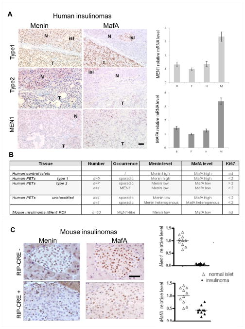
MENIN and MAFA are co-expressed in native β cells. In this study, we evaluated the expression of these two proteins by IHC in 15 insulin-expressing PETs (14 sporadic cases and 1 MEN1-related). Different MENIN expression patterns were observed in the 14 sporadic cases. In 5 of these cases, MENIN was detectable in all tumour cells, with a high expression level similar to that detected in non-tumoral tissue; in parallel, MAFA was detected in all tumour cells with the same apparent high expression level as in adjacent normal Langerhans islets (Fig. 1A, Type 1). In seven additional sporadic cases, the majority of tumour cells displayed low but detectable MENIN expression levels compared with the non-tumoral tissue, and the MAFA expression level was also low (Fig. 1A, Type 2). In the final two sporadic cases, MENIN was low or heterogeneously expressed in tumour cells, and MAFA expression was heterogeneous in one case and high in the other (Fig. 1B, “unclassified”). In the MEN-1 related case, MENIN was undetectable, whereas MAFA was detectable in a minority of tumour cells, with low and heterogeneous intensity (Fig. 1A, MEN1).
Figure 1. Analysis of MENIN and MAFA expression in PETs by IHC and RT-qPCR.
(A) Left: Representative data on human insulin-expressing PETs. Tumour cells (T), adjacent non-tumoral tissue (N) and normal Langerhans islets (isl) are indicated. Scale bar, 50 μm. Right: RT-qPCR analysis of MEN1 and MAFA mRNA levels in a series of four human insulinomas (B, F, H and M). (B) Table with recapitulative data on tumours. (C) Representative data on mouse insulinomas. Left: Normal islets are from a control Men1F/FRipCre− mouse and an insulinoma from a 12-month-old mutant Men1F/F-RipCre+ mouse. Scale bars, 50 μm. Right: RT-qPCR analysis of Men1 and MafA mRNA levels in age-matched control islets from Men1F/F-RipCre− mice (△, n = 10) and insulinomas developed in Men1F/F-RipCre+ mice (▲, n = 10). All experiments were performed in triplicate. The bars represent the median.
There was no correlation between the functional status of the tumour and MENIN/MAFA expression; the proportion of cases with abnormal MENIN/MAFA expression was comparable in both functioning and non-functioning tumours (7/10 and 3/5, respectively). Interestingly, the three malignant cases were all associated with low MENIN/MAFA expression. In addition, the Ki67 proliferation index was <2% in all cases with normal MENIN/MAFA expression but >2% in 8 of the 10 cases with abnormal MENIN/MAFA expression. RT-qPCR analysis on a series of four human insulinoma confirmed a strong correlation between the MENIN and MAFA mRNA levels (Fig. 1A, right panel).
MAFA expression is reduced in mouse insulinoma
We next determined if MAFA expression was also compromised after Men1 inactivation in islet β cells in mice. Specific Men1 disruption mediated by Rat insulin promoter (RIP)-driven Cre in floxed Men1 mice led to full-penetrance insulinoma development at about 10 to 12 months, as we described previously (Bertolino et al. 2003). All tested mouse Men1 insulinomas (n=10) displayed marked reduction in MAFA staining compared with normal islet β cells. This result was confirmed by RT-PCR analysis of MAFA mRNA levels, with >50% reduction found in islets from the mutant mice (Fig. 1C).
MENIN level affects MAFA protein expression following acute glucose stimulation
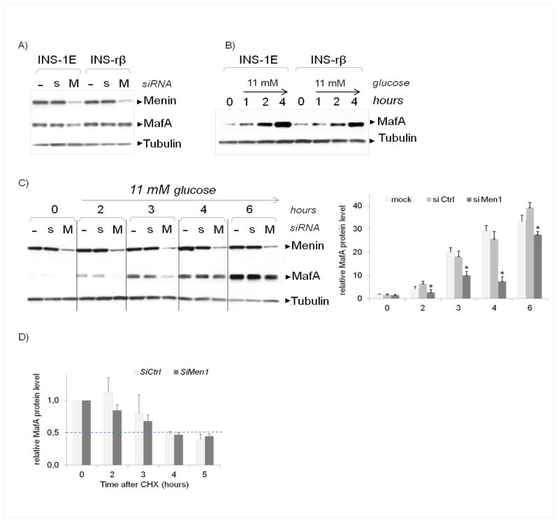
We next analysed the effects of MENIN downregulation on MAFA protein expression in two insulinoma cell lines, INS-1E and INS-rβ. We found that siRNA-mediated inhibition of MENIN induced a slight decrease in MAFA levels detectable by immunoblotting (IB) in INS-1E, but this protein decrease was not always reproducible in basal culture conditions, as shown in Fig. 2A in INS-rβ cells.
Figure 2. Effect of MENIN inhibition on MAFA protein levels and stability in cell lines.
(A) IB of MAFA expression in basal culture conditions: INS-1E and INS–rβ cells were transfected with either non-targeted (s) or anti-Men1 (M) siRNA pools or with transfection agent only (−), then maintained in 5 mM glucose medium and harvested 48 h after transfection. Each analysis was performed with 30 μg of total protein. Each membrane was cut in half at the 60-kDa ladder band; the upper section was used to detect MENIN, and the lower section was used to detect MAFA (between 45 and 55 kDa) and the loading control tubulin (around 55 kDa). The image is representative of at least four experiments. (B) IB of MAFA following acute glucose stimulation. Two days after seeding, the cells were starved overnight in 0.1 mM glucose and then stimulated with 11 mM glucose medium before being harvested at several time points. As mentioned in Fig. 2A, 30 μg total protein was analysed Lane 0: after overnight 0.1 mM glucose starvation before glucose stimulation. C) Left: IB of MAFA following MENIN inhibition and acute glucose stimulation. Two days after seeding, cells were transfected as described for Fig. 2A; after 36 h, they were starved overnight in 0.1 mM glucose and then stimulated in 11 mM glucose medium as described for Fig. 2B, before being harvested and analysed by IB as described above. The image is representative of at least four experiments. Right: Quantitative analysis was performed using Bio-Rad Quantity One software (version 4.6.7). The results represent the relative MAFA signal normalised to the tubulin signal at each time point. *p<0.05. (D) Effect of MENIN inhibition on MAFA protein stability. INS-1E cells were transfected as described above. After 48 h, cells were treated with CHX and harvested at the indicated time points before quantitative analysis of the IB was performed as described above. The results represent the MAFA signal normalised to the tubulin signal at each time point. To better visualise any differences or similarities, we arbitrarily set 1 as the value for MAFA mRNA level at T0 both in siCtrl and siMen1 conditions. The dotted lines represent half of the protein quantity. The experiments were repeated in triplicate.
We therefore tested the effect of MENIN inhibition on MAFA expression during acute glucose stimulation. In INS-1E, and INS-rβ cells, acute stimulation with 11 mM glucose medium resulted in rapid and robust accumulation of MAFA protein, with maximum protein levels reached after 4 h (Fig. 2B). A reduction in MENIN level greatly reduced MAFA accumulation, whereas MAFB protein level increased in parallel, both in INS-1E and in INS-rβ cells (Fig. 2C, D; Supplementary Fig. S1).
To determine if MENIN knockdown might be accelerating MAFA degradation, nascent protein synthesis was blocked by CHX treatment in control and MENIN siRNA-treated INS cells. The half-life of the MAFA protein was about 4 h in INS-1E (Fig. 2D) and INS-rβ cells (Supplementary Fig. S1B) and was unchanged after MENIN down-regulation.
MENIN inhibition alters MAFA transcription
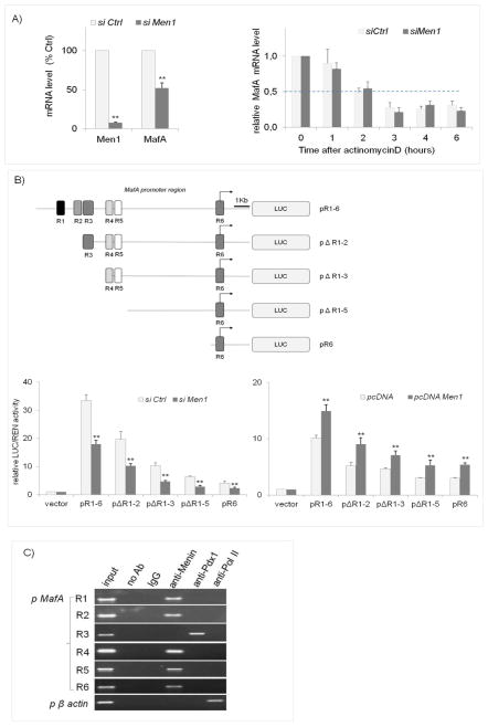
We then tested whether down-regulation of expression of MENIN might alter MAFA mRNA levels. Transfection of INS-1E cells with anti-Men1 siRNA was found to reduce both endogenous Men1 and MafA mRNA levels (by about 92% and 50%, respectively) under basal culture conditions with 5 mM glucose (Fig. 3A, left) and also during glucose stimulation (Supplementary Fig. S2A). Comparable results were obtained using INS-rβ cells (Supplementary Fig. S2B). To determine if MENIN levels were affecting MafA mRNA stability, RNA synthesis was blocked by actinomycin D treatment in control and anti-Men1 siRNA-transfected INS-1E cells. The half-life (i.e. about 2 hours) of MafA mRNA was unaffected by MENIN knockdown in either INS-1E cells (Fig. 3A, right) or INS-rβ cells (Supplementary Fig. S2C).
Figure 3. Effect of MENIN on MAFA gene transcription under basal culture conditions.
(A) Left panel: INS-1E cells were transfected with either non-targeted (siCtrl) or anti-Men1 siRNA pools as described in Figure 2A. After 48 h, total RNA was extracted. Gene expression was measured by RT-qPCR. The relative quantity of mRNA in INS-1E cells is indicated as a percentage. **Statistical significance was defined as p<0.05. Right panel: The effect on MAFA mRNA stability. INS-1E cells were transfected as described above. After 48 h, the cells were treated with actinomycin D and harvested at the indicated time points. The relative levels of MAFA mRNA were measured by RT-qPCR as described above. The time point T0 was arbitrarily set to 1, independently of each condition. (B) Top: Schematic representation of the MAFA promoter regions inserted upstream of a luciferase-coding sequence (LUC) as previously described (Raum et al. 2006). Bottom: The effect of MENIN on MAFA promoter activity. INS-1E cells were co-transfected with the plasmids described above and with either anti-Men1 siRNA pool (left panel) or the WT MENIN expression vector (right panel). LUC and REN activity were measured in the cell lysate 48 h after transfection. The relative LUC/REN activity shown is the result of three independent experiments. **Statistical significance was defined as p<0.05. (C) MENIN binding to the MAFA promoter region. ChIP was performed from INS-1E cells using anti-MENIN, anti-Pdx-1 and anti-PolII antibodies. The recovered chromatin was subjected to PCR analysis using primers directed against the R1 to R6 MAFA promoter regions to identify the bound promoter fragments. The image is representative of at least three experiments. Anti-polII antibodies and β-actin primers were provided in the ActiveMotif kit to serve as a positive control for ChIP experiments.
To analyse if MENIN directly modulates MafA transcription, we evaluated the effect of altering MENIN expression level on the activity of various 5′-flanking MafA promoter-driven luciferase reporter genes. The MafA promoter fragments examined spanned the six conserved regions controlling islet β-cell-specific expression (termed R to R6 (Raum et al. 2006)). MENIN level modulated the activity of all these regions in both underexpression and overexpression studies (Fig. 3B).
Using ChIP experiments in INS-1E cells, we could recover MENIN bound to the R1, R2, R4, R5 and R6 regions, but it did not appear to bind to R3 (Fig. 3C). This inability to detect binding to R3 was not due to a technical problem, as PDX-1 was observed as described previously (Raum et al. 2006). Unexpectedly, no signal was detected with anti-PolII antibody using R6 primers, in contrast to the β-actin primers provided as a positive ChIP control in the kit. The related technical reasons for this inability to detect PolII binding to R6 remain unclear.
MENIN inhibition impairs expression of β-cell differentiation markers
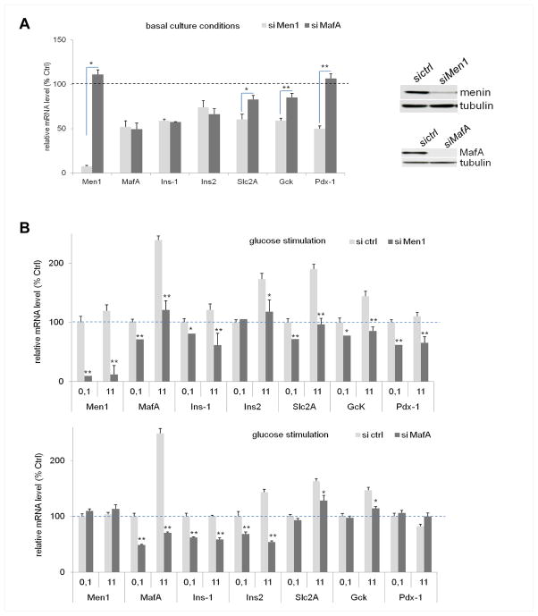
We next analysed the effect of either MENIN or MAFA inhibition on several MAFA-regulated genes critical to β-cell differentiation and function, including Ins-1/Ins-2, Slc2a2, Gck and Pdx-1 (Matsuoka et al. 2007; Wang et al. 2007). The expression of these differentiation markers was differentially affected by either Men1 or MafA siRNA-mediated knockdown in INS-1E cells, although MafA levels were inhibited to a similar level in both cases (Fig. 4). Under basal culture conditions, MENIN inhibition resulted in a decrease not only in MafA but also in Ins1/Ins2, Scl2a, Gck and Pdx-1 (to about 50% of control). Interestingly, if the MENIN level was kept stable, direct inhibition of MafA affected Ins-1/Ins-2 in an identical manner to that described above but had a reduced effect on Slc2a2 and Gck (>80% of control) and no effect on Pdx-1 (Fig. 4A). Similar gene-regulation results were obtained following 11-mM glucose stimulation of INS-1E cells (Fig. 4B).
Figure 4. Effect of MENIN versus MAFA inhibition on differentiation genes.
(A) Under basal 5 mM glucose culture conditions, the expression levels of the Men1, MAFA, Ins1/Ins2, Slc2A, Gck and Pdx-1 genes were measured using RT-qPCR in INS-1E cells after transfection with either anti-Men1 or anti-MAFA siRNA pools. Cells were maintained for 48 h in standard culture conditions. Inset: effective downregulation of MENIN and MAFA was determined by IB as in Fig. 2A. The relative quantity of each mRNA is indicated as a percentage of the respective control values (i.e. mRNA value, arbitrary fixed to 100%, of cells transfected with non-targeted siRNA and represented here by the dotted line). *Statistical significance was defined as p<0.1. **Statistical significance was defined as p<0.05. (A) Following 11 mM glucose stimulation, the cells were transfected with either anti-Men1 (top panel) or anti-MAFA (bottom panel) siRNA pools as described above and then starved overnight in 0.1 mM glucose (delineated as 0.1 on diagram) and stimulated in 11 mM glucose medium (11 on diagram). RT-qPCR analysis was performed as described above. The relative quantity of each mRNA is indicated as a percentage of the respective control value fixed to 100% (i.e. the mRNA value for cells transfected with non-targeted siRNA maintained in 0.1 mM glucose). *Statistical significance was defined as p<0.1. **Statistical significance was defined as p<0.05.
MEN1-related missense MENIN proteins lose their regulatory effects on MAFA expression
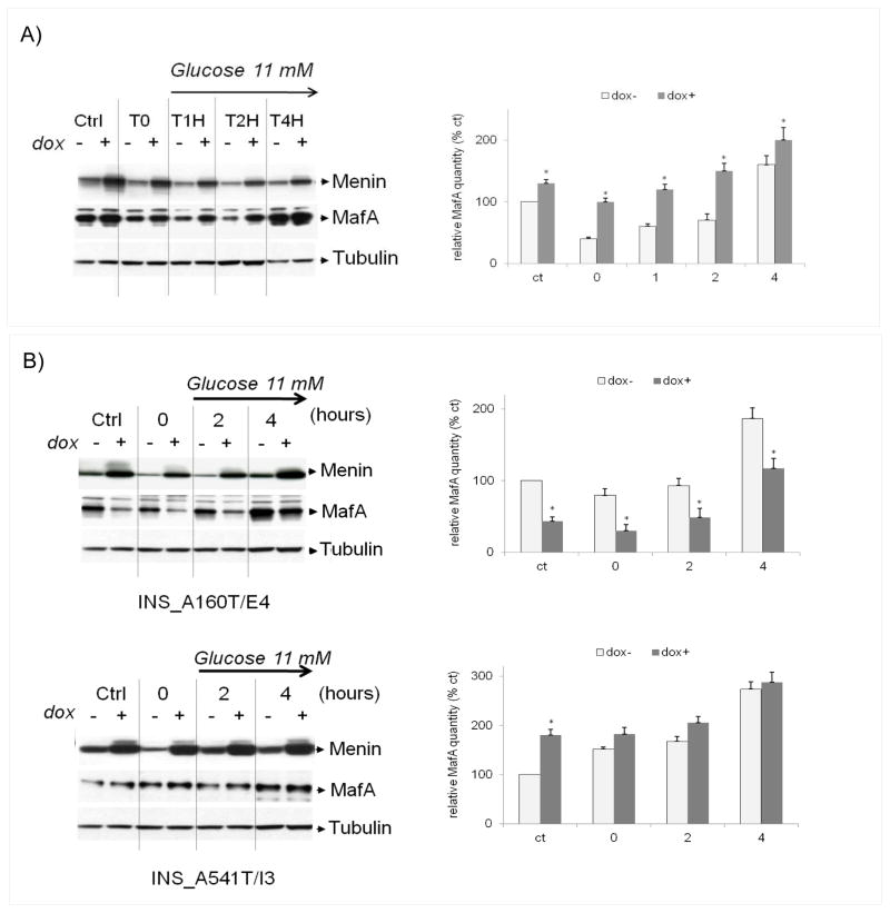
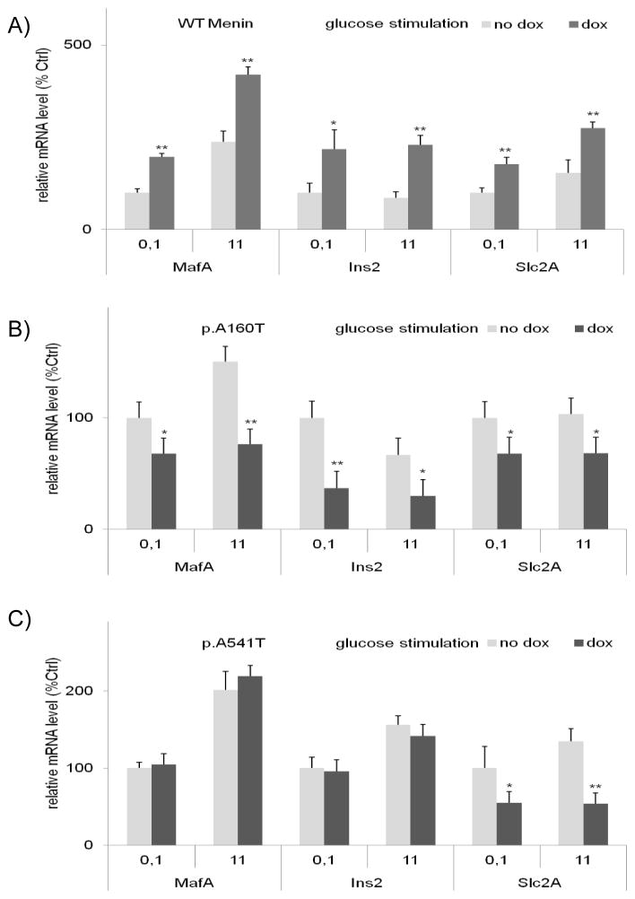
Two different missense versions of MENIN found in patients with MEN1 syndrome were next analysed for their ability to influence glucose-activated MAFA expression. We used INS-rβ cells in which MENIN knockdown had been found to affect MafA levels in response to glucose stimulation (Fig. S1A). As expected, conditional WT MENIN overexpression amplified MAFA protein and mRNA stimulation in INS_WT/B7 cells (Figs. 5A and 6A), but overexpression of the MENIN p.A160T and p.A541T variants did not increase MAFA expression (Figs. 5B and 6B, C). In addition, the effects of these two variants on MAFA were different. The p.A160T protein actually decreased MAFA protein accumulation (Fig. 5B) and had the same effect on Ins2 and Slc2A2 mRNA levels (Fig. 6B). By contrast, MENIN p.A541T did not have either an activating or a repressive effect on glucose-induced MafA or Ins2 mRNA expression, although Slc2A levels were decreased (Figs. 5 and 6). No reproducible data were obtained with the Ins-1 gene in these experiments.
Figure 5. The effect of overexpression of either WT MENIN or MEN1 disease-related variants on MAFA protein expression.
Following overnight glucose starvation (hour 0), INS-rβ cells overexpressing WT or variant MENIN after a doxycyclin treatment (dox −/+) were incubated in 11 mM glucose for 1 to 4 h as described in Fig. 2B. Under the Ctrl conditions, the cells were maintained in 5 mM glucose medium for the entire experiment. (A) IB of MAFA in INS_WT/B7 cells, a representative cell clone overexpressing WT MENIN. (B) IB of MAFA in INS_A160T/E4 and INS_A541T/I3 cells, representative cell clones overexpressing variant MENIN. The image is representative of at least four experiments on two independent cell clones for WT and variant MENIN proteins.
Figure 6. The effect of overexpression of either WT MENIN or disease-related variants on target genes mRNA levels.
Total RNA was extracted in parallel with the experiments shown in Figure 5. The expression levels of MafA, Ins2 and Slc2A mRNA were measured by real-time RT-qPCR as described above in INS_WT/B7 cells (A), INS_A160T/E4 (B) and INS_A541T/I3 (C) cells. The experiments were repeated four times. The relative quantity of each mRNA is indicated as a percentage of the respective control value fixed to 100% (i.e mRNA value for cells without overexpression of MENIN (no doxycyclin treatment) maintained in 0.1 mM glucose). *Statistical significance was defined as p<0.1. **Statistical significance was defined as p<0.05.
MENIN and MAFA modulate the glucose-evoked proliferation rate in insulinoma cell lines
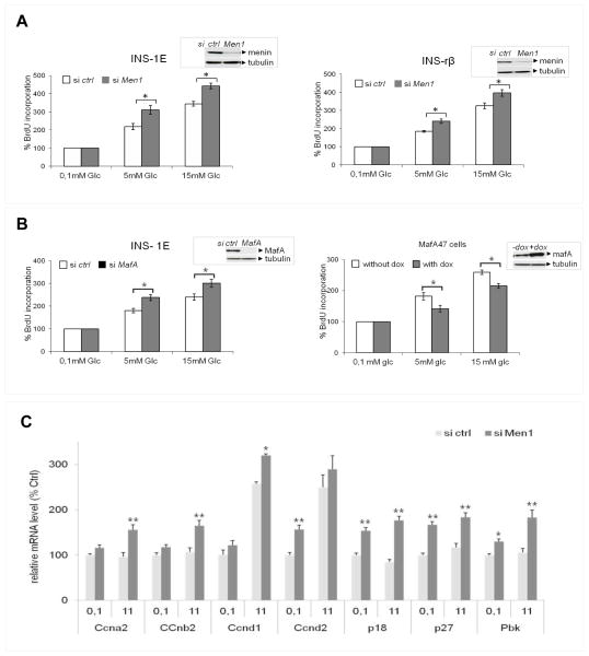
To test whether downregulation of differentiation markers might be associated with dysregulation of the differentiation/proliferation balance, we analysed the effects of MENIN inhibition on proliferation in insulinoma cell lines. Glucose has potent β-cell mitogen activity both in vitro and in vivo (Martens and Pipeleers 2009), hence short-term stimulation (up to 24 h) can stimulate the β-cell proliferation rate as measured by bromodeoxyuridine (BrdU) incorporation. As expected, the basal proliferation rate of INS-1E and INS-rβ cells greatly increased at 24 h after variation of glucose concentrations (the concentration of fetal calf serum remaining constant) from 0.1 to 5 and 15 mM (Fig. 7A). Interestingly, inhibition of MENIN expression resulted in greater BrdU incorporation after 5- and 15-mM glucose stimulation in both INS-1E and INS-rβ cells.
Figure 7. Alteration of MENIN and MAFA expression modified cell proliferation rate NewFigure.
(A, B) BrdU incorporation was measured by a colorimetric assay following manufacturer’s instructions (Roche Diagnostics GmbH, Mannheim, Germany) after incubation in 0.1, 5 and 15 mM glucose in cells transfected with non-targeting (sictrl) or anti-Men1 (siMen1) siRNA (A) or anti-MAFA siRNA (B, left) or in cells overexpressing MAFA after doxycyclin treatment (B, right). Results are expressed as a percentage of the reference value, arbitrarily chosen as the optical density (OD) value obtained for cells incubated in 0.1 mM glucose medium without siRNA transfection. Results represent the mean of at least four experiments. *Statistical significance was defined as p<0.05, Insets: Efficiency of siRNA transfection or doxycyclin induction was controlled by IB. (C) The cells transfected with anti-Men1 were starved overnight in 0.1 mM glucose (delineated as 0.1 on diagram) and stimulated in 11 mM glucose medium (11 on diagram) as described in Fig. 2C. Expression of proliferation-related genes was analysed by RT-qPCR as described above. The relative quantity of each mRNA is indicated a percentage of the respective control value fixed to 100% (i.e. the mRNA value for cells maintained in 0.1 mM glucose). *Statistical significance was defined as p<0.1. **Statistical significance was defined as p<0.05.
Because MENIN inhibition decreased MAFA expression, we then tested whether direct siRNA-mediated inhibition of MAFA could modify the proliferation rate in insulinoma cell lines. Direct MAFA knockdown was correlated with increased BrdU incorporation rate following glucose stimulation both with 5 and 15 mM glucose (Fig. 7B, left panel). Similar results were obtained using INS-rβ cells (data not shown). In a parallel set of experiments, we used MAFA47 cells (Wang et al. 2007) that overexpressed MAFA after doxycyclin treatment (Fig. 7B, right panel) and observed that, remarkably, the glucose-induced BrdU incorporation rate decreased as MAFA expression increased.
Finally, we measured the expression of candidate genes whose expression had previously been found to be related to β-cell proliferation in response to either glucose stimulation, MENIN expression level or both (Cozar-Castellano et al. 2006; Yang et al. 2010; Yesil and Lammert 2008). We found that following glucose stimulation, the basal mRNA level of the start-cyclin genes Ccnd1 and Ccnd2 had a significant increase not modified after MENIN inhibition (Fig. 7C). By contrast, MENIN inhibition significantly stimulated expression of the cyclin kinase inhibitor genes p18 and p27, of the mitoticcyclins a2 and b2 (Ccna2 and, Ccnb2) and of the mitotic PDZ-binding kinase (Pbk), whose expression was not affected after glucose stimulation. Similar results were obtained in INS-rβ cells (data not shown). Unexpectedly, expression of these proliferation genes was not modified following direct MAFA inhibition by anti-MAFA siRNA (data not shown). Double knockdown by anti-Men1 and anti-MafA siRNA reproduced the anti-Men1 siRNA effect (data not shown).
Discussion
Our data show for the first time that MENIN directly regulates the expression of MAFA, a TF that is vital for maintaining endocrine β-cell differentiation and function. Consequently, loss of MENIN alters both MAFA expression and the endocrine β-cell differentiation/proliferation balance. These data indicate a potentially new oncosuppressive mechanism in insulinoma that is mediated through the direct deregulation of the terminal differentiation pathway.
Our first relevant observation is that MENIN directly regulates MAFA, both in vivo and in vitro. A prevalent co-repression of these two proteins was evident in human insulinomas. Owing to the scarcity of these tumours, large series are difficult to obtain. Nevertheless, a convincing confirmation came from our Men1-mice insulinoma analysis, which reproduced these observations. In vitro analyses showed that MENIN directly regulated MafA transcription, thus affecting MAFA protein and mRNA levels. We show that MENIN modulate the transcriptional activity of the large MafA R1-6 promoter fragment, which ensures specific pancreatic expression and mediates glucose-induced MAFA expression in vivo (Raum et al. 2006). MENIN also regulated all the promoter sub-fragments tested. We found that MENIN bound to multiple DNA sequences in this R1 to R6 promoter fragment, including the R1, R2, R4, R5 and R6 regions, but not R3. Thus, MENIN may regulate MafA transcription via distinct molecular mechanisms in a promoter region-dependent fashion. Firstly, MENIN could act through a direct mechanism, as suggested by its binding to the R1, R2, R4, R5 and R6 promoter regions, and secondly, it could also operate through an indirect mechanism, by regulating expression of other TFs that themselves bind to the R3 region. PDX-1 might constitute such a candidate partner; its binds specifically to the R3 region (Raum et al. 2006), and we observed here that Pdx-1 mRNA levels were decreased by MENIN inhibition.
It is known that MAFA protein expression is tightly controlled in response to glucose stimulation, but the mechanisms of this are poorly understood. Numerous post-translational modifications modulate MAFA stability and activity (Eychene et al. 2008). Our data show that MENIN is essential for MAFA protein accumulation in response to glucose, partly through transcriptional mechanisms, but additional mechanisms cannot be excluded. MENIN levels were previously reported to be significantly reduced by long-term high-glucose exposure of both INS-1 cells and rat primary islets (Zhang et al. 2012). In our experiments, we did not find any modification of endogenous MENIN protein or mRNA levels following glucose stimulation. These discrepancies could be attributable to different experimental conditions. In the current study, only acute (<6 h) stimulation with 11 mM glucose was tested; the half-life of the WT MENIN protein is >8 h (Canaff et al. 2012; Yaguchi et al. 2004), and that of its mRNA is around 3 h in INS-1 cells (personal observations).
Our second relevant observation is that MENIN expression is crucial for MAFA-mediated β-cell differentiation, as its downregulation had a dramatic negative effect on the expression of major MAFA-regulated differentiation markers such as Slc2a2, Gck and especially Pdx-1. Indeed, MENIN inhibition had greater deleterious effects than found with MAFA inhibition on these markers. The transcriptional properties of MENIN could be directly or indirectly responsible for these observations. MENIN can regulate transcription by interacting directly with TFs or through several epigenetic mechanisms (Dreijerink et al. 2009; Yang and Hua 2007). We cannot exclude that MENIN could participate through such mechanisms to regulate transcription not only of MafA but also of a larger series of differentiation-related β-cell genes, including those studied here. Consequently, differentiation functions must be altered. In particular, MENIN expression affected Slc2a2 gene transcription and missense MENIN proteins were unable to simulate the Sclc2a2 mRNA level, emphasising a potential key functional link. Thus, because Slc2A2 is the gene encoding for the glucose transporter GLUT2, modulation of the number of GLUT2 molecules at the cell surface could directly affect glucose uptake and consequently downstream differentiation/proliferation signalling in β cells. More generally, MENIN inhibition seems to block maintenance of the differentiation process; such a mechanism could contribute to tumorigenesis, in parallel with re-expression of early markers (MAFB and HLXB9) and stimulation of proliferation/survival (Lejonklou et al. 2009; Lu et al. 2012; Shi et al. 2013). A very tightly controlled and interconnected regulation of MAFA and MAFB has been described in developing β cells, with their levels being in inverse proportion to one another (Artner et al. 2010) and MAFB expression in mice being restricted to immature β cells (Nishimura et al. 2006). These previous observations suggest that the effect of MENIN on MAFA expression level can directly or indirectly influence MAFB expression. In line with this, we found that MENIN knockdown simultaneously downregulated MAFA and upregulated MAFB in both cell lines analysed here. These observations corroborate our previously published data showing that, in adult β cells, decreased expression level of MENIN is correlated with an increased expression of MAFB in insulinoma and in the tested cell lines (Lu et al. 2012). By regulating the MAFA/MAFB ratio, MENIN could thus influence the differentiation/proliferation balance in β cells. In line with this, our data showed an increase in proliferation rate following MENIN inhibition in the insulinoma cells analysed. A relevant observation is that isolated inhibition of MAFA also stimulated the proliferation rate, suggesting that MAFA could possess an unsuspected tumour suppressor function in β cells. MENIN inhibition affected candidate proliferation genes that had previously been implicated in rodent β-cell proliferation, but interestingly, MAFA repression did not have any effect on these genes, suggesting the implication of other MAFA-regulated proliferation pathways. Large MAF proteins were previously shown to act as tumour suppressors or oncogenes depending on the cell type, although nothing is known concerning this aspect in β cells (Eychene et al. 2008).
In addition, increases in both Ki67 expression and malignancy in human insulinomas with concomitant MENIN and MAFA repression support this hypothesis. Finally, the relevance of the MENIN/MAFA/MAFB axis in tumorigenesis is supported by our data, showing that in insulinoma cell lines, the p.A160T or p.A541T protein counterparts of MENIN found in patients with MEN1 were not able to exert a similar influence to WT MENIN on MAFA regulation. We found different functional consequences depending on the type of mutation; p.A160T had a dominant-negative effect that mimicked the effect of anti-Men1 siRNA, while p.A541T mutation abolished the stimulatory effect of WT MENIN. Unexpectedly, the p.A541T variant (dBSNP: rs 2959656), the only abnormality identified in a French cohort of patients with MEN1, is also present in a percentage of healthy individuals. Because the frequency of p.A541T in the normal population is higher than expected for a rare disease, it is thus considered a polymorphism. We previously reported that this controversial p.A541T variant did display a disease-related mutant behaviour in a series of functional and biochemical experiments (Bazzi et al. 2008). The data from the current study are therefore in agreement with our previous observations.
In conclusion, we show for the first time that MENIN loss is associated with downregulation of MAFA, a crucial TF for maintaining endocrine differentiation in mature β cells. Interestingly, the previously identified TF MAFB and HLXB9 are reportedly upregulated with MENIN loss (Lu et al. 2012; Shi et al. 2013). They are known to play crucial roles during endocrine pancreatic development (Hang and Stein 2011; Li and Edlund 2001) and to control proliferation/survival in specific cells (Lu et al. 2012; Shi et al. 2013). The downregulation of MAFA associated with MENIN loss has a dramatic effect on the differentiation/proliferation balance in insulinoma cells. Previous studies reported the crucial role of MENIN in the regulation of Pit-1 TF, a major regulator of terminal differentiation in pituitary cells, but without elucidating the molecular basis (Lacerte et al. 2004). Tissue specificity of tumours may thus result from a complex network of complementary effects on β-cell differentiation factors following MENIN loss. Whether such a cooperative oncosuppressive mechanism exists in other foregut NETs deserves to be explored.
Supplementary Material
Acknowledgments
Funding
Grant support: This study was supported by the Ligue contre le cancer Comité de la Loire (to MCB) et Comité du Rhône (to CXZ), Association pour Recherche sur le Cancer (to ZH, CV and CXZ), Agence Nationale de la Recherche (grant SVSE2 ANR 10 BLAN 1240 04 (to CXZ and MC-B) and National Institute of Health (USA) (DK50203 to RS).
We are grateful to the Ligue contre le cancer, Comité de la Loire et Comité du Rhône, the Association de Recherche sur le Cancer (ARC) and the Agence Nationale de la Recherche (ANR) for their financial support. We thank ARC for its financial support to ZH and CV. We gratefully acknowledge H Wang, C Wollheim, P Maechler and their colleagues for the kind gift of the INS-rβ, INS-1E and MafA47 cell lines.
Footnotes
Declaration of interest
The authors have no potential conflicts of interest to disclose.
Author contributions
MC-B is the guarantor of this work and as such, had full access to all of the data in this study and assumes responsibility for the integrity of the data and accuracy of the data analysis. ZH, CV AB-L, WB, JL and RB performed the experiments. CXZ, CP, PB, CR, JYS and AC contributed to valuable discussions. MC-B, CV and ZH wrote the manuscript. CP and RS reviewed and edited the manuscript RS provided MAFA promoter and luciferase plasmids.
References
- Agarwal SK, Kester MB, Debelenko LV, Heppner C, Emmert-Buck MR, Skarulis MC, Doppman JL, Kim YS, Lubensky IA, Zhuang Z, et al. Germline mutations of the MEN1 gene in familial multiple endocrine neoplasia type 1 and related states. Hum Mol Genet. 1997;6:1169–1175. doi: 10.1093/hmg/6.7.1169. dda154 [pii] [DOI] [PubMed] [Google Scholar]
- Aramata S, Han SI, Kataoka K. Roles and regulation of transcription factor MafA in islet beta-cells. Endocr J. 2007;54:659–666. doi: 10.1507/endocrj.kr-101. JST.JSTAGE/endocrj/KR-101 [pii] [DOI] [PubMed] [Google Scholar]
- Artner I, Hang Y, Mazur M, Yamamoto T, Guo M, Lindner J, Magnuson MA, Stein R. MafA and MafB regulate genes critical to beta-cells in a unique temporal manner. Diabetes. 2010;59:2530–2539. doi: 10.2337/db10-0190. [DOI] [PMC free article] [PubMed] [Google Scholar]
- Asfari M, Janjic D, Meda P, Li G, Halban PA, Wollheim CB. Establishment of 2-mercaptoethanol-dependent differentiated insulin-secreting cell lines. Endocrinology. 1992;130:167–178. doi: 10.1210/endo.130.1.1370150. [DOI] [PubMed] [Google Scholar]
- Bazzi W, Renon M, Vercherat C, Hamze Z, Lacheretz-Bernigaud A, Wang H, Blanc M, Roche C, Calender A, Chayvialle JA, et al. MEN1 missense mutations impair sensitization to apoptosis induced by wild-type MENIN in endocrine pancreatic tumor cells. Gastroenterology. 2008;135:1698–1709. e1692. doi: 10.1053/j.gastro.2008.07.031. [DOI] [PubMed] [Google Scholar]
- Bertolino P, Tong WM, Herrera PL, Casse H, Zhang CX, Wang ZQ. Pancreatic beta-cell-specific ablation of the multiple endocrine neoplasia type 1 (MEN1) gene causes full penetrance of insulinoma development in mice. Cancer Res. 2003;63:4836–4841. [PubMed] [Google Scholar]
- Bustin SA, Benes V, Garson JA, Hellemans J, Huggett J, Kubista M, Mueller R, Nolan T, Pfaffl MW, Shipley GL, et al. The MIQE guidelines: minimum information for publication of quantitative real-time PCR experiments. Clin Chem. 2009;55:611–622. doi: 10.1373/clinchem.2008.112797. [DOI] [PubMed] [Google Scholar]
- Canaff L, Vanbellinghen JF, Kanazawa I, Kwak H, Garfield N, Vautour L, Hendy GN. Menin missense mutants encoded by the MEN1 gene that are targeted to the proteasome: restoration of expression and activity by CHIP siRNA. J Clin Endocrinol Met. 2012;97:E282–291. doi: 10.1210/jc.2011-0241. [DOI] [PubMed] [Google Scholar]
- Corbo V, Dalai I, Scardoni M, Barbi S, Beghelli S, Bersani S, Albarello L, Doglioni C, Schott C, Capelli P, et al. MEN1 in pancreatic endocrine tumors: analysis of gene and protein status in 169 sporadic neoplasms reveals alterations in the vast majority of cases. Endocr Relat Cancer. 2010;17:771–783. doi: 10.1677/ERC-10-0028. [DOI] [PubMed] [Google Scholar]
- Cozar-Castellano I, Fiaschi-Taesch N, Bigatel TA, Takane KK, Garcia-Ocana A, Vasavada R, Stewart AF. Molecular control of cell cycle progression in the pancreatic beta-cell. Endocr Rev. 2006;27:356–370. doi: 10.1210/er.2006-0004. [DOI] [PubMed] [Google Scholar]
- Crabtree JS, Scacheri PC, Ward JM, McNally SR, Swain GP, Montagna C, Hager JH, Hanahan D, Edlund H, Magnuson MA, et al. Of mice and MEN1: Insulinomas in a conditional mouse knockout. Mol Cell Biol. 2003;23:6075–6085. doi: 10.1128/MCB.23.17.6075-6085.2003. [DOI] [PMC free article] [PubMed] [Google Scholar]
- de Herder WW, Niederle B, Scoazec JY, Pauwels S, Kloppel G, Falconi M, Kwekkeboom DJ, Oberg K, Eriksson B, Wiedenmann B, et al. Well-differentiated pancreatic tumor/carcinoma: insulinoma. Neuroendocrinology. 2006;84:183–188. doi: 10.1159/000098010. [DOI] [PubMed] [Google Scholar]
- Dreijerink KM, Lips CJ, Timmers HT. Multiple endocrine neoplasia type 1: a chromatin writer’s block. J Intern Med. 2009;266:53–59. doi: 10.1111/j.1365-2796.2009.02115.x. [DOI] [PubMed] [Google Scholar]
- Eychene A, Rocques N, Pouponnot C. A new MAFia in cancer. Nat Rev Cancer. 2008 doi: 10.1038/nrc2460. [DOI] [PubMed] [Google Scholar]
- Fontaniere S, Tost J, Wierinckx A, Lachuer J, Lu J, Hussein N, Busato F, Gut I, Wang ZQ, Zhang CX. Gene expression profiling in insulinomas of Men1 beta-cell mutant mice reveals early genetic and epigenetic events involved in pancreatic beta-cell tumorigenesis. Endocr Relat Cancer. 2006;13:1223–1236. doi: 10.1677/erc.1.01294. [DOI] [PubMed] [Google Scholar]
- Fromaget M, Vercherat C, Zhang CX, Zablewska B, Gaudray P, Chayvialle JA, Calender A, Cordier-Bussat M. Functional characterization of a promoter region in the human MEN1 tumor suppressor gene. J Mol Biol. 2003;333:87–102. doi: 10.1016/j.jmb.2003.08.001. S0022283603010040 [pii] [DOI] [PubMed] [Google Scholar]
- Gracanin A, Dreijerink KM, van der Luijt RB, Lips CJ, Hoppener JW. Tissue selectivity in multiple endocrine neoplasia type 1-associated tumorigenesis. Cancer Res. 2009;69:6371–6374. doi: 10.1158/0008-5472.CAN-09-0678. [DOI] [PubMed] [Google Scholar]
- Hang Y, Stein R. MafA and MafB activity in pancreatic beta cells. Trends in endocrinology and metabolism: TEM. 2011;22:364–373. doi: 10.1016/j.tem.2011.05.003. [DOI] [PMC free article] [PubMed] [Google Scholar]
- Hussein N, Casse H, Fontaniere S, Morera AM, Asensio MJ, Bakeli S, Lu JL, Coste I, Di Clemente N, Bertolino P, et al. Reconstituted expression of MENIN in Men1-deficient mouse Leydig tumour cells induces cell cycle arrest and apoptosis. Eur J Cancer. 2007;43:402–414. doi: 10.1016/j.ejca.2006.08.038. [DOI] [PubMed] [Google Scholar]
- Jiao Y, Shi C, Edil BH, de Wilde RF, Klimstra DS, Maitra A, Schulick RD, Tang LH, Wolfgang CL, Choti MA, et al. DAXX/ATRX, MEN1, and mTOR pathway genes are frequently altered in pancreatic neuroendocrine tumors. Science. 2011;331:1199–1203. doi: 10.1126/science.1200609. [DOI] [PMC free article] [PubMed] [Google Scholar]
- Karnik SK, Chen H, McLean GW, Heit JJ, Gu X, Zhang AY, Fontaine M, Yen MH, Kim SK. MENIN controls growth of pancreatic beta-cells in pregnant mice and promotes gestational diabetes mellitus. Science. 2007;318:806–809. doi: 10.1126/science.1146812. [DOI] [PubMed] [Google Scholar]
- Lacerte A, Lee EH, Reynaud R, Canaff L, De Guise C, Devost D, Ali S, Hendy GN, Lebrun JJ. Activin inhibits pituitary prolactin expression and cell growth through Smads, Pit-1 and MENIN. Molecular endocrinology. 2004;18:1558–1569. doi: 10.1210/me.2003-0470. [DOI] [PubMed] [Google Scholar]
- Lejonklou MH, Edfeldt K, Johansson TA, Stalberg P, Skogseid B. Neurogenin 3 and neurogenic differentiation 1 are retained in the cytoplasm of multiple endocrine neoplasia type 1 islet and pancreatic endocrine tumor cells. Pancreas. 2009;38:259–266. doi: 10.1097/MPA.0b013e3181930818. [DOI] [PubMed] [Google Scholar]
- Li H, Edlund H. Persistent expression of Hlxb9 in the pancreatic epithelium impairs pancreatic development. Dev Biol. 2001;240:247–253. doi: 10.1006/dbio.2001.0440. [DOI] [PubMed] [Google Scholar]
- Liu Y, Mziaut H, Ivanova A, Solimena M. beta-Cells at the crossroads: choosing between insulin granule production and proliferation. Diabetes Obes Metab. 2009;11(Suppl 4):54–64. doi: 10.1111/j.1463-1326.2009.01107.x. [DOI] [PubMed] [Google Scholar]
- Livak KJ, Schmittgen TD. Analysis of relative gene expression data using real-time quantitative PCR and the 2(-Delta Delta C(T)) Method. Methods. 2001;25:402–408. doi: 10.1006/meth.2001.1262. S1046-2023(01)91262-9 [pii] [DOI] [PubMed] [Google Scholar]
- Lu J, Hamze Z, Bonnavion R, Herath N, Pouponnot C, Assade F, Fontaniere S, Bertolino P, Cordier-Bussat M, Zhang CX. Reexpression of oncoprotein MafB in proliferative beta-cells and Men1 insulinomas in mouse. Oncogene. 2012;31:3647–3654. doi: 10.1038/onc.2011.538. [DOI] [PubMed] [Google Scholar]
- Martens GA, Pipeleers D. Glucose, regulator of survival and phenotype of pancreatic beta cells. Vitam Horm. 2009;80:507–539. doi: 10.1016/S0083-6729(08)00617-1. [DOI] [PubMed] [Google Scholar]
- Matsuoka TA, Kaneto H, Stein R, Miyatsuka T, Kawamori D, Henderson E, Kojima I, Matsuhisa M, Hori M, Yamasaki Y. MafA regulates expression of genes important to islet beta-cell function. Mol Endocrinol. 2007;21:2764–2774. doi: 10.1210/me.2007-0028. [DOI] [PubMed] [Google Scholar]
- Merglen A, Theander S, Rubi B, Chaffard G, Wollheim CB, Maechler P. Glucose sensitivity and metabolism-secretion coupling studied during two-year continuous culture in INS-1E insulinoma cells. Endocrinology. 2004;145:667–678. doi: 10.1210/en.2003-1099. [DOI] [PubMed] [Google Scholar]
- Metz DC, Jensen RT. Gastrointestinal neuroendocrine tumors: pancreatic endocrine tumors. Gastroenterology. 2008;135:1469–1492. doi: 10.1053/j.gastro.2008.05.047. [DOI] [PMC free article] [PubMed] [Google Scholar]
- Modlin IM, Oberg K, Chung DC, Jensen RT, de Herder WW, Thakker RV, Caplin M, Delle Fave G, Kaltsas GA, Krenning EP, et al. Gastroenteropancreatic neuroendocrine tumours. Lancet Oncol. 2008;9:61–72. doi: 10.1016/S1470-2045(07)70410-2. [DOI] [PubMed] [Google Scholar]
- Nishimura W, Kondo T, Salameh T, El Khattabi I, Dodge R, Bonner-Weir S, Sharma A. A switch from MafB to MafA expression accompanies differentiation to pancreatic beta-cells. Dev Biol. 2006;293:526–539. doi: 10.1016/j.ydbio.2006.02.028. [DOI] [PMC free article] [PubMed] [Google Scholar]
- Pouponnot C, Sii-Felice K, Hmitou I, Rocques N, Lecoin L, Druillennec S, Felder-Schmittbuhl MP, Eychene A. Cell context reveals a dual role for Maf in oncogenesis. Oncogene. 2006;25:1299–1310. doi: 10.1038/sj.onc.1209171. [DOI] [PubMed] [Google Scholar]
- Raum JC, Gerrish K, Artner I, Henderson E, Guo M, Sussel L, Schisler JC, Newgard CB, Stein R. FoxA2, Nkx2.2, and PDX-1 regulate islet beta-cell-specific mafA expression through conserved sequences located between base pairs -8118 and -7750 upstream from the transcription start site. Mol Cell Biol. 2006;26:5735–5743. doi: 10.1128/MCB.00249-06. [DOI] [PMC free article] [PubMed] [Google Scholar]
- Rindi G, Wiedenmann B. Neuroendocrine neoplasms of the gut and pancreas: new insights. Nature reviews Endocrinology. 2012;8:54–64. doi: 10.1038/nrendo.2011.120. [DOI] [PubMed] [Google Scholar]
- Schnepp RW, Mao H, Sykes SM, Zong WX, Silva A, La P, Hua X. MENIN induces apoptosis in murine embryonic fibroblasts. J Biol Chem. 2004;279:10685–10691. doi: 10.1074/jbc.M308073200. [DOI] [PMC free article] [PubMed] [Google Scholar]
- Shi K, Parekh VI, Roy S, Desai SS, Agarwal SK. The embryonic transcription factor Hlxb9 is a menin interacting partner that controls pancreatic beta-cell proliferation and the expression of insulin regulators. Endocr Relat Cancer. 2013;20:111–122. doi: 10.1530/ERC-12-0077. [DOI] [PMC free article] [PubMed] [Google Scholar]
- Shin JJ, Gorden P, Libutti SK. Insulinoma: pathophysiology, localization and management. Future oncology. 2010;6:229–237. doi: 10.2217/fon.09.165. [DOI] [PMC free article] [PubMed] [Google Scholar]
- Wang H, Brun T, Kataoka K, Sharma AJ, Wollheim CB. MAFA controls genes implicated in insulin biosynthesis and secretion. Diabetologia. 2007;50:348–358. doi: 10.1007/s00125-006-0490-2. [DOI] [PMC free article] [PubMed] [Google Scholar]
- Wang H, Iynedjian PB. Modulation of glucose responsiveness of insulinoma beta-cells by graded overexpression of glucokinase. Proc Natl Acad Sci U S A. 1997;94:4372–4377. doi: 10.1073/pnas.94.9.4372. [DOI] [PMC free article] [PubMed] [Google Scholar]
- Wautot V, Vercherat C, Lespinasse J, Chambe B, Lenoir GM, Zhang CX, Porchet N, Cordier M, Beroud C, Calender A. Germline mutation profile of MEN1 in multiple endocrine neoplasia type 1: search for correlation between phenotype and the functional domains of the MEN1 protein. Hum Mutat. 2002;20:35–47. doi: 10.1002/humu.10092. [DOI] [PubMed] [Google Scholar]
- Wu T, Hua X. Menin represses tumorigenesis via repressing cell proliferation. Am J Cancer Res. 2011;1:726–739. [PMC free article] [PubMed] [Google Scholar]
- Yaguchi H, Ohkura N, Takahashi M, Nagamura Y, Kitabayashi I, Tsukada T. Menin missense mutants associated with multiple endocrine neoplasia type 1 are rapidly degraded via the ubiquitin-proteasome pathway. Mol Cell Biol. 2004;24:6569–6580. doi: 10.1128/MCB.24.15.6569-6580.2004. [DOI] [PMC free article] [PubMed] [Google Scholar]
- Yang Y, Cvekl A. Large Maf Transcription Factors: Cousins of AP-1 Proteins and Important Regulators of Cellular Differentiation. The Einstein journal of biology and medicine: EJBM. 2007;23:2–11. doi: 10.23861/ejbm20072347. [DOI] [PMC free article] [PubMed] [Google Scholar]
- Yang Y, Gurung B, Wu T, Wang H, Stoffers DA, Hua X. Reversal of preexisting hyperglycemia in diabetic mice by acute deletion of the Men1 gene. Proc Natl Acad Sci U S A. 2010;107:20358–20363. doi: 10.1073/pnas.1012257107. [DOI] [PMC free article] [PubMed] [Google Scholar]
Associated Data
This section collects any data citations, data availability statements, or supplementary materials included in this article.


