Abstract
Over the past decades, stress/rest myocardial perfusion SPECT (MPS) has been utilized as a standard modality for the diagnosis, risk stratification and prognostic assessment of coronary artery disease (CAD). In addition to the perfusion information, MPS can also provide functional information of the left ventricle, including volume, ejection fraction, wall motion and dyssynchrony. This article introduces the incremental value of these non-perfusion parameters as markers and prognosticators of CAD.
Keywords: coronary artery disease, additional markers, myocardial perfusion SPECT
INTRODUCTION
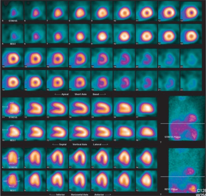
Myocardial perfusion SPECT (MPS) is an imaging procedure that utilizes an intravenously administered radiotracer to delineate the distribution of blood flow in the myocardium during exercise or pharmacological stimulation and at rest using tomographic camera. Patients with hemodynamically significant coronary artery stenosis will have a myocardial region of diminished radiotracer concentration in areas of diseased vessels, which constitute the basis of diagnosing coronary artery disease (CAD) with MPS. If the perfusion abnormality shown on MPS is worse at stress than at rest, the so-called reversible defect, the myocardial segment is most likely due to ischemia (Fig. 1). If the perfusion abnormality remains unchanged from stress to rest (fixed defect), the lesion usually indicates scarred myocardium (Fig. 2). In current clinical practice, the major indications of MPS are used for evaluation of patients with suspected or known CAD, including assessment of the presence, location, extent and severity of myocardial ischemia and scar, determination of the functional significance of anatomic stenosis detected by invasive or CT coronary angiography, and assessment of myocardial viability in ischemic cardiomyopathy[1]–[3].
Fig. 1. Representative images of a patient with severe and extensive myocardial ischemia (reversible perfusion defects) at the apex, mid-basal anterolateral, mid-basal inferoseptal and apical-mid-basal inferior segments.
Fig. 2. Representative images of a patient with scarred myocardium (fixed perfusion defects) at the apex, mid-basal anteroseptal, and mid-basal inferolateral segments.
Plenty of published studies had confirmed the diagnostic accuracy of MPS in detecting obstructive CAD when using the diameter stenosis of diseased vessels demonstrated on coronary angiography[4]. As a functional modality for assessing the hemodynamic significance of coronary lesion, MPS had also established its role in risk stratification and prognostic assessment in patients with CAD[5]–[7]. The patients with normal or near-normal results on MPS would have an annual cardiac event rate of less than 1 percent. In addition, those showing normal perfusion or only mild ischemia on MPS could be safely managed with conservative medical treatment rather than invasive revascularizations. For those with moderate to severe ischemia on MPS, however, revascularization significantly improves the survival in comparison with medication only[8]–[9]. Therefore, MPS has been clinically considered as a gatekeeper of invasive procedure in the management of CAD.
ECG-GATED MYOCARDIAL PERFUSION SPECT
In gated image acquisition of MPS, ECG provides the R wave trigger to the acquisition computer, with 2 successive R wave peaks on the ECG for defining a cardiac cycle. Reconstruction of 3D images of the left-ventricular (LV) myocardium at different time points over the cardiac cycle allows calculation of end-systolic volume (ESV), end-diastolic volume (EDV) and ejection fraction (EF). Cinematic display of MPS images at different cardiac cycles enable readers to assess wall motion (WM) and wall thickening (WT) in all segments of the myocardium (Fig. 3). The application of ECG-gating technique in MPS enables the simultaneous assessment of LV perfusion and function in a single imaging modality. These functional parameters not only provide invaluable information on diagnosis and prognosis of cardiac disease but also are useful for differentiation of attenuation artifacts from true myocardial injuries[10]. Gated MPS now has become the standard procedure for myocardial perfusion imaging in the nuclear medicine labs worldwide.
Fig. 3. Example results of gated MPS images processed by a commercial program in a patient with heart failure.
The images reveal poor LV function with an EF of 36%, and impaired regional wall motion in anterior, septal and inferior walls.
LIMITATION OF MPS
The mechanism of myocardial perfusion imaging (MPI) in detecting coronary artery disease (CAD) is primarily based on the detection of perfusion abnormalities of LV myocardium caused by the coronary flow gradients between normal and diseased arteries, which results in differences in tracer uptake in respective myocardial territories. In patients with multivessel CAD, however, the perfusion abnormalities may be unremarkable, or even be missed due to relatively balanced hypoperfusion between the stenosed vessels[11]. A pervious study found that the performance of detecting flow-limited CAD with MPS was very limited in patients with stenoses in multiple coronary vessels; thereby, the diagnostic sensitivity was as low as only 48%[12].
Fig. 4 shows the perfusion images from an 82 year-old male patient who presented with typical angina pectoris and was referred for stress MPS test for diagnosis and risk stratification of suspected CAD. Only unremarkably reversible perfusion abnormality was noted in the apical anterior segment, and conservative management was initially suggested based on the result of MPS. However, he was still referred to receive invasive coronary angiography due to exacerbating symptoms. The angiography revealed an extensive and severe CAD in multi-coronary vessels, including 60% stenosis in the left main artery, 95% in the proximal left anterior descending artery and 70% in the right coronary artery. This patient was thereafter referred for receiving invasive revascularization procedure of coronary artery bypass grafting.
Fig. 4. Example MPS images of balanced ischemia in a patient with multi-vessel CAD.
The images show only minimally reversible perfusion abnormality in apical anterior segment of LV, which apparently underestimates the severity of CAD.
TRANSIENT ISCHEMIC DILATION OF THE LV
Transient ischemic dilation (TID) refers to a significant enlargement in LV size on the stress images compared to the rest images on MPS. The presence of TID, either in exercise or pharmacologic stress protocols, has already been found to be highly indicative of severe and extensive CAD and also predictive of high cardiac-event risk[13]–[15]. Abdidov et al. further studied the prognostic value of TID in patients with normal MPS[16]. They found that an entirely normal stress MPS did not always imply an excellent prognosis. They concluded that TID was an independent and incremental prognostic marker of total cardiac event even for patients with otherwise normal MPS, and the interpreter should be very cautious in making low-risk prognostic statements, especially in patients with typical angina, the elderly, and diabetics[16].
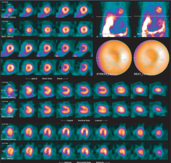
Fig. 5 shows the perfusion images of a 63 year-old male patient who presented with exertional shortness of breath and was referred for stress MPS as a work-up of suspected CAD. There is completely normal perfusion in all myocardial areas on both stress and rest images, except for an apparent TID. Based on the normal perfusion and the ignorance of prognostic significance of TID, the patient was managed with conservative medical treatment only without further imaging studies. Unfortunately, acute myocardial infarction involving the anterior wall developed one and a half years later. An emergent invasive coronary angiography revealed total occlusion of left anterior descending artery, 60% stenosis in left circumflex artery and 70% stenosis in right coronary artery.
Fig. 5. Example case of a patient with normal perfusion but significant transient ischemic dilation (TID).
This patient developed a cardiac event of acute myocardial infarction with confirmed three-vessel CAD by invasive coronary angiography after one and a half years after MPS study.
There is still no definite conclusion about the underlying mechanisms of TID. The most widely accepted explanation is myocardial thinning resulting from stress-induced diffuse subendocardial hypoperfusion, which produces a visually larger LV cavity[17]–[19]. At rest, the subendocardium is better perfused and the LV cavity appears smaller. Thus TID of this mechanism is “apparent,” not “true.”
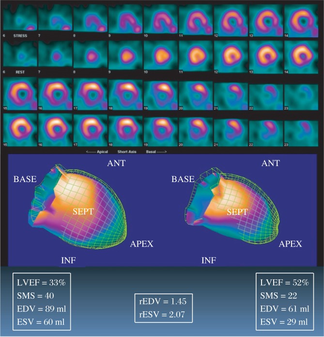
Alternatively, other investigators had also postulated that TID was secondary to “true” dilation of LV[13]. Hung et al. conducted an interesting study, which assessed the relationship of TID and stress-induced changes of functional variables using early post-stress and rest Tl-201 gated MPS[20]. They found that TID was significantly correlated with stress-induced ischemic stunning, as manifested by dropping LVEF and worsening of regional WM at stress. The enlargement of ESV at stress was more profound than that of EDV. A representative case was demonstrated on Fig. 6. A 61-year-old female patient was found to have 3-vessel disease (80% stenosis in the proximal left anterior descending, 100% in the mid left circumflex and 100% in the right coronary arteries) on invasive coronary angiography. The MPS images revealed reversible perfusion defects involving the apex, septum, inferior wall and lateral wall, and also prominent TID. Functional analyses of gated MPS revealed a severely depressed LVEF of 33% on stress and returned to 52% on rest. The regional WM abnormality was also more severe at stress than at rest, and the enlargement of ESV was significantly greater than that of EDV.
Fig. 6. An representative case of severe TID in a patient with 3-vessel disease.
The images show extensive reversible perfusion abnormalities on MPS. The volumes are 66 mL on stress images and 41 mL on rest with a ratio, 1.61 (much greater than the reference cut-off value of 1.19[20]). In addition to a significant drop in LVEF from 52% to 33% (rest to stress), reversible regional wall motion abnormality is also noted with significant increase of summed motion scores (SMS) from 22 to 40. The end-systolic volume (ESV) is severely enlarged from 29 mL to 60 mL with a ratio of 2.07; however, the end-diastolic volume (EDV) is modestly enlarged from 61 mL to 89 mL (ratio: 1.45).
Although TID has already been confirmed to be a highly specific marker of “extensive and severe CAD”, TID might also be related to microvascular disease[21], which usually refers to the patients with chest pain but normal coronary angiograms (CPNCA). CPNCA is used to be clinically called as “microvascular angina” or “syndrome X”, which was found to have stress-induced diffuse subendocardial hypoperfusion as evidenced on vasodilator-stress and rest cardiac MRI[22]. Fig. 7 shows the images of an 82 year-old female patient, who presented with typical angina pectoris and was referred for MPS as a work-up of suspected CAD. Homogenous perfusion was noted on both stress and rest images, but she was still referred for invasive coronary angiography due to the appearance of remarkable TID on MPS. This case was finally concluded as “microvascular disease” because no significant diameter stenosis was found in the epicardial coronary arteries on angiography. In order to avoid unnecessary invasive diagnostic procedure in “microvascular angina”, multi-slice CT coronary angiography might be an optimal choice of imaging modalities for cases of TID on MPS with normal or near-normal perfusion[23].
Fig. 7. Representative images showing transient ischemic dilation (TID) related to “microvascular angina”.
The images show significant enlargement of the inner cavity of the LV on stress as compared to rest MPS. In contrary to the TID shown on Fig. 6, which has consistently enlargements for both inner and outer surfaces, the size of the LV based on epicardial borders is almost identical between stress and rest in the current case. The pathophysiological mechanism of “TID” in this case should be stress-induced diffuse subendocardial hypoperfusion, resulting in “visually” enlargement of LV cavity.
SRESS-INDUCED ISCHEMIC STUNNING
“Myocardial stunning”, defined as prolonged global and/or regional myocardial dysfunction after stress-induced ischemia, has been well described on stress/rest gated MPS[24]–[25]. It might be manifested as transient post-stress decline of LVEF or reversible regional wall motion abnormality on gated MPS and were found to correlate with the severity of stress-induced perfusion defects. Plenty of studies further demonstrated that stress-induced stunning shown on gated MPS was correlated with severe and extensive CAD[26]–[29]. It should be noted that the manifestations of ischemic stunning might be different when using different kinds of radiotracers (e.g. Tl-201 or Tc-99m sestamibi) or different types of stress (e.g. exercise or vasodilator).
Gated SPECT myocardial perfusion imaging provides information on perfusion at the time of tracer injection and function during image acquisitions. Although stress-induced myocardial stunning might persist for 30 to 240 minutes[30], or even more than 24 hours[31] after stress, most of its effects resolve during the first 30 to 60 minutes[30]. Accordingly, the detection of stress-induced stunning on gated MPS depends greatly on the time interval between the end of stress and start of image acquisition. Image acquisition usually started 60 minutes after stress with the use of Tc-99m sestamibi, but started within 5-10 minutes after stress with Tl-201. Thus, gated MPS using Tl-201 as the radiotracer might be more sensitive for detecting stress-induced ischemic stunning.
It is well-known that exercise-stress (such as treadmill test) used for MPS may cause true myocardial ischemia and thus induce ischemic stunning in patients with CAD during MPS. In contrary, vasodilator-stress does not increase myocardial oxygen demand as much as exercise stress and is believed not to cause ischemia as often as exercise[32]. Using the diameter stenosis shown on invasive coronary angiography as the reference standard, Hung et al. found that dropping of LVEF more than 6% after dipyridamole (a commonly used vasodilator-stress agent) stress was a highly specific indicator of high-grade coronary stenosis (a positive predictive value of 90%), but a sensitivity of only 35%[33]. In addition to the incremental diagnostic value in severe CAD, ischemic stunning shown on Tl-201 gated MPS has also been found to be an independent prognostic factor of predicting major adverse cardiac event[34].
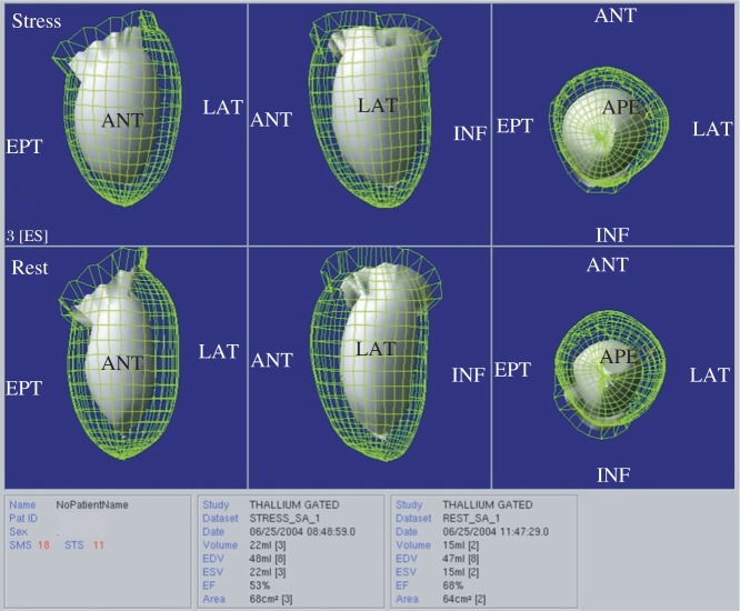
Fig. 8 shows the images of the same patient with triple vessel disease in Fig. 4. Although the perfusion images shown on MPS are near normal, the functional images show significant stress-induced stunning, as manifested by both dropping of LVEF and worsening of regional wall motion after vasodilator stress. This case indicates the incremental value of ischemic stunning in the detection of severe and extensive CAD with gated MPS.
Fig. 8. Representative functional images of ischemic stunning, in which wired grid and solid surface represent endocardial surfaces at end-diastole and end-systole, respectively.
The images show mild to moderate hypokinesis involving apical-mid anterior wall, septum and inferior wall on stress images. On rest images, the anterior wall and septum are normokinetic, and apical inferior wall is mildly hypokinetic. Worsening of regional wall motion after dipyridamole-stress is observed in apical mid anterior wall, septum and apical inferior wall. The LVEF of stress and rest are 53% and 68%, respectively. These findings indicate ischemic stunning.
STRESS-INDUCED DYSSYNCHRONY
Phase analysis for gated MPS was first developed by Chen et al. in 2005 for measuring LV dyssynchrony[35]. Previous studies have shown that quantitative indices given by phase analysis, such as phase standard deviation (PSD) and phase histogram bandwidth (PHB), were correlative well with LV dyssynchrony measured by tissue Doppler imaging[36]–[38], highly reproductive and repeatable[39],[40], and predictive of response to cardiac resynchronization therapy (CRT) in heart failure patients[41]. All of the above studies were done using Tc-99m labeled radiotracers (sestamibi or tetrofosmin). As we mentioned previously, gated MPS with Tc-99m tracers usually acquired data one hour or more post injection. They represented late post-stress function, which is close to resting function. Therefore, a study using Tc-99m sestamibi MPS reported that the presence of even large reversible defects did not alter LV dyssynchrony from rest to stress[42].
Chen et al. first validated the performance of Tl-201 gated MPS for phase analysis and found good correlation with Tc-99m sestamibi[43]. The same group further investigated whether stress-induced myocardial ischemia is associated with LV mechanical dyssynchrony[44]. This study demonstrated that stress-induced myocardial ischemia caused dyssynchronous contraction in the ischemic region, deteriorating LV synchrony. Normal myocardium had more synchronous contraction at stress, but scarred myocardium had no change. Example images are shown on Fig. 9. The different dyssynchrony pattern between ischemic and normal myocardium at early post-stress may aid diagnosis coronary artery disease using Tl-201 gated MPS. Further study is warranted to assess the incremental value of stress-induced dyssynchrony over conventional assessment of Tl-201 gated SPECT MPI in the diagnosis of CAD, especially in the settings of multi-vessel disease with balanced ischemia.
Fig. 9. Representative patients with ischemic (A), infarcted (B), or normal myocardium (C).
Myocardial ischemia deteriorates LV dyssynchrony and caused dropping of LVEF. Improved LV synchrony is noted for normal myocardium. In the patient with myocardial infarction, no change in LV synchrony is noted in the infarcted area, but the global indices of LV dyssynchrony (PSD and PHB) improves due to better synchrony in areas with normal myocardium.
CONCLUSION
MPS has been an established imaging modality, which provides very useful diagnostic and prognostic information for patients with suspected or known CAD. It has also been clinically accepted as the gatekeeper of further invasive procedures in the management of CAD. However, its limitations to underestimate the severity and/or extent of myocardial ischemia in patients with multi-vessel disease should be noticed carefully. Additional markers shown on MPS, such as TID, ischemic stunning or worsening dyssynchrony caused by stress, are all useful to help reduce or avoid this rare but serious clinical setting.
References
- 1.Strauss HW, Miller DD, Wittry MD, Cerqueira MD, Garcia EV, Iskandrian AS, et al. Procedure guideline for myocardial perfusion imaging. Society of Nuclear Medicine. J Nucl Med. 1998;39:918–23. [PubMed] [Google Scholar]
- 2.Anagnostopoulos C, Harbinson M, Kelion A, Kundley K, Loong CY, Notghi A, et al. Procedure guidelines for radionuclide myocardial perfusion imaging. Nucl Med Commun. 2003;24:1105–19. doi: 10.1097/01.mnm.0000095842.16659.4d. [DOI] [PubMed] [Google Scholar]
- 3.Strauss HW, Miller DD, Wittry MD, Cerqueira MD, Garcia EV, Iskandrian AS, et al. Procedure guideline for myocardial perfusion imaging 3.3. J Nucl Med Technol. 2008;36:155–61. doi: 10.2967/jnmt.108.056465. [DOI] [PubMed] [Google Scholar]
- 4.Underwood SR, Anagnostopoulos C, Cerqueira M, Ell PJ, Flint EJ, Harbinson M, et al. Myocardial perfusion scintigraphy: the evidence. Eur J Nucl Med Mol Imaging. 2004;31:261–91. doi: 10.1007/s00259-003-1344-5. [DOI] [PMC free article] [PubMed] [Google Scholar]
- 5.Berman DS, Hachamovitch R, Kiat H, Cohen I, Cabico JA, Wang FP, et al. Incremental value of prognostic testing in patients with known or suspected ischemic heart disease: a basis for optimal utilization of exercise technetium-99m sestamibi myocardial perfusion single-photon emission computed tomography. J Am Coll Cardiol. 1995;26:639–47. doi: 10.1016/0735-1097(95)00218-S. [DOI] [PubMed] [Google Scholar]
- 6.Hachamovitch R, Berman DS, Shaw LJ, Kiat H, Cohen I, Cabico JA, et al. Incremental prognostic value of myocardial perfusion single photon emission computed tomography for the prediction of cardiac death: differential stratification for risk of cardiac death and myocardial infarction. Circulation. 1998;97:535–43. doi: 10.1161/01.cir.97.6.535. [DOI] [PubMed] [Google Scholar]
- 7.Shaw LJ, Iskandrian AE. Prognostic value of gated myocardial perfusion SPECT. J Nucl Cardiol. 2004;11:171–85. doi: 10.1016/j.nuclcard.2003.12.004. [DOI] [PubMed] [Google Scholar]
- 8.Hachamovitch R, Hayes SW, Friedman JD, Cohen I, Berman DS. Comparison of the short-term survival benefit associated with revascularization compared with medical therapy in patients with no prior coronary artery disease undergoing stress myocardial perfusion single photon emission computed tomography. Circulation. 2003;107:2900–7. doi: 10.1161/01.CIR.0000072790.23090.41. [DOI] [PubMed] [Google Scholar]
- 9.Mahmarian JJ, Shaw LJ, Filipchuk NG, Dakik HA, Iskander SS, Ruddy TD, et al. A multinational study to establish the value of early adenosine technetium-99m sestamibi myocardial perfusion imaging in identifying a low-risk group for early hospital discharge after acute myocardial infarction. J Am Coll Cardiol. 2006;48:2448–57. doi: 10.1016/j.jacc.2006.07.069. [DOI] [PubMed] [Google Scholar]
- 10.Go V, Bhatt MR, Hendel RC. The diagnostic and prognostic value of ECG-gated SPECT myocardial perfusion imaging. J Nucl Med. 2004;45:912–21. [PubMed] [Google Scholar]
- 11.Berman DS, Kang X, Slomka PJ, Gerlach J, de Yang L, Hayes SW, et al. Underestimation of extent of ischemia by gated SPECT myocardial perfusion imaging in patients with left main coronary artery disease. J Nucl Cardiol. 2007;14:521–8. doi: 10.1016/j.nuclcard.2007.05.008. [DOI] [PubMed] [Google Scholar]
- 12.Bateman TM, Heller GV, McGhie AI, Friedman JD, Case JA, Bryngelson JR, et al. Diagnostic accuracy of rest/stress ECG-gated Rb-82 myocardial perfusion PET: comparison with ECG-gated Tc-99m sestamibi SPECT. J Nucl Cardiol. 2006;13:24–33. doi: 10.1016/j.nuclcard.2005.12.004. [DOI] [PubMed] [Google Scholar]
- 13.Weiss AT, Berman DS, Lew AS, Nielsen J, Potkin B, Swan HJ, et al. Transient ischemic dilation of the left ventricle on stress thallium-201 scintigraphy: a marker of severe and extensive coronary artery disease. J Am Coll Cardiol. 1987;9:752–9. doi: 10.1016/s0735-1097(87)80228-0. [DOI] [PubMed] [Google Scholar]
- 14.Mazzanti M, Germano G, Kiat H, Friedman J, Berman DS. Identification of severe and extensive coronary artery disease by automatic measurement of transient ischemic dilation of the left ventricle in dual-isotope myocardial perfusion SPECT. J Am Coll Cardiol. 1996;27:1612–20. doi: 10.1016/0735-1097(96)00052-6. [DOI] [PubMed] [Google Scholar]
- 15.McClellan JR, Travin MI, Herman SD, Baron JI, Golub RJ, Gallagher JJ, et al. Prognostic importance of scintigraphic left ventricular cavity dilation during intravenous dipyridamole technetium-99m sestamibi myocardial tomographic imaging in predicting coronary events. Am J Cardiol. 1997;79:600–5. doi: 10.1016/s0002-9149(96)00823-5. [DOI] [PubMed] [Google Scholar]
- 16.Abidov A, Bax JJ, Hayes SW, Hachamovitch R, Cohen I, Gerlach J, et al. Transient ischemic dilation ratio of the left ventricle is a significant predictor of future cardiac events in patients with otherwise normal myocardial perfusion SPECT. J Am Coll Cardiol. 2003;42:1818–25. doi: 10.1016/j.jacc.2003.07.010. [DOI] [PubMed] [Google Scholar]
- 17.Takeishi Y, Tono-oka I, Ikeda K, Komatani A, Tsuiki K, Yasui S . Dilatation of the left ventricular cavity on dipyridamole thallium-201 imaging: a new marker of triple-vessel disease. Am Heart J. 1991;121:466–75. doi: 10.1016/0002-8703(91)90713-r. [DOI] [PubMed] [Google Scholar]
- 18.Marcassa C, Galli M, Baroffio C, Campini R, Giannuzzi P. Transient left ventricular dilation at quantitative stress-rest sestamibi tomography: clinical, electrocardiographic, and angiographic correlates. J Nucl Cardiol. 1999;6:397–405. doi: 10.1016/s1071-3581(99)90005-3. [DOI] [PubMed] [Google Scholar]
- 19.Iskandrian AS, Heo J, Nguyen T, Beer S, Cave V, Cassel D, et al. Left ventricular dilatation and pulmonary thallium uptake after single-photon emission computer tomography using thallium-201 during adenosine-induced coronary hyperemia. Am J Cardiol. 1990;66:807–11. doi: 10.1016/0002-9149(90)90356-6. [DOI] [PubMed] [Google Scholar]
- 20.Hung GU, Lee KW, Chen CP, Lin WY, Yang KT. Relationship of transient ischemic dilation in dipyridamole myocardial perfusion imaging and stress-induced changes of functional parameters evaluated by Tl-201 gated SPECT. J Nucl Cardiol. 2005;12:268–75. doi: 10.1016/j.nuclcard.2005.03.004. [DOI] [PubMed] [Google Scholar]
- 21.Hansen CL, Goldstein RA, Berman DS, Churchwell KB, Cooke CD, Corbett JR, et al. Myocardial perfusion and function single photon emission computed tomography. J Nucl Cardiol. 2006;13:e97–120. doi: 10.1016/j.nuclcard.2006.08.008. [DOI] [PubMed] [Google Scholar]
- 22.Panting JR, Gatehouse PD, Yang GZ, Grothues F, Firmin DN, Collins P, et al. Abnormal subendocardial perfusion in cardiac syndrome X detected by cardiovascular magnetic resonance imaging. N Engl J Med. 2002;346:1948–53. doi: 10.1056/NEJMoa012369. [DOI] [PubMed] [Google Scholar]
- 23.Cannon RO., 3rd Microvascular angina and the continuing dilemma of chest pain with normal coronary angiograms. J Am Coll Cardiol. 2009;54:877–85. doi: 10.1016/j.jacc.2009.03.080. [DOI] [PMC free article] [PubMed] [Google Scholar]
- 24.Johnson LL, Verdesca SA, Aude WY, Xavier RC, Nott LT, Campanella MW, et al. Postischemic stunning can affect left ventricular ejection fraction and regional wall motion on poststress gated sestamibi tomograms. J Am Coll Cardiol. 1997;30:1641–8. doi: 10.1016/s0735-1097(97)00388-4. [DOI] [PubMed] [Google Scholar]
- 25.Hashimoto J, Kubo A, Iwasaki R, Iwanaga S, Mitamura H, Ogawa S, et al. Gated single-photon emission tomography imaging protocol to evaluate myocardial stunning after exercise. Eur J Nucl Med. 1999;26:1541–6. doi: 10.1007/s002590050492. [DOI] [PubMed] [Google Scholar]
- 26.Emmett L, Iwanochko RM, Freeman MR, Barolet A, Lee DS, Husain M. Reversible regional wall motion abnormalities on exercise technetium-99m-gated cardiac single photon emission computed tomography predict high-grade angiographic stenoses. J Am Coll Cardiol. 2002;39:991–8. doi: 10.1016/s0735-1097(02)01707-2. [DOI] [PubMed] [Google Scholar]
- 27.Yamagishi H, Shirai N, Yoshiyama M, Teragaki M, Akioka K, Takeuchi K, et al. Incremental value of left ventricular ejection fraction for detection of multivessel coronary artery disease in exercise (201)Tl gated myocardial perfusion imaging. J Nucl Med. 2002;43:131–9. [PubMed] [Google Scholar]
- 28.Shirai N, Yamagishi H, Yoshiyama M, Teragaki M, Akioka K, Takeuchi K, et al. Incremental value of assessment of regional wall motion for detection of multivessel coronary artery disease in exercise (201)Tl gated myocardial perfusion imaging. J Nucl Med. 2002;43:443–50. [PubMed] [Google Scholar]
- 29.Sharir T, Bacher-Stier C, Dhar S, Lewin HC, Miranda R, Friedman JD, et al. Identification of severe and extensive coronary artery disease by postexercise regional wall motion abnormalities in Tc-99m sestamibi gated single-photon emission computed tomography. Am J Cardiol. 2000;86:1171–5. doi: 10.1016/s0002-9149(00)01206-6. [DOI] [PubMed] [Google Scholar]
- 30.Ambrosio G, Betocchi S, Pace L, Losi MA, Perrone-Filardi P, Soricelli A, et al. Prolonged impairment of regional contractile function after resolution of exercise-induced angina. Evidence of myocardial stunning in patients with coronary artery disease. Circulation. 1996;94:2455–64. doi: 10.1161/01.cir.94.10.2455. [DOI] [PubMed] [Google Scholar]
- 31.Lee DS, Yeo JS, Chung JK, Lee MM, Lee MC. Transient prolonged stunning induced by dipyridamole and shown on 1- and 24-hour poststress 99mTc-MIBI gated SPECT. J Nucl Med. 2000;41:27–35. [PubMed] [Google Scholar]
- 32.Samady H, Wackers FJ, Joska TM, Zaret BL, Jain D. Pharmacologic stress perfusion imaging with adenosine: role of simultaneous low-level treadmill exercise. J Nucl Cardiol. 2002;9:188–96. doi: 10.1067/mnc.2002.119973. [DOI] [PubMed] [Google Scholar]
- 33.Hung GU, Lee KW, Chen CP, Yang KT, Lin WY. Worsening of left ventricular ejection fraction induced by dipyridamole on Tl-201 gated myocardial perfusion imaging predicts significant coronary artery disease. J Nucl Cardiol. 2006;13:225–32. doi: 10.1007/BF02971247. [DOI] [PubMed] [Google Scholar]
- 34.Shen TY, Chang MC, Hung GU, Kao CH, Hsu BL. Prognostic Value of Functional Variables as Assessed by Gated Tl-201 Myocardial Perfusion SPECT for Major Adverse Cardiac Events in Patients with Coronary Artery Disease. Acta Cardiologica Sinica. 2013;29:243–250. [PMC free article] [PubMed] [Google Scholar]
- 35.Chen J, Garcia EV, Folks RD, Cooke CD, Faber TL, Tauxe EL, et al. Onset of left ventricular mechanical contraction as determined by Phase analysis of ECG-gated myocardial Perfusion SPECT imaging: development of a diagnostic tool for assessment of cardiac mechanical dyssynchrony. J Nucl Cardiol. 2005;12:687–95. doi: 10.1016/j.nuclcard.2005.06.088. [DOI] [PubMed] [Google Scholar]
- 36.Henneman MM, Chen J, Ypenburg C, Dibbets P, Stokkel M, van der Wall EE, et al. Phase analysis of gated myocardial Perfusion SPECT compared to tissue Doppler imaging for the assessment of left ventricular dyssynchrony. J Am Coll Cardiol. 2007;49:1708–14. doi: 10.1016/j.jacc.2007.01.063. [DOI] [PubMed] [Google Scholar]
- 37.Marsan NA, Henneman MM, Chen J, Ypenburg C, Dibbets P, Ghio S, et al. Real-time 3-dimensional echocardiography as a novel approach to quantify left ventricular dyssynchrony: a comparison study with phase analysis of gated myocardial perfusion single photon emission computed tomography. J Am Soc Echocardiogr. 2008;21:801–7. doi: 10.1016/j.echo.2007.12.006. [DOI] [PMC free article] [PubMed] [Google Scholar]
- 38.Marsan NA, Henneman MM, Chen J, Ypenburg C, Dibbets P, Ghio S, et al. Left ventricular dyssynchrony assessed by two 3-dimensional imaging modalities: phase analysis of gated myocardial perfusion SPECT and tri-plane tissue Doppler imaging. Eur J Nucl Med Mol Imaging. 2008;35:166–73. doi: 10.1007/s00259-007-0539-6. [DOI] [PMC free article] [PubMed] [Google Scholar]
- 39.Trimble MA, Velazquez EJ, Adams GL, Honeycutt EF, Pagnanelli RA, Barnhart HX, et al. Repeatability and reproducibility of phase analysis of gated SPECT myocardial perfusion imaging used to quantify cardiac dyssynchrony. Nucl Med Commun. 2008;29:374–81. doi: 10.1097/MNM.0b013e3282f81380. [DOI] [PMC free article] [PubMed] [Google Scholar]
- 40.Lin X, Xu H, Zhao X, Folks RD, Faber TL, Garcia EV, et al. Repeatability of left ventricular dyssynchrony and function parameters in serial gated myocardial perfusion SPECT studies. J Nucl Cardiol. 2010;17:811–6. doi: 10.1007/s12350-010-9238-y. [DOI] [PMC free article] [PubMed] [Google Scholar]
- 41.Henneman MM, Chen J, Dibbets P, Stokkel M, Bleeker GB, Ypenburg C, van der Wall EE, Schalij MJ, Garcia EV, Bax JJ. Can LV dyssynchrony as assessed with phase analysis on gated myocardial Perfusion SPECT predict response to CRT? J Nucl Med. 2007;48:1104–11. doi: 10.2967/jnumed.107.039925. [DOI] [PubMed] [Google Scholar]
- 42.Aljaroudi W, Koneru J, Heo J, Iskandrian AE. Impact of ischemia on left ventricular dyssynchrony by phase analysis of gated single photon emission computed tomography myocardial perfusion imaging. J Nucl Cardiol. 2011;18:36–42. doi: 10.1007/s12350-010-9296-1. [DOI] [PubMed] [Google Scholar]
- 43.Chen CC, Huang WS, Hung GU, et al. Left Ventricular Dyssynchrony Evaluated by Tl-201 Gated SPECT Myocardial Perfusion Imaging: A Comparison with Tc-99m Sestamibi. Nucl Med Commun. 2013;34:229–32. doi: 10.1097/MNM.0b013e32835c91b9. [DOI] [PMC free article] [PubMed] [Google Scholar]
- 44.Chen CC, Shen TY, Chang MC, et al. Stress-induced Myocardial Ischemia is Associated with Early Post-stress Left Ventricular Mechanical Dyssynchrony as Assessed by Phase Analysis of Tl-201 Gated SPECT Myocardial Perfusion Imaging. Eur J Nucl Med Mol Imaging. 2012;39:1904–9. doi: 10.1007/s00259-012-2208-7. [DOI] [PMC free article] [PubMed] [Google Scholar]


