Abstract
Purpose:
To compare visual acuity with two visual acuity charts in preschool children.
Materials and Methods:
Visual acuity measurement with Lea symbols and Bailey-Lovie tumbling E chart was performed on children between 3 and 6 years of age. Visual acuity data from the two charts were analyzed with Bland-Altman plot to determine the limits of agreement. The Wilcoxon signed test was performed in children aged 3-4 years and in children aged 5-6 years separately to evaluate the influence of age. The inter-eye difference between the two charts were further analyzed with the paired t-test. A p value > 0.05 was considered statistically significant.
Results:
A total of 47 children were enrolled for the study. The average logarithm of the Minimum Angle of Resolution (LogMAR) monocular visual acuity with Lea symbols (0.17 ± 0.13) was better than the Bailey-Lovie tumbling E chart (0.22 ± 0.14). The mean difference between Bailey-Lovie tumbling E chart and Lea symbol chart was 0.05 ± 0.12 in logMAR units. A second analysis eliminating outliers showed the same result but lower differences (n = 43, 0.05 ± 0.05 logMAR units). Visual acuity results between the two charts in children aged 3-4 years showed a significant difference (p = 0.000), but not for children aged 5-6 years (p = 0.059). Inter-eye differences between the two charts was not statistically significant (p = 0.77).
Conclusion:
Bailey-Lovie tumbling E chart is comparable to the Lea symbols chart in pre-school children. But preference should be given to Lea symbols for children aged 3-4 years as the symbols are more familiar than a directional test for this age group.
Keywords: Bailey-Lovie tumbling E chart, Lea symbols, visual acuity
INTRODUCTION
Visual acuity assessment in children is one of the important ocular examinations for early detection of pediatric disorders including amblyopia.[1] It is also an important indicator of normal development of the eye. Various charts have already been in use for this purpose. Visual acuity measurement in preschool children can be a tedious process due to the uncooperative behavior with relevance to the age group. Charts such as Lea symbols, HOTV, landolt C, and tumbling E are commonly used to measure vision.[2,3,4]
Lea symbol chart is well accepted for visual acuity measurement worldwide and this can be used to test visual acuity in young children.[5] It uses four symbols (square, circle, house, and heart) and it is based on the logarithm of the Minimum Angle of Resolution (LogMAR) principle with lines that progress in 0.1 log unit steps and optotypes that are spaced proportionally. The Bailey-Lovie chart is commonly used for measurement of visual acuity in adults and it is constructed with proportional spacing of optotypes.[6] In addition to the letter acuity chart, the Bailey-Lovie tumbling E chart is also available for acuity measurement. Though many studies have been published on visual acuity measurements with tumbling E chart,[4,7,8] the comparison of this chart constructed based on logMAR principle has not been previously reported. Previous studies have used the Snellen E chart which has a number of well documented limitations.[9] In this study, we compared the visual acuity measurement between Lea symbol and Bailey-Lovie E chart in a group of preschool children in a day care center at Manipal University, India.
MATERIALS AND METHODS
The study protocol was approved by institutional review board at the Manipal College of Allied Health Sciences (MCOAHS), Manipal University and the study followed the tenets of the Declaration of Helsinki. This prospective, cross sectional study was conducted at the day care center located within the university campus. Permission from authorities was obtained to conduct the study in day care center and informed written consent was obtained from the parents before the commencement of the study.
A pilot study was conducted initially at the day care center on children between 3 and 6 years of age. The sample size for this study was calculated based on a 20% noncompliance rate that gave a sample size of 60 children. We recruited 63 children (36 males and 27 females) for our study. Children with poor cooperation and unreliable answers were excluded from the study. Distance visual acuity was measured monocularly using both charts. The order of testing (Lea or Bailey-Lovie E) was randomized across the subjects and visual acuity of both eyes was recorded with the right eye tested first in all children. Spectacle wearers were allowed to use spectacles while performing the visual acuity and the testing distance was 3 meters. In order to familiarize the children with the optotypes, all the children underwent a pretest at near which was conducted by the examiner (second author) with Lea symbols and E cards. During the visual acuity measurement, left eye was patched first and the children were asked to identify optotypes on the top line of the chart. If the child was able to identify three or more optotypes, then acuity measurement was with the identification of three optotypes in each subsequent line until one wrong response was made by the subject in one line. Then the examiner showed the remaining two symbols of the same line and if the child was able to answer at least three out of five symbols correctly, then he would proceed to the next line. If not, then the previous line plus a value of -0.02 log unit for each optotype that was identified correctly beyond that acuity level was taken as the visual acuity. The same procedure was repeated in the left eye. The time gap between the visual acuity measurements with two charts was not greater than 10–15 minutes.
All visual acuity data are presented in LogMAR units. Bland-Altman plots were used to determine the limits of agreement between the two charts. Intraclass correlation was also obtained to determine the agreement between the two tests.
The Wilcoxon signed rank test was performed for the monocular visual acuity obtained with two charts in children between 3 and 4 years old and in children between 5 and 6 years old separately. Inter-eye differences in visual acuity between the two charts was obtained and the difference between the results of the two charts was also determined with the paired t-test. A p value > 0.05 was considered statistically significant. The data were analyzed with SPSS 16.0 software (IBM Corp., Armonk, NY, USA) and the data for the right eye was only considered for the analysis except for the estimation of inter-eye differences between the two charts.
RESULTS
Of the 63 children in the day care center, 47 (75%) children were eligible for the study. The mean age of the study population was 4.3 ± 1.2 years.
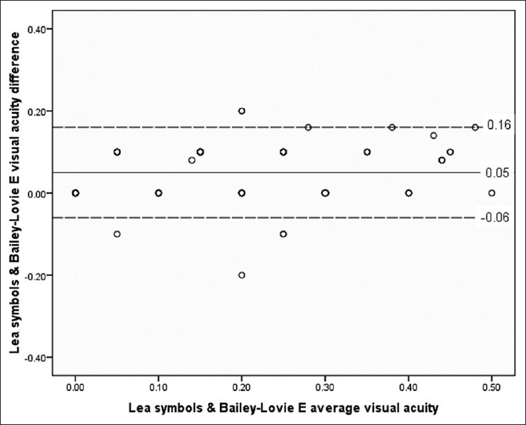
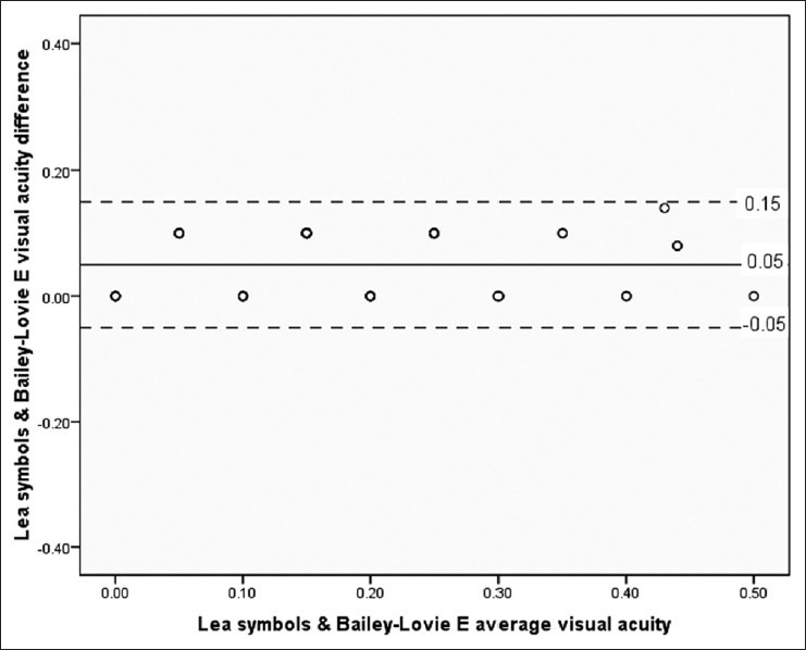
Mean, median, standard deviation (SD), and range of visual acuity with Lea symbols and Bailey-Lovie E chart in 3-4 years and 5-6 years are provided separately in Tables 1 and 2. The mean visual acuity was 0.17 ± 0.13 (range, 0.0-0.50), median 0.10, for the Lea symbols. The mean visual acuity was 0.22 ± 0.14 (range, 0.0-0.50), and the median was 0.20 for the Bailey-Lovie E chart. The mean difference between the Bailey-Lovie tumbling E chart and Lea symbol chart [Figure 1] was 0.05 ± 0.12 logMAR units with the upper and lower limits of agreement at 0.16 and -0.06, respectively. When the outliers observed in Figure 1 are removed and the analysis repeated [Figure 2], the mean difference was 0.05 ± 0.05 logMAR units with the upper and lower limits of agreement 0.15 and -0.05 (n = 43), respectively. The intraclass correlation was strong and statistically significant between the two visual acuity charts (alpha = 0.943, 95% CI: 0.592-0.965).
Table 1.
Mean, median, standard deviation (SD), and range of the visual acuity with Lea symbols

Table 2.
Mean, median, standard deviation (SD) and range of the visual acuity with Bailey-Lovie E chart

Figure 1.

Bland-Altman plots of Bailey-Lovie tumbling E and Lea symbols
Figure 2.

Bland-Altman plots between Bailey-Lovie tumbling E and Lea symbols after removing outliers
A statistically significant difference was found in visual acuity between the two charts in children aged 3 to 4 years (n = 28, p = 0.000); it was not significant in children aged 5-6 years (n = 19, p = 0.059). The inter-eye difference between Bailey-Lovie tumbling E and Lea symbols was not statistically significant (p = 0.77 paired t-test).
DISCUSSION
The results of this study showed that monocular visual acuity obtained with the Bailey-Lovie E chart is comparable to that of Lea symbols chart in the preschool age group. The intraclass correlation between the two charts was also comparable. Though the mean monocular visual acuity with Lea symbols was better than Bailey-Lovie tumbling E chart, the limits of agreement between the two charts were within clinically acceptable levels. A visual acuity difference of more than two lines (0.2 logMAR) is considered clinically significant.[10] In our study, the visual acuity difference between the two charts was (0.05 ± 0.12) within a two line difference. This was even more evident after the removal of outliers from the plot [Figure 2]. Both the plots in this study showed that the level of difference in the visual acuity between the two charts were unrelated to the level of average visual acuity.
We also separately analyzed the visual acuity results between charts in children aged 3-4 years and 5-6 years and found a statistically significant difference only for the 3-4 year aged group between the two charts was (p = 0.000). One advantage of Lea symbols chart is that it can be applicable even in young children and extracts better responses from them.[2] The tumbling E chart and the Landolt C chart are widely used charts for measuring visual acuity in children in various areas.[7,11] However the outcome indicates that in younger children the difference in monocular visual acuity between the two charts is significant. Similar to the previous studies our study also confirmed that visual acuity is almost fully developed in children aged 5-6 years.[12,13]
For successful management of amblyopia, early diagnosis of amblyopia is imperative. Hence, inter-eye difference in measurement has to be accurate for this purpose. This study showed that the inter-eye difference between the two charts was not statistically significant (p = 0.77), which means the Bailey-Lovie E chart also provided a similar inter-eye difference as the Lea symbols chart. However, both the charts are based on the logMAR principle and they are much more sensitive for detecting inter-eye differences in visual acuity.[14,15]
The research in vision in preschoolers (VIP) study compared visual acuity results between Lea symbols and Bailey-Lovie letter chart in individuals between 4.5 60 years old.[16] They[16] found that there was no absolute inter-eye difference measured with two acuity charts. They also showed that the monocular visual acuity with Lea symbol was better than Bailey-Lovie letter chart. The reason being that there is 25% chance of guessing the correct symbol with Lea symbol compared to the 10% chance of guessing with Bailey-Lovie letter chart (consisted of 10 letters).[16] However, in our study, both the Lea and E charts have a 25% chance of guessing since both consisted of four different optotypes. Therefore the chance of guessing cannot explain better monocular visual acuity with Lea symbols. The probable reason for this could be due to the familiarity with the symbols as compared with a directional test in the younger age group. Moreover, the abstraction of Lea symbols is minimal, hence, it is readily comprehensible.[17]
In our study, out of the 63 children, 16 were unable to complete the visual acuity assessment, particularly with the tumbling E chart. The reason for this good success rate in visual acuity assessment in this age group could be due to the pretest conducted by the examiner. However, despite pretesting, it was observed that the success rate with tumbling E was not as good as the Lea symbols (14 children were unable to complete the visual acuity assessment compared to three with Lea symbols). It also should be noted that the study was carried out in a single day care center. Though success rate with Lea symbols has been previously reported,[18] it would be ideal to carry out this comparison between the two charts in each age group separately in a larger population. This comparison will also help to establish the visual acuity norms with each chart in each age group separately.
In summary, we examined the effectiveness of Bailey-Lovie E chart in comparison with Lea symbols chart. The results indicated that Bailey-Lovie E chart is interchangeable with Lea symbols chart in children aged 5–6 years but not in children who are 3–4 years old.
Footnotes
Source of Support: Nil
Conflict of Interest: None declared.
REFERENCES
- 1.Lorenz B, Brodsky MC. Pediatric ophthalmology, neuro ophthalmology, genetics. 1st ed. Heidelberg: Springer; 2010. [Google Scholar]
- 2.Becker R, Hübsch S, Gräf MH, Kaufmann H. Examination of young children with Lea symbols. Br J Ophthalmol. 2002;86:513–6. doi: 10.1136/bjo.86.5.513. [DOI] [PMC free article] [PubMed] [Google Scholar]
- 3.Cyert L, Schmidt P, Maguire M, Moore B, Dobson V, Quinn G. Threshold visual acuity testing of preschool children using the crowded HOTV and Lea Symbols acuity tests. J AAPOS. 2003;7:396–9. doi: 10.1016/s1091-8531(03)00211-8. [DOI] [PubMed] [Google Scholar]
- 4.Lai HY, Wang HZ, Hsu HT. Development of visual acuity in preschool children as measured with Landolt C and Tumbling E charts. J AAPOS. 2011;15:251–5. doi: 10.1016/j.jaapos.2011.03.010. [DOI] [PubMed] [Google Scholar]
- 5.Hyvärinen L, Näsänen R, Laurinen P. New visual acuity test for pre-school children. Acta Ophthalmol (Copenh) 1980;58:507–11. doi: 10.1111/j.1755-3768.1980.tb08291.x. [DOI] [PubMed] [Google Scholar]
- 6.Bailey IL, Lovie JE. New design principles for visual acuity letter charts. Am J Optom Physiol Opt. 1976;53:740–5. doi: 10.1097/00006324-197611000-00006. [DOI] [PubMed] [Google Scholar]
- 7.Lai YH, Hsu HT, Wang HZ, Chang SJ, Wu WC. The visual status of children ages 3 to 6 years in the vision screening program in Taiwan. J AAPOS. 2009;13:58–62. doi: 10.1016/j.jaapos.2008.07.006. [DOI] [PubMed] [Google Scholar]
- 8.Grimm W, Rassow B, Wesemann W, Saur K, Hilz R. Correlation of optotypes with the Landolt ring-a fresh look at the comparability of optotypes. Optom Vis Sci. 1994;71:6–13. doi: 10.1097/00006324-199401000-00002. [DOI] [PubMed] [Google Scholar]
- 9.Hussain B, Saleh GM, Sivaprasad S, Hammod CJ. Changing from snellen to logMAR: Debate or Delay? Clin Exp Ophthalmol. 2006;34:6–8. doi: 10.1111/j.1442-9071.2006.01135.x. [DOI] [PubMed] [Google Scholar]
- 10.Lovie-Kitchin J. Validity and reliability of visual acuity measurements. Ophthalmic Physiol Opt. 1988;8:363–70. doi: 10.1111/j.1475-1313.1988.tb01170.x. [DOI] [PubMed] [Google Scholar]
- 11.He M, Xu J, Yin Q, Ellwein LB. Need and challenges of refractive correction in urban Chinese school children. Optom Vis Sci. 2005;82:229–34. doi: 10.1097/01.opx.0000159362.48835.16. [DOI] [PubMed] [Google Scholar]
- 12.Pan Y, Tarczy-Hornoch K, Cotter SA, Wen G, Borchert MS, Azen SP, et al. Visual acuity norms in pre-school children: The multi-ethnic pediatric eye disease study. Optom Vis Sci. 2009;86:607–12. doi: 10.1097/OPX.0b013e3181a76e55. [DOI] [PMC free article] [PubMed] [Google Scholar]
- 13.Dobson V, Clifford-Donaldson CE, Green TK, Miller JM, Harvey EM. Normative monocular visual acuity for early treatment diabetic retinopathy study charts in emmetropic children 5 to 12 years of age. Ophthalmology. 2009;116:1397–401. doi: 10.1016/j.ophtha.2009.01.019. [DOI] [PMC free article] [PubMed] [Google Scholar]
- 14.Simmers AJ, Gray LS, Spowart K. Screening for amblyopia: a comparison of paediatric letter tests. Br J Ophthalmol. 1997;81:465–9. doi: 10.1136/bjo.81.6.465. [DOI] [PMC free article] [PubMed] [Google Scholar]
- 15.McGraw PV, Winn B, Gray LS, Elliot DB. Improving the reliability of visual acuity measures in young children. Ophthal Physiol Opt. 2000;20:173–84. [PubMed] [Google Scholar]
- 16.Dobson V, Maguire M, Bixler DO, Quinn G, Ying GS. Visual acuity results in school-aged children and adults: Lea symbols chart versus Bailey-Lovie chart. Optom Vis Sci. 2003;80:650–54. doi: 10.1097/00006324-200309000-00010. [DOI] [PubMed] [Google Scholar]
- 17.Gräf MH, Becker R, Kaufmann H. Lea symbols: Visual acuity assessment and detection of amblyopia. Br J Ophthalmol. 2000;238:53–8. doi: 10.1007/s004170050009. [DOI] [PubMed] [Google Scholar]
- 18.Hered RW, Murphy S, Clancy M. Comparison of the HOTV and Lea Symbols charts for preschool vision screening. J Pediatr Ophthalmol Strabismus. 1997;34:24–8. doi: 10.3928/0191-3913-19970101-06. [DOI] [PubMed] [Google Scholar]


