Abstract
Abstract
The interactions between phonons and electrons induced by the dopants or the substrate of graphene in spectroscopic investigation reveal a rich source of interesting physics. Raman spectra and surface-enhanced Raman spectra of supported and suspended monolayer graphenes were measured and analyzed systemically with different approaches. The weak Raman signals are greatly enhanced by the ability of surface-enhanced Raman spectroscopy which has attracted considerable interests. The technique is regarded as wonderful and useful tool, but the dopants that are produced by depositing metallic nanoparticles may affect the electron scattering processes of graphene. Therefore, the doping and substrate influences on graphene are also important issues to be investigated. In this work, the peak positions of G peak and 2D peak, the I2D/IG ratios, and enhancements of G and 2D bands with suspended and supported graphene flakes were measured and analyzed. The peak shifts of G and 2D bands between the Raman and SERS signals demonstrate the doping effect induced by silver nanoparticles by n-doping. The I2D/IG ratio can provide a more sensitive method to carry out the doping effect on the graphene surface than the peak shifts of G and 2D bands. The enhancements of 2D band of suspended and supported graphenes reached 138, and those of G band reached at least 169. Their good enhancements are helpful to measure the optical properties of graphene. The different substrates that covered the graphene surface with doping effect are more sensitive to the enhancements of G band with respect to 2D band. It provides us a new method to distinguish the substrate and doping effect on graphene.
PACS
78.67.Wj (optical properties of graphene); 74.25.nd (Raman and optical spectroscopy); 63.22.Rc (phonons in graphene)
Keywords: Suspended graphene, Raman spectroscopy, SERS
Background
Graphene, the thinnest sp2 allotrope of carbon arranged in a honeycomb lattice, has attracted many attentions because its unique and novel electrical and optical properties [1-3]. The wonderful and remarkable carrier transport properties of suspended graphene compared with supported graphene have been studied [4-9]. The performances of dopants, the effects of defects in graphene, and the phonon modes of suspended and supported graphenes vary but can be well understood using Raman spectroscopy [10-12]. Raman spectroscopy and surface-enhanced Raman spectroscopy (SERS) have been extensively applied to understand the vibration properties of materials [13-18], and they are regarded as powerful techniques in characterizing the band structure and detail of phonon graphene interaction [19-24]. The ability of SERS, a wonderful and useful technique used to enhance weak Raman signals, has attracted considerable attention. In previous SERS measurements, however, the doping induced by metallic nanoparticles on graphene deposition may affect the electron scattering processes of graphene. Otherwise, the metallic nanoparticles on graphene are also used as an electrode in graphene-based electronic devices [25,26]. Therefore, the effect of charged dopants and the substrate which affected graphene are both important issues to be investigated. In this work, the supported and suspended monolayer graphene samples were fabricated by micromechanical cleavage method. They were identified as the monolayer graphene by the optical microscopy and Raman spectroscopy because of the color contrast, various bandwidth, and peak position of 2D band with different graphene layers [27,28]. The Raman and SERS signals of suspended and supported graphenes can be measured and analyzed systematically. The peak positions of G and 2D bands, the I2D/IG ratio, and enhancements of G and 2D bands were obtained, respectively. With our analysis, details about the effects of charged impurities and substrate can be realized. The peak shift of G and 2D bands and the I2D/IG ratio are useful to demonstrate the dopants and substrate effects on the graphene. The well-enhanced G and 2D bands are obtained to enhance the weak Raman signals. Moreover, the enhancements of G band with respect to 2D band are found to be more sensitive to various substrate influences on the graphene surface. This paper provides a new approach to investigate the substrate and doping effect on graphene.
Methods
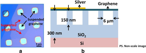
Suspended graphene was fabricated by mechanical exfoliation of graphene flakes onto an oxidized silicon wafer. The optical image of suspended and supported graphenes and the illustration of their coverage by silver nanoparticles are shown in Figure 1. Orderly arranged squares with areas of 6 μm2 were first defined by photolithography on an oxidized silicon wafer with an oxide thickness of 300 nm. Reactive ion etching was then used to etch the squares to a depth of 150 nm. Highly ordered pyrolytic graphite was consequently cleaved with the protection of scotch tape to enable the suspended graphene flakes to be deposited over the indents. To study the SERS, silver nanoparticles were deposited on the graphene flake at a deposition rate of 0.5 nm/min by a thermal deposition system. A 5-nm-thick layer of silver nanoparticles on the graphene flake was thus formed. To measure the graphene flake, a micro-Raman microscope (Jobin Yvon iHR550; HORIBA, Ltd., Minami-ku, Kyoto, Japan) was utilized to obtain the Raman and SERS signals of monolayer graphene. The monolayer graphene was identified through optical observation with various color contrast and by Raman spectroscopy with the different shape bandwidths and peak positions of 2D band under different graphene layers. During spectroscopic measurement, a 632-nm He-Ne laser was used as the excitation source; the power was monitored and controlled under 0.5 mW to avoid the heating of the graphene surface.
Figure 1.

Optical image of suspended and supported graphenes and their coverage by silver nanoparticles. Optical image of suspended and supported graphenes (a) and their illustrations covered by silver nanoparticles (b).
Results and discussion
To explore the SERS on graphene, the interactions between metallic nanoparticles and graphene surface has to be presumably understood. This is because the plasmonic resonances of nanoparticles with different shapes and sizes can affect the interactions between them, and then change the SERS signals [18,29-33]. Therefore, the morphologies of silver nanoparticles that covered the suspended and supported graphenes are to be investigated. The images of silver nanoparticles that covered suspended and supported graphenes were obtained by the scanning electron microscopy (SEM) and are shown in Figure 2a, b, c. The average size of silver nanoparticles were determined by the histogram analysis [34], of which the suspended graphene is 25.4 ± 2.2 nm and the supported graphene is 25.2 ± 2.4 nm. No clear size difference has been found between supported and suspended graphene flakes. In addition, their shapes are found in random form. It can also be seen that the silver nanoparticles deposited on the suspended and supported graphenes are in indistinguishable shape. Silver nanoparticles are therefore not contributing to any SERS variation.
Figure 2.

SEM images. (a) Supported and suspended graphenes which was identified as monolayer graphene. (b) Suspended graphene. (c) Supported graphene
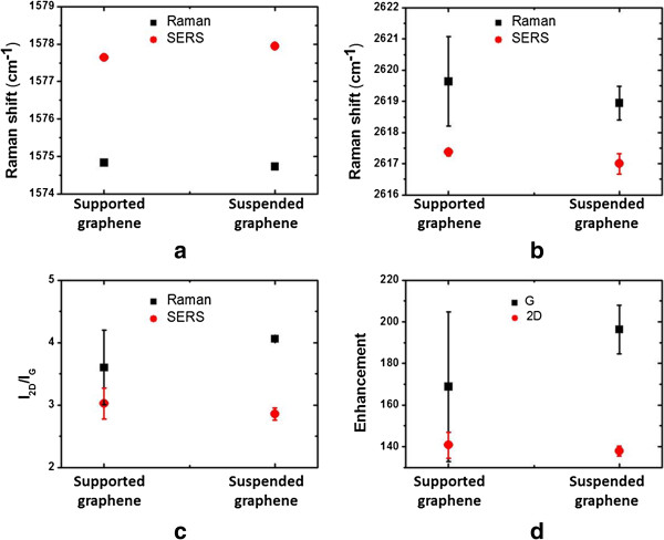
According to previous work, the peak positions and I2D/IG ratios of G and 2D bands were important indicators of doping effect on graphene [35-40], in which the I2D/IG ratio is particularly more sensitive than the peak shifts to the doping effect. A lower I2D/IG ratio is related to more charged impurities in graphene. The Raman and SERS signals of the suspended and the supported graphenes are shown in Figure 3a, b, c, d. The peak positions of G and 2D bands are presented in Figure 3a, b. Both the peak positions of G and 2D bands are indistinguishable between the suspended and supported graphenes, which reveals the difference in substrates which do not affect the graphene emission spectra. The G peak position of suspended and supported graphenes under Raman signals is both upshifted with respect to SERS signals, while the 2D peak under Raman signals is both downshifted with respect to SERS signals. According to previous work [35-37,39], the upshifting of G peak and the downshifting of 2D peak is caused by n-doping, as the silver nanoparticles were depositing on the graphene. The experimental results of this work have had a significant agreement with the previous research.
Figure 3.
Peak positions. (a) G band and (b) 2D band of suspended and supported graphenes with Raman and SERS signals. (c) I2D/IG ratios of suspended and supported graphenes with Raman and SERS signals. (d) Enhancements of G and 2D bands of suspended and supported graphenes.
In order to minimize the random errors, each Raman spectra data point was obtained by five-time repetitions. As presented in Figure 3c, the I2D/IG ratio of suspended graphene under Raman signals is 4.1 ± 0.1 and larger than supported graphene which is 3.6 ± 0.5, while the I2D/IG ratio of suspended graphene on the SERS signals is around 2.9 ± 0.1 and smaller than supported graphene which is 3.0 ± 0.2. The result disclosed the substrate effect on the supported graphene is stronger than the suspended graphene. To our understanding, this above effect can be reasonable because SiO2/Si substrate has a direct contact on the graphene surface so the silver nanoparticles deposition causes stronger doping effect on the suspended graphene than supported graphene.
To understand the Raman and SERS signals, the enhancements of G and 2D bands with the suspended and supported graphenes are shown in Figure 3d, respectively. The enhancement is defined as the integrated intensities of SERS over Raman signals for the G and 2D bands, respectively. In our analysis, the enhancement of G band on supported graphene is 169.3 ± 20.1 and smaller than suspended graphene which is 196.2 ± 8.3, while the enhancement of 2D band with supported graphene which is 141.1 ± 4.3 is similar with suspended graphene which is 138.6 ± 1.6. The high enhancements of G and 2D bands are useful to enhance weak Raman signals, and the enhancements of G band with suspended and supported graphenes are both stronger than those of 2D band. Otherwise, the enhancement of G band is reduced obviously as silver nanoparticles deposited on suspended graphene, revealing that the enhancement of G band is sensitive to substrate effect on graphene with respect to 2D band. Based on the results, the doping effects with various substrates are obviously related to the enhancement of G band.
Conclusions
In our work, Raman and SERS signals of supported and suspended monolayer graphenes were measured systematically. The peak positions of G and 2D bands and the I2D/IG ratios were varied. The enhancement effect of suspended and supported graphenes was calculated and analyzed. The peak shifts of G and 2D bands and their Raman spectra and I2D/IG of SERS signals are found very useful in the investigation of the substrate and doping effect on the optical properties of graphene. The enhancements of G and 2D bands have been found to cause the great improvement of weak Raman signals. Otherwise, the more sensitive enhancements of G band with respect to 2D band are related to the doping effect with various substrates that covered the graphene surface. The optical emission spectra of suspended and supported graphenes have provided us a with new identification approach to understand the substrate and doping effect on graphene.
Competing interests
The authors declare that they have no competing interests.
Authors' contributions
C-H and B-JL carried on the experimental parts: the acquisition of data and analysis and interpretation of data. C-H also had been involved in drafting the manuscript. H-YL and C-HH analyzed and interpreted the data. They also had been involved in revising the manuscript. F-YS and W-HW (Institute of Atomic and Molecular Sciences, Academia Sinica) prepared the samples, suspended graphene using by micromechanical method, and captured the OM and AFM images. C-YL have made substantial contributions to the conception and design of the study and revising it critically for important intellectual content. H-CC, the corresponding author, had made substantial contributions to the conception and design of the study and had been involved in drafting the manuscript and revised it critically for important intellectual content. All authors read and approved the final manuscript.
Contributor Information
Cheng-Wen Huang, Email: chengwen620@gmail.com.
Bing-Jie Lin, Email: kingstart13@yahoo.com.tw.
Hsing-Ying Lin, Email: acsiolin@gmail.com.
Chen-Han Huang, Email: chenhan512@gmail.com.
Fu-Yu Shih, Email: fu.yu.shih@gmail.com.
Wei-Hua Wang, Email: wwang@sinica.edu.tw.
Chih-Yi Liu, Email: liu.chihyi@gmail.com.
Hsiang-Chen Chui, Email: hcchui@mail.ncku.edu.tw.
Acknowledgement
We wish to acknowledge the support of this work by the National Science Council, Taiwan under contact nos. NSC 98-2112-M-006-004-MY3, NSC 101-2112-M-006-006, and NSC 102-2622-E-006-030-CC3.
References
- Novoselov KS, Geim AK, Morozov SV, Jiang D, Zhang Y, Dubonos SV, Grigorieva IV, Firsov AA. Electric field effect in atomically thin carbon films. Science. 2004;8:666–669. doi: 10.1126/science.1102896. [DOI] [PubMed] [Google Scholar]
- Geim AK, Novoselov KS. The rise of graphene. Nat Mater. 2007;8:183–191. doi: 10.1038/nmat1849. [DOI] [PubMed] [Google Scholar]
- Geim AK. Graphene: status and prospects. Science. 2009;8:1530–1534. doi: 10.1126/science.1158877. [DOI] [PubMed] [Google Scholar]
- Du X, Skachko I, Barker A, Andrei EY. Approaching ballistic transport in suspended graphene. Nat Nanotechnol. 2008;8:491–495. doi: 10.1038/nnano.2008.199. [DOI] [PubMed] [Google Scholar]
- Du X, Skachko I, Duerr F, Luican A, Andrei EY. Fractional quantum Hall effect and insulating phase of Dirac electrons in graphene. Nature. 2009;8:192–195. doi: 10.1038/nature08522. [DOI] [PubMed] [Google Scholar]
- Bolotin KI, Ghahari F, Shulman MD, Stormer HL, Kim P. Observation of the fractional quantum Hall effect in graphene. Nature. 2009;8:196. doi: 10.1038/nature08582. [DOI] [PubMed] [Google Scholar]
- Bolotin KI, Sikes KJ, Hone J, Stormer HL, Kim P. Temperature-dependent transport in suspended graphene. Phys Rev Lett. 2008;8:096802. doi: 10.1103/PhysRevLett.101.096802. [DOI] [PubMed] [Google Scholar]
- Chen SY, Ho PH, Shiue RJ, Chen CW, Wang WH. Transport/magnetotransport of high-performance graphene transistors on organic molecule-functionalized substrates. Nano Lett. 2012;8:964–969. doi: 10.1021/nl204036d. [DOI] [PubMed] [Google Scholar]
- Rouhi N, Wang YY, Burke PJ. Ultrahigh conductivity of large area suspended few layer graphene films. Appl Phys Lett. 2012;8:263101. doi: 10.1063/1.4772797. [DOI] [Google Scholar]
- Compagnini G, Forte G, Giannazzo F, Raineri V, La Magna A, Deretzis I. Ion beam induced defects in graphene: Raman spectroscopy and DFT calculations. J Mol Struct. 2011;8:506–509. doi: 10.1016/j.molstruc.2010.12.065. [DOI] [Google Scholar]
- Sahoo S, Palai R, Katiyar RS. Polarized Raman scattering in monolayer, bilayer, and suspended bilayer graphene. J Appl Phys. 2011;8:044320. doi: 10.1063/1.3627154. [DOI] [Google Scholar]
- Cancado LG, Jorio A, Ferreira EHM, Stavale F, Achete CA, Capaz RB, Moutinho MVO, Lombardo A, Kulmala TS, Ferrari AC. Quantifying defects in graphene via Raman spectroscopy at different excitation energies. Nano Lett. 2011;8:3190–3196. doi: 10.1021/nl201432g. [DOI] [PubMed] [Google Scholar]
- Suëtaka W. Surface Infrared and Raman Spectroscopy: Methods and Applications. New York: Plenum; 1995. [Google Scholar]
- Wang JK, Tsai CS, Lin CE, Lin JC. Vibrational dephasing dynamics at hydrogenated and deuterated semiconductor surfaces: symmetry analysis. J Chem Physics. 2000;8:5041–5052. doi: 10.1063/1.1289928. [DOI] [Google Scholar]
- Kneipp K, Moskovits M, Kneipp H. Surface-Enhanced Raman Scattering: Physics and Applications. Berlin and Heidelberg: Springer; 2006. [Google Scholar]
- Wang HH, Liu CY, Wu SB, Liu NW, Peng CY, Chan TH, Hsu C-F, Wang J-K, Wang Y-L. Highly Raman-enhancing substrates based on silver nanoparticle arrays with tunable sub-10 nm gaps. Adv Mater. 2006;8:491–495. doi: 10.1002/adma.200501875. [DOI] [Google Scholar]
- Liu CY, Dvoynenko MM, Lai MY, Chan TH, Lee YR, Wang JK, Wang YL. Anomalously enhanced Raman scattering from longitudinal optical phonons on Ag-nanoparticle-covered GaN and ZnO. Appl Phys Lett. 2010;8:033109. doi: 10.1063/1.3291041. [DOI] [Google Scholar]
- Huang CH, Lin HY, Chen ST, Liu CY, Chui HC, Tzeng YH. Electrochemically fabricated self-aligned 2-D silver/alumina arrays as reliable SERS sensors. Opt Express. 2011;8:11441–11450. doi: 10.1364/OE.19.011441. [DOI] [PubMed] [Google Scholar]
- Ferrari AC, Meyer JC, Scardaci V, Casiraghi C, Lazzeri M, Mauri F, Piscanec S, Jiang D, Novoselov KS, Roth S, Geim AK. Raman spectrum of graphene and graphene layers. Phys Rev Lett. 2006;8:187401. doi: 10.1103/PhysRevLett.97.187401. [DOI] [PubMed] [Google Scholar]
- Malard LM, Pimenta MA, Dresselhaus G, Dresselhaus MS. Raman spectroscopy in graphene. Physics-Rep Rev Sec Physics Lett. 2009;8:51–87. [Google Scholar]
- Gao LB, Ren WC, Liu BL, Saito R, Wu ZS, Li SS, Jiang C, Li F, Cheng H-M. Surface and interference coenhanced Raman scattering of graphene. ACS Nano. 2009;8:933–939. doi: 10.1021/nn8008799. [DOI] [PubMed] [Google Scholar]
- Schedin F, Lidorikis E, Lombardo A, Kravets VG, Geim AK, Grigorenko AN, Novoselov KS, Ferrari AC. Surface-enhanced Raman spectroscopy of graphene. Acs Nano. 2010;8:5617–5626. doi: 10.1021/nn1010842. [DOI] [PubMed] [Google Scholar]
- Wu D, Zhang F, Liu P, Feng X. Two-dimensional nanocomposites based on chemically modified graphene. Chem-a Eur J. 2011;8:10804–10812. doi: 10.1002/chem.201101333. [DOI] [PubMed] [Google Scholar]
- Huang CW, Lin BJ, Lin HY, Huang CH, Shih FY, Wang WH, Liu CY, Chui HC. Observation of strain effect on the suspended graphene by polarized Raman spectroscopy. Nanoscale Res Lett. 2012;8:533. doi: 10.1186/1556-276X-7-533. [DOI] [PMC free article] [PubMed] [Google Scholar]
- Casiraghi C, Pisana S, Novoselov KS, Geim AK, Ferrari AC. Raman fingerprint of charged impurities in graphene. Appl Phys Lett. 2007;8:233108. doi: 10.1063/1.2818692. [DOI] [Google Scholar]
- Ni ZH, Yu T, Luo ZQ, Wang YY, Liu L, Wong CP, Miao J, Huang W, Shen ZX. Probing charged impurities in suspended graphene using Raman spectroscopy. Acs Nano. 2009;8:569–574. doi: 10.1021/nn900130g. [DOI] [PubMed] [Google Scholar]
- Gupta A, Chen G, Joshi P, Tadigadapa S, Eklund PC. Raman scattering from high-frequency phonons in supported n-graphene layer films. Nano Lett. 2006;8:2667–2673. doi: 10.1021/nl061420a. [DOI] [PubMed] [Google Scholar]
- Graf D, Molitor F, Ensslin K, Stampfer C, Jungen A, Hierold C, Wirtz L. Spatially resolved Raman spectroscopy of single- and few-layer graphene. Nano Lett. 2007;8:238–242. doi: 10.1021/nl061702a. [DOI] [PubMed] [Google Scholar]
- Mock JJ, Barbic M, Smith DR, Schultz DA, Schultz S. Shape effects in plasmon resonance of individual colloidal silver nanoparticles. J Chem Physics. 2002;8:6755–6759. doi: 10.1063/1.1462610. [DOI] [Google Scholar]
- Duan GT, Cai WP, Luo YY, Li ZG, Li Y. Electrochemically induced flowerlike gold nanoarchitectures and their strong surface-enhanced Raman scattering effect. Appl Phys Lett. 2006;8:211905. doi: 10.1063/1.2392822. [DOI] [Google Scholar]
- Tiwari VS, Oleg T, Darbha GK, Hardy W, Singh JP, Ray PC. Non-resonance: SERS effects of silver colloids with different shapes. Chem Physics Lett. 2007;8:77–82. doi: 10.1016/j.cplett.2007.07.106. [DOI] [Google Scholar]
- Huang CH, Lin HY, Lin CH, Chui HC, Lan YC, Chu SW. The phase-response effect of size-dependent optical enhancement in a single nanoparticle. Opt Express. 2008;8:9580–9586. doi: 10.1364/OE.16.009580. [DOI] [PubMed] [Google Scholar]
- Zhang JT, Li XL, Sun XM, Li YD. Surface enhanced Raman scattering effects of silver colloids with different shapes. J Physical Chem B. 2005;8:12544–12548. doi: 10.1021/jp050471d. [DOI] [PubMed] [Google Scholar]
- Huang CW, Lin HY, Huang CH, Shiue RJ, Wang WH, Liu CY, Chui H-C. Layer-dependent morphologies of silver on n-layer graphene. Nanoscale Res Lett. 2012;8:618. doi: 10.1186/1556-276X-7-618. [DOI] [PMC free article] [PubMed] [Google Scholar]
- Lee J, Novoselov KS, Shin HS. Interaction between metal and graphene: dependence on the layer number of graphene. Acs Nano. 2011;8:608–612. doi: 10.1021/nn103004c. [DOI] [PubMed] [Google Scholar]
- Pisana S, Lazzeri M, Casiraghi C, Novoselov KS, Geim AK, Ferrari AC, Mauri F. Breakdown of the adiabatic Born-Oppenheimer approximation in graphene. Nat Mater. 2007;8:198–201. doi: 10.1038/nmat1846. [DOI] [PubMed] [Google Scholar]
- Shi YM, Dong XC, Chen P, Wang JL, Li LJ. Effective doping of single-layer graphene from underlying SiO2 substrates. Physical Rev B. 2009;8:115402. [Google Scholar]
- Basko DM, Piscanec S, Ferrari AC. Electron–electron interactions and doping dependence of the two-phonon Raman intensity in graphene. Phys Rev. 2009;8:165413. [Google Scholar]
- Das A, Pisana S, Chakraborty B, Piscanec S, Saha SK, Waghmare UV, Novoselov KS, Krishnamurthy HR, Geim AK, Ferrari AC, Sood AK. Monitoring dopants by Raman scattering in an electrochemically top-gated graphene transistor. Nat Nanotechnol. 2008;8:210–215. doi: 10.1038/nnano.2008.67. [DOI] [PubMed] [Google Scholar]
- Stampfer C, Molitor F, Graf D, Ensslin K, Jungen A, Hierold C, Ensslin K. Raman imaging of doping domains in graphene on SiO(2) Appl Phys Lett. 2007;8:241907. doi: 10.1063/1.2816262. [DOI] [Google Scholar]


