Abstract
CD56 positive B-cell lymphoma is very rare. We experienced a case of CD56 positive diffuse large B-cell lymphoma, occurred in a young child. A 5-year-old girl complained with snoring and open mouth breathing. No any abnormality in laboratory or physical examination was present, except enlarged both tonsils. Bilateral tonsillectomy was performed. Cut sections of right tonsil showed a 2 cm size, solid mass. On microscopically, large monomorphic lymphoid cells were diffusely proliferated and showed positivity for CD20 and CD56 and negative for Epstein-Barr virus (EBV) polymerase chain reaction (PCR). Monoclonality was observed on immunoglobulin heavy chain gene rearrangement. This is a unique case with incidentally found and occurred in a young child.
Keywords: CD56, diffuse large B cell lymphoma, immunohistochemistry
Introduction
CD56, the neural cell adhesion molecule (NCAM), is a member of the immunoglobulin superfamily, is expressed on neuroendocrine cells, natural killer cell (NK) cells and a subset of T-cells [1,2]. CD56 expression has been reported in mainly NK/T-cell lymphoma, multiple myeloma, acute myeloid leukemia, and myeloproliferative disorder [3,4]. CD56 expression in B-cell lymphomas has been reported to be <0.5% of all B-cell lymphomas [5]. To the best of our knowledge, there are 10 reports, including 35 cases of CD56 positive diffuse large B-cell lymphoma (CD56 positive DLBCL) in English literatures. We report here a case of CD56 positive DLBCL occurred in a young child with review of the literatures.
Case report
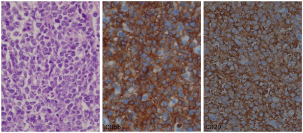
A 5-year-old girl visited for snoring and open mouth breathing for 3 years. She did not manifest symptoms, such as fever, night sweats, lymphadenopathy or weight loss. She has been suffered from frequently relapsed tonsillitis. The laboratory examination did not show any abnormality. Bilateral tonsillectomy was performed. Right tonsil was larger than that of left, measuring 2.5x2.5x1.8 cm. Cut sections of right tonsil showed a relatively well defined homogenously yellow solid mass, measuring 2x2 cm. On microscopically, large monomorphic atypical lymphoid cells were diffusely proliferated and showed strong positivity for CD20, Bcl-2, Bcl-6, Mum1, and CD56 and were negative for CD3, CD5, CD7, CD10, and EBV PCR (Figure 1). Immunoglobulin heavy chain gene rearrangement showed monoclonality (Agilent 2100 Bioanalyser, Agilent Technologies, Waldbronn, Germany). She has been treated with six courses of COPAD (cyclophosphamide, vincristine, prednisolone and doxorubicin) chemotherapy. On recent follow-up, there is no recurrent or any other organ involvement and complete remission has been maintained.
Figure 1.

Microscopic finding shows diffuse large monomorphic lymphoid cells proliferation. The cells are diffusely positive for CD56 and CD20 (x400).
Discussion
CD56 expression is an unusual feature in B-cell lymphomas. The CD56 expression rate has been described 0.5 to 5.5% of all B-cell lymphomas [5]. CD56 positive B-cell lymphoma has been reported 1.2% to 7% of DLBCL [4,6,7]. We found 10 reports, which included 35 cases of CD56 positive DLBCL [4-13]. CD56 positive DLBCL is more frequent in male (male to female ratio = 3:1) and ranged in age from 15 to 87 years (mean age: 56.5 years). Clinical data was available in 31 cases. Sixteen cases (51.6%) were occurred in lymph node, 3 cases (9.7%) were occurred in nasopharynx or Waldeyer’s ring, 3 cases in ileum, 3 cases in stomach, and each one in the nasal cavity, spine, brain, intradural and extramedullary mass, abdomen and subcutaneous tissue. Kern et al. have demonstrated that CD56 expression is associated with lymphomas with predominant extranodal involvement [7]. Extranodal involvement of CD56 positive DLBCL has been reported 45% to 60% [4,5]. The neoplastic lymphocytes were composed of centroblasts (8/19 cases, 42.1%), centroblasts/centrocytes (5/19, 26.3%), immunoblasts (1/19, 5.3%), and large atypical cells (5/19, 26.3%). On immunohistochemistry, the cells were positive for CD10 (22/29 cases, 75.9%), bcl-2 (18/26, 69.2%), and bcl-6 (17/19, 89.5%). According to the immunohistochemical results, 22 of 25 cases (88%) expressed germinal center immunophenotype and the other 3 cases showed non-germinal center phenotype. CD56 positive DLBCL expresses CD10 and bcl-6 more frequently than general DLBCL, is associated with germinal centre origin. This case showed non-germinal center phenotype.
The biologic function of CD56 in B-cell ontogeny is not clearly defined. Reynaud et al. have described that early B-cells have the capacity to differentiate to CD56 positive NK cells [14]. Isobe et al. have described that CD56 expression may be related with growth, expansion and dissemination of lymphoma, not be essential for lymphomagenesis [11]. Stacchini et al. have also reported that CD56 plays a role in lymphoma growing and spreading [4]. However, the prognostic significance of CD56 expression on DLBCL is still unknown. For clarify the prognostic significance and biologic function of CD56 expression in DLBCL, further cases collection is necessary.
We report a case of CD56 positive DLBCL, which is incidentally found and is occurred in a young age. She has been maintained a complete remission state.
Disclosure of conflict of interest
None.
References
- 1.Griffin JD, Hercend T, Beveridge R, Schlossman SF. Characterization of an antigen expressed by human natural killer cells. J Immunol. 1983;130:2947–2951. [PubMed] [Google Scholar]
- 2.Cunningham BA, Hemperly JJ, Murray BA, Prediger EA, Brackenbury R, Edelman GM. Neural cell adhesion molecule: Structure, immunoglobulin-like domains, cell surface modulation, and alternative RNA splicing. Science. 1987;236:799–806. doi: 10.1126/science.3576199. [DOI] [PubMed] [Google Scholar]
- 3.Van Camp B, Durie BG, Spier C, De Waele M, Van Riet I, Vela E, Frutiger Y, Richter L, Grogan TM. Plasma cells in multiple myeloma express a natural killer cell-associated antigen: CD56 (NKH-1; leu-19) Blood. 1990;76:377–82. [PubMed] [Google Scholar]
- 4.Stacchini A, Barreca A, Demurtas A, Aliberti S, di Celle PF, Novero D. Flow cytometric detection and quantification of CD56 (neural cell adhesion molecule, NCAM) expression in diffuse large B cell lymphomas and review of the literature. Histopathology. 2012;60:452–9. doi: 10.1111/j.1365-2559.2011.04098.x. [DOI] [PubMed] [Google Scholar]
- 5.Weisberger J, Gorczyca W, Kinney MC. CD56-positive large B-cell lymphoma. Appl Immunohistochem Mol Morphol. 2006;14:369–74. doi: 10.1097/01.pai.0000208279.66189.43. [DOI] [PubMed] [Google Scholar]
- 6.Gomyo H, Kajimoto K, Miyata Y, Maeda A, Mizuno I, Yamamoto K, Obayashi C, Hanioka K, Murayama T. CD56-positive diffuse large B-cell lymphoma: Possible association with extranodal involvement and bcl-6 expression. Hematology. 2010;15:157–161. doi: 10.1179/102453309X12583347113573. [DOI] [PubMed] [Google Scholar]
- 7.Kern WF, Spier CM, Miller TP, Grogan TM. NCAM (CD56)-positive malignant lymphoma. Leuk Lymphoma. 1993;12:1–10. doi: 10.3109/10428199309059565. [DOI] [PubMed] [Google Scholar]
- 8.Suzuki R, Yamamoto K, Seto M, Kagami Y, Ogura M, Yatabe Y, Suchi T, Kodera Y, Morishima Y, Takahashi T, Saito H, Ueda R, Nakamura S. CD7+ and CD56+ myeloid/natural killer cell precursor acute leukemia: A distinct hematolymphoid disease entity. Blood. 1997;90:2417–28. [PubMed] [Google Scholar]
- 9.Sekita T, Tamaru JI, Isobe K, Harigaya K, Masuoka S, Katayama T, Kobayashi M, Mikata A. Diffuse large B cell lymphoma expressing the natural killer cell marker CD56. Pathol Int. 1999;49:752–8. doi: 10.1046/j.1440-1827.1999.00929.x. [DOI] [PubMed] [Google Scholar]
- 10.Muroi K, Omine K, Kuribara R, Uchida M, Izumi T, Hatake K, Miura Y. CD56 expression in B-cell lymphoma. Leuk Res. 1998;22:201–202. doi: 10.1016/s0145-2126(97)00153-7. [DOI] [PubMed] [Google Scholar]
- 11.Isobe Y, Sugimoto K, Takeuchi K, Ando J, Masuda A, Mori T, Oshimi K. Neural cell adhesion molecule (CD56)-positive B-cell lymphoma. Eur J Haematol. 2007;79:166–9. doi: 10.1111/j.1600-0609.2007.00893.x. [DOI] [PubMed] [Google Scholar]
- 12.Chen B, Sun WY, Luo J, Zhang G. [CD56-positive diffuse large B-cell lymphoma: report of a case] . Zhonghua Bing Li Xue Za Zhi. 2010;39:343–4. [PubMed] [Google Scholar]
- 13.Hammer RD, Vnencak-Jones CL, Manning SS, Glick AD, Kinney MC. Microvillous lymphomas are B-cell neoplasms that frequently express CD56. Mod Pathol. 1998;11:239–46. [PubMed] [Google Scholar]
- 14.Reynaud D, Lefort N, Manie E, Coulombel L, Levy Y. In vitro identification of human pro-B cells that give rise to macrophages, natural killer cells, and T cells. Blood. 2003;101:4313–21. doi: 10.1182/blood-2002-07-2085. [DOI] [PubMed] [Google Scholar]


