Abstract
Background
Iatrogenic spondylolisthesis is a challenging condition for spinal surgeons. Posterior surgery in these cases is complicated by poor anatomical landmarks, scar tissue adhesion of muscle and dural structures and difficult access to the intervertebral disc. Anterior interbody fusion provides an alternative treatment method, allowing indirect foraminal decompression, reliable disc clearance and implantation of large surface area implants.
Materials and methods
A retrospective chart review of patients with iatrogenic spondylolisthesis including pre- and post-operative Oswestry Disability Index (ODI) and Visual Analogue Scale (VAS) scores was performed. Imaging criteria were pelvic incidence, overall lumbar lordosis and segmental lordosis. In addition, the fusion rate was investigated after 6 months.
Results
Six consecutive patients treated between 2008 and 2011 (4 female, 2 male, mean age 61 ± 7.1 years) were identified. The initially performed surgeries included decompression with or without discectomy; posterior instrumented and non-instrumented fusion. The olisthetic level was in all cases at the decompressed level. All patients were revised with stand-alone anterior interbody fusion devices at the olisthetic level filled with BMP 2. Average ODI dropped from 49 ± 11 % pre-operatively to 26.0 ± 4.0 at 24 months follow-up. VAS average dropped from 7 ± 1 to 2 ± 0. Mean total lordosis of 39.8 ± 2.8° increased to 48.5 ± 4.9° at pelvic incidences of 48.8 ± 6.8° pre-operatively. Mean segmental lordosis at L4/5 improved from 10.5 ± 6.7° to 19.0 ± 4.9° at 24 months. Mean segmental lordosis in L5/S1 increased from 15.1 ± 7.4° to 23.2 ± 5.6°. Cage subsidence due to severe osteoporosis occurred in one case after 5 months, and hence there was no further follow-up. Fusion was confirmed in all other patients.
Conclusion
Anterior interbody fusion offers good stabilisation and restoration of lordosis in iatrogenic spondylolisthesis and avoids the well-known problems associated with reentering the spinal canal for revision fusions. In this group, ODI and VAS scores were improved.
Keywords: Iatrogenic spondylolisthesis, Acquired spondylolisthesis, Revision surgery, Anterior lumbar interbody fusion
Introduction
Iatrogenic or acquired spondylolisthesis occurs in 3.7–20 % of cases after posterior decompression or fusion [1–9]. In particular, mechanical overload, devascularisation and decortication of the pars interarticularis seem to be important factors in developing a spondylolisthesis after spinal fusion [4, 5]. In decompressive surgery, extensive bony resection can lead to post-operative lumbar instability and thus to spondylolisthesis [3, 9–11].
Patients suffering from acquired spondylolisthesis after spinal surgery often present with increased back pain and new or deteriorating sciatic symptoms. Posterior lumbar interbody or screw-rod fusions were deemed the gold standard in treating these conditions. The main goal is to increase rotational and longitudinal stiffness over the affected area with slip reduction and to extend decompression if necessary [12, 13]. Posterior re-operations can be challenging due to altered and poor anatomical landmarks as well as scar tissue, leading to injuries to neural structures. The repeat stripping of paraspinal musculature could contribute to the so-called “fusion disease” with persistent back pain [14]. In addition, the anterior column, responsible for 80 % of the weight loading, is not addressed biomechanically with posterior fusion devices, leading to decreased fusion rates and a loss of correction in slip angle [14, 15].
Anterior lumbar interbody fusion (ALIF) offers a virgin approach to the lumbar spine in these cases and addresses the anterior column directly. This procedure offers a complete discectomy, restoration of lumbar lordosis without harming neurological structures and indirect foraminal decompression. Disadvantages include a higher risk of severe vascular and intraperitoneal injuries, limited direct access to neural structures, as well as potential sympathetic plexus lesions [16–18]. Encouraging results have been published in long-term follow-up for isthmic spondylolisthesis cases [19, 20]. In this study, stand-alone anterior interbody fusion was used as salvage option in patients with iatrogenic spondylolisthesis.
Materials and methods
Patients who received ALIF for iatrogenic spondylolisthesis between 2008 and 2011 were identified. Outcome measures included the Oswestry Disability Score (ODI) and Visual Analogue Scale (VAS). These two scales were recorded pre-operatively and at follow-up 6, 12 and 24 months after surgery.
The radiological investigation included pre-operative MRI scans and standard anterior-posterior and lateral X-rays. The grade of slippage was evaluated according to Meyerding [21], while pelvic incidence was recorded pre-operatively. At 6, 12 and 24 months follow-up, a standard X-ray was acquired. Total lumbar lordosis, measured from the endplate of S1 to the superior endplate of L1, and the segmental lordosis of affected levels (from inferior to superior endplate), were measured pre-operatively and for all follow-ups. The fusion rate was evaluated using plain radiographs. Where unclear, additional computed tomography (CT) was obtained. Fusion was defined as obvious bridging of trabeculae across the disc space.
Results
Six consecutive patients (Table 1) were included (4 female and 2 male) with a mean age of 61 ± 7.1 years (range 52–71 years). Three patients had a decompression and discectomy as initial spinal surgery, while one patient was treated with decompression alone. The remaining two patients had fusion procedures in addition to decompression (one instrumented, one non-instrumented). Details regarding the prior surgeries can be found in Table 1. The time in between the first spinal surgery and treatment of iatrogenic spondylolisthesis ranged from 12 to 144 months. All patients presented with sciatic symptoms and back pain.
Table 1.
Patients’ demographics
| Patient | Gender | Age | Time between surgeries (months) | Previous surgery | Level of surgery | Spondylolisthesis level | Grade |
|---|---|---|---|---|---|---|---|
| 1 | M | 57 | 12 | Decompression | L4/5 | L4 | 2 |
| Discectomy | L4 | ||||||
| 2 | F | 58 | 144 | Decompression | L4/5, L5/S1 | L4 | 2 |
| Instrumented Fusion | L5/S1 | ||||||
| 3 | F | 71 | 17 | Decompression | L4/5 | L4 | 1 |
| Non-instrumented Fusion | L5/S1 | ||||||
| 4 | F | 61 | 16 | Decompression | L3/4, L4/5 | L5 | 1 |
| Discectomy | L4 | ||||||
| 5 | F | 68 | 96 | Decompression | L3/4, L4/5 | L4 | 1 |
| 6 | M | 52 | 124 | Decompression | L5/S1 | L5 | 2 |
| Discectomy | L5 |
Spondylolisthesis grade according to the Meyerding classification [21]
Revision surgery consisted of stand-alone anterior interbody fusion cages filled with recombinant bone morphogenic protein 2 (SnyCage® Synthes Paoli, PA, USA; InFuse® Medtronic Minneapolis, MN, USA). Two patients were treated with one level ALIF, three with two levels and one patient with three levels anterior fusion. The levels in addition to the olisthetic level were included to improve overall sagittal balance on an individual basis.
The mean operative time including anaesthesia was 223 ± 26 min (range 200–300 min) with a mean blood loss of 284 ± 76.6 ml (range 200–400 ml). A summary of the surgical data is presented in Table 2.
Table 2.
Surgical data
| Patient | ALIF level | Duration (min) | Blood loss (ml) | Intra-operative complications | Spondylolisthesis grade post-op |
|---|---|---|---|---|---|
| 1 | L4/5, L5/S1 | 270 | 270 | None | 1 |
| 2 | L4/5 | 200 | 200 | None | 2 |
| 3 | L4/5 | 210 | 210 | None | 1 |
| 4 | L3/4, L4/5, L5/S1 | 300 | 285 | Venous bleeding | 1 |
| 5 | L3/4, L4/5 | 235 | 340 | None | 1 |
| 6 | L4/5, L5/S1 | 210 | 400 | Peritoneal tear | 1 |
| Mean (Standard deviation) | 223 (± 26) | 284 (± 76.6) |
ALIF anterior lumbar interbody fusion; spondylolisthesis grade according to Meyerding [21]
Complications
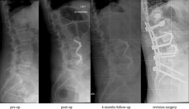
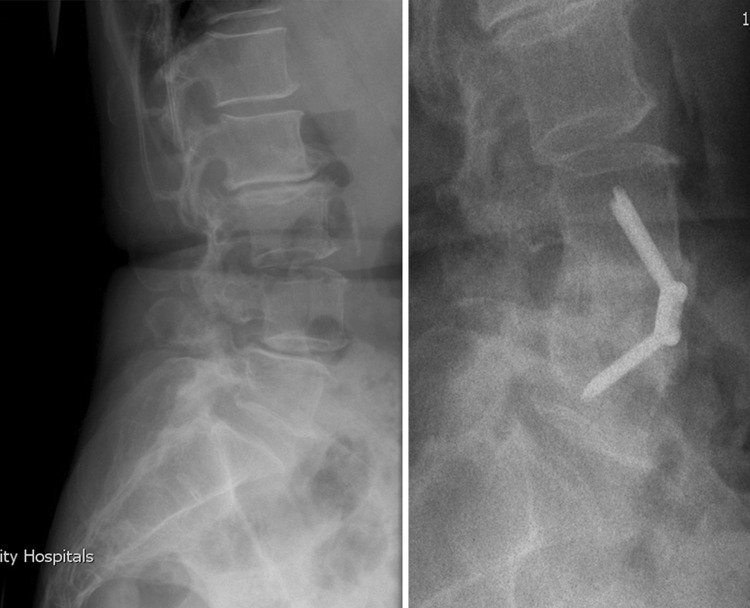
Patient 5 showed a cage subsidence into the vertebral body of L3 after 6 months due to severe osteoporosis. No further follow-up was therefore possible. A revision posterior instrumented fusion and cement augmentation was later performed (Fig. 1).
Fig. 1.
Patient 5 showing cage migration in the vertebral body L3 after 6 months requiring posterior instrumented fusion and kyphoplasty
In one patient, an injury of an unusual large middle sacral vein occurred which was managed with vessel ligation. One accidental opening of the peritoneum occurred intra-operatively.
ODI and VAS results
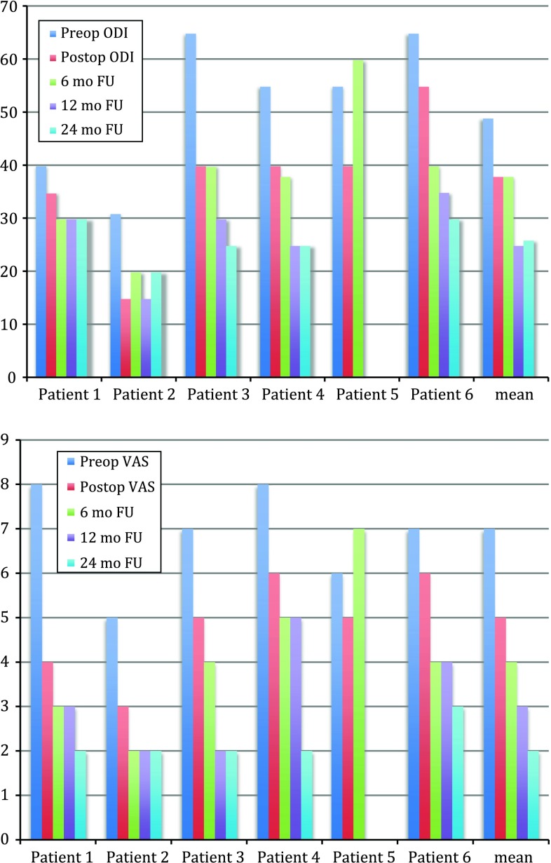
The mean ODI pre-operatively was 49 ± 12 % and dropped to 38 ± 12 % post-operatively. At the first follow-up, the ODI remained at a mean of 38 ± 12 %, but improved after 12 months to 25 ± 7 %. At the last follow-up 24 months after anterior lumbar interbody fusion, the ODI was similarly low with a mean value of 26 ± 3.7 %.
The VAS showed a decrease from 7 ± 1 to 5 ± 1 directly after surgery. After 6 months, a mean score of 4 ± 2 was recorded. The 12-month follow-up showed an improvement to a mean of 3 ± 1.2. Again, the score was slightly improved to 2.2 ± 0.4 after 24 months. The individual results are shown in Fig. 2.
Fig. 2.
a ODI outcome measurements for individual patients and mean values. b VAS outcome measurements for individual patients and mean values
Clinical symptoms
All patients had back pain and sciatic symptoms prior to anterior interbody fusion. Three patients described their back pain as severe, two as moderate and one had only mild back pain. Radiculopathy of L5 occurred in four and S1 sciatica in two patients pre-operatively.
Three patients showed immediate improvement of back pain and radiculopathy after surgery. After 6 months, four patients reported relief of back pain and five reported an improvement of their sciatica. Only Patient 5 showed a worsening of back pain and radiculopathy due to cage subsidence.
After 1 year, all patients reported occasional back pain and two patients had no sciatica at all. No radiculopathy was reported at the last follow-up by any patients, while back pain was unchanged to the 1-year follow-up.
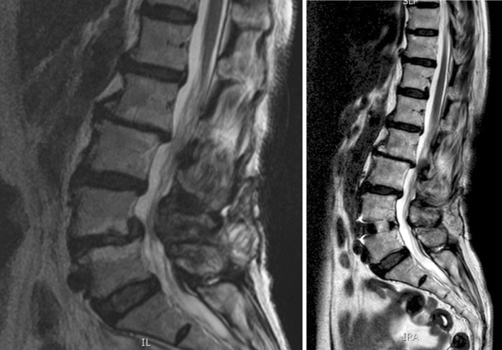
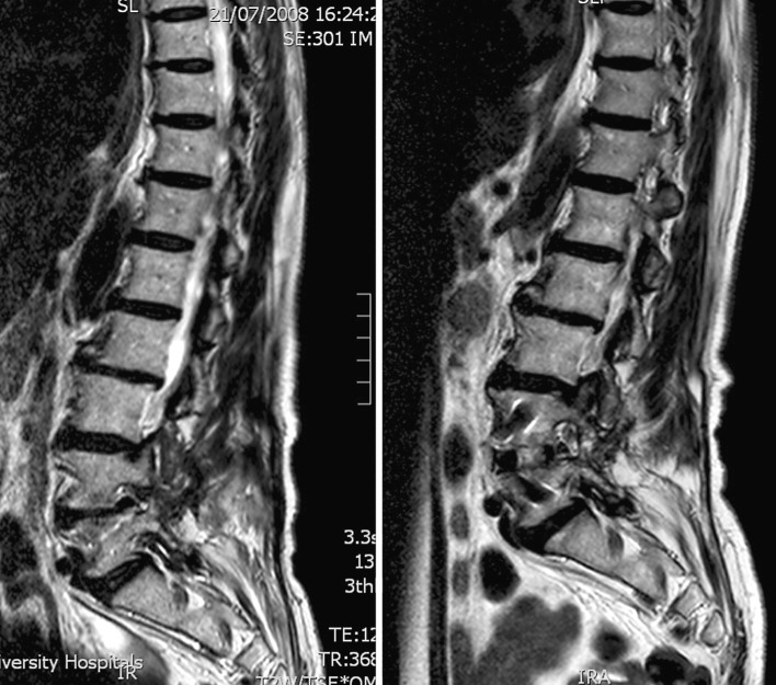
The relief of sciatica and back pain seem to be an effect of the indirect decompression of the implant. Figures 3 and 4 show the pre-operative and post-operative MRI scans at mid-sagittal plain and at the level of the neuroforamen of patient 3.
Fig. 3.
Pre- and post-operative MRI scans of patient 3 demonstrating the effect of indirect decompression in the mid-sagittal plain
Fig. 4.
Pre- and post-operative MRI scans of patient 3 demonstrating the effect of indirect decompression at the foraminal plain
Radiological outcome
The pelvic incidence measured pre-operatively had a mean value of 48.8 ± 6.8° (range 38.5°–58°).
The total lordosis was increased after the procedure from a mean of 39.8 ± 2.8 (range 36°–44.3°) to 48.5 ± 4.9° (range 38.4°–55.6°). Table 3 summarizes the values for each patient.
Table 3.
Radiological outcome measurements
| Patient | PI | TL | SL L3/4 |
SL L4/5 |
SL L5/S1 |
F | ||||||||
|---|---|---|---|---|---|---|---|---|---|---|---|---|---|---|
| Pre-op | Post-op | 24 mo FU | Pre-op | Post-op | 24 mo FU | Pre-op | Post-op | 24 mo FU | Pre-op | Post-op | 24 mo FU | |||
| 1 | 56.4 | 37.3 | 38.4 | 38.4 | 14.2 | 15.8 | 15.8 | 12.1 | 18.5 | 18.5 | X-ray | |||
| 2 | 38.5 | 44.3 | 48.3 | 48.3 | 9.1 | 24.0 | 24.0 | X-ray/ CT |
||||||
| 3 | 47.1 | 38.6 | 48.0 | 48.0 | 20.9 | 24.2 | 24.2 | X-ray | ||||||
| 4 | 43.4 | 41.7 | 55.6 | 55.6 | 1.5 | 5.0 | 5.0 | 3.5 | 12.3 | 12.3 | 25.8 | 30.1 | 30.1 | CT |
| 5a | 49.5 | 40.7 | 41.0 | 4.3 | 5.3 | 1.3 | 10.0 | No | ||||||
| 6 | 58.0 | 36.0 | 38.5 | 38.5 | 14.0 | 19.8 | 19.8 | 8.6 | 17.4 | 17.4 | X-ray/ CT |
|||
PI pelvic incidence, TL total lordosis, SL segmental lordosis, F fusion
aCage migration after 5 months
The mean L4/5 segmental lordosis increased from 10.5 ± 6.7° (range 1.3°–20.9°) pre-operatively to 19.0 ± 4.9 (range 12.3°–24.2°) at the last follow-up of 24 months.
The L5/S1 segmental lordosis increased as well, from a mean of 15.5 ± 7.4 (range 8.6–25.8) prior to surgery to 23.2 ± 5.6° (range 17.4°–30.1°) after 2 years (Table 3).
Immediate slippage reduction was achieved in two patients (Patient 1 and 6) from Grade 2 to 1, without reduction loss over the follow-up period. In the majority of the patients, the olisthesis was fused in situ without further slippage at 6, 12 and 24-month follow-up (Table 2).
Fusion was confirmed in five patients through obvious osseous bridging of the ALIF implant on plain X-ray or CT (Fig. 5).
Fig. 5.
Example of successful fusion after 6 months: patient 2 with pre-operative and follow-up x-ray
Discussion
Iatrogenic or acquired spondylolisthesis is an uncommon, but well-recognised complication after posterior spinal fusion or decompression. As early as 1963, Harris et al. [5] had published a case series of six patients suffering from acquired spondylolisthesis after fusion of the lower lumbar spine. Their biomechanical work revealed that a rotational moment combined with flexion extension movement might cause a fatigue fracture through the pars interarticularis of the uppermost vertebra of the fused segments. Brunet and Wiley added the concept of adjacent level disc degeneration as a supplementary factor for iatrogenic spondylolisthesis [4]. The authors described the phenomenon of this pathology as an exclusive event after interlaminar fusion, whereas posterolateral and anterior fusions rarely seemed to be the cause. They postulated that decortication of the pars might lead to a deterioration of mechanical properties in the adjacent level of fusion. This is unlikely to be the case if anterior or posterolateral fusion techniques are used. A posterolateral bonegraft may reinforce the pars interarticularis instead of weakening through decortication. However, iatrogenic spondylolisthesis may occur even if posterolateral fusion is used [7].
In anterior interbody fusion, the bonegraft is put closer to the rotational axis thereby neutralising the intervertebral movement more effectively. Iatrogenic spondylolisthesis has been shown to negatively influence outcome.
In the study by Frymoyer et al. [8], acquired spondylolisthesis was the only predictable and demonstrable source of failure after spinal fusion in a long-term follow-up of 45 patients.
In cases of acquired slippage after posterior decompression for degenerative disc disease or spinal stenosis, the amount of bone resection seems to influence the occurrence of spinal instability. Bisschop et al. [22] discovered that laminectomy significantly reduced the shear stiffness in the affected motion segment. In addition, shear yield force and shear force leading to failure dropped after laminectomy. They therefore concluded that these factors could influence the disc geometry and enhance further spinal instability.
The clinical study of Johnsson et al. [1], however, could not find a correlation between extension of resection and slippage, but a higher olisthesis was seen in patients with a poor clinical outcome. Suzuki et al. showed [3] in their investigation of 35 patients after decompression without fusion that in cases of acquired spondylolisthesis, the decompression was greater and the number of laminae operated on was higher. Decompression over 75 % in width over three laminae was identified as a risk factor. Celik et al. [10] observed an increased spinal instability after total laminectomy, leading to a bilateral microdecompressive approach to reduce this problem. Hong et al. confuted this fact in their study [11], showing that bilateral decompression also increases the translational stress, thereby increasing the acquired listhesis. In their opinion, a unilateral decompression might reduce the risk of late instability. Maurer et al. [9] postulated a similar theory in their case report, where stress fractures across the pars interarticularis were seen in patients after posterior fusion when a unilateral laminectomy had been performed.
The treatment of iatrogenic spondylolisthesis, however, is still challenging. In a functional outcome analysis, L’Heureux et al. [15] found that a significant functional improvement was seen in patients treated surgically for iatrogenic spondylolisthesis. Hombold treated 12 cases with posterior fusion in his study [6].
The main goal of spondylolysis fixation is to restore motional stiffness and avoid rotational instability. Posterior fusion techniques, however, need a renewed stripping of the paraspinal muscles, which might contribute to ongoing back pain as described by Zdeblick [14]. In addition, scar tissue and altered anatomical landmarks make the posterior approach especially demanding. Kwon et al. [12] showed a decreased fusion rate with posterior lumbar interbody fusion due to insufficient treatment of the anterior column.
Anterior interbody fusion addresses the anterior column directly. With this method, a complete discectomy can be achieved with an indirect decompression. Ishihara et al. [20] successfully treated patients with stand-alone anterior lumbar interbody fusion in cases of isthmic spondylodesis with a minimum 10-year follow-up. The authors observed good clinical results although the low-back pain score decreased over the time with loss of correction and recurrence of slippage. The loss of correction might have been due to the bonegraft harvesting at the proximate vertebral body.
Barrick et al. [19] used anterior interbody fusion in cases of recurrent back pain after posterior interbody fusion or posterolateral fusion. This study reveals better outcome regarding discogenic pain even at the level of posterolateral fusion with confirmed solid spondylodesis.
These investigations encourage the usage of anterior interbody fusion in cases of iatrogenic spondylolisthesis, but no literature is yet published using these devices as a salvage option. In the present study, six consecutive patients were treated with this method; five had a very satisfactory result after a follow-up of 24 months. The two main problems, severe back pain and sciatica, were improved dramatically. All five patients had no sciatic symptoms and even back pain, reported as severe in three patients, were successfully treated. Only one patient with severe osteoporosis showed cage subsidence after 5 months.
In the remaining five patients, the total lordosis as well as the segmental lordosis were improved, resulting in a better sagittal balance restoration.
This investigation is limited by its retrospective nature and relatively small case number. As this is a cohort collected in a large tertiary referral centre over several years, it may not be possible to conduct a more formal randomised controlled trial. Anterior spinal surgery requires the availability of appropriately trained surgeons that are capable of dealing with potential complications. The two access-related complications could be controlled by the performing surgeon (senior author) who was fellowship trained to perform anterior surgery. However, major vessel complications or perforated intraperitoneal organs should be treated by vascular or visceral surgeons, which might limit the utilisation of anterior interbody fusion devices in departments with unavailable vascular or general surgery support. As seen in one patient of the study, osteoporosis can strongly influence the outcome of anterior interbody fusion. The lack of sufficient structural bone quality might not be able to support the rigid implant leading to cage subsidence in the adjacent vertebral body. Pre-operative bone densitometry could help to avoid this problem.
Conclusion
This study provides encouraging results for stand-alone anterior interbody fusion as a salvage procedure for iatrogenic spondylolisthesis. However, careful patient selection and specific anterior surgical training are needed to avoid severe complications, which might limit the usage of anterior fusion devices in smaller hospitals. Furthermore, long-term follow-up observations are needed to control recurrent slippage and clinical symptoms in these patients. Due to the small numbers of acquired spondylolisthesis, randomised controlled trials are unlikely to be successful. In order to evaluate the efficiency of this treatment method, multicenter studies could solve the problem.
Conflict of interest
No benefits or funds in any form have been received or will be received from a commercial party related directly or indirectly to the subject of this article.
Contributor Information
M. A. König, Email: matthias.a.koenig@gmail.com
B. M. Boszczyk, Phone: +44-115-9249924, Phone: +44-115-9262410, FAX: +44-115-9709991, Email: bronek.boszczyk@nuh.nhs.uk
References
- 1.Johnsson KE, Willner S, Johnsson K. Postoperative instability after decompression for lumbar spinal stenosis. Spine. 1986;11(2):107–110. doi: 10.1097/00007632-198603000-00001. [DOI] [PubMed] [Google Scholar]
- 2.Lee CK. Lumbar spinal instability (olisthesis) after extensive posterior spinal decompression. Spine. 1983;8(4):429–433. doi: 10.1097/00007632-198305000-00014. [DOI] [PubMed] [Google Scholar]
- 3.Suzuki K, Ishida Y, Ohmori K. Spondylolysis after posterior decompression of the lumbar spine. 35 patients followed for 3-9 years. Acta Orthop Scand. 1993;64(1):17–21. doi: 10.3109/17453679308994519. [DOI] [PubMed] [Google Scholar]
- 4.Brunet JA, Wiley JJ. Acquired spondylolysis after spinal fusion. J Bone Jt Surg Br. 1984;66(5):720–724. doi: 10.1302/0301-620X.66B5.6501368. [DOI] [PubMed] [Google Scholar]
- 5.Harris RI, Wiley JJ. Acquired spondylolysis as a sequel to spine fusion. J Bone Jt Surg Am. 1963;45:1159–1170. [PubMed] [Google Scholar]
- 6.Rombold C. Spondylolysis: a complication of spine fusion. J Bone Jt Surg Am. 1965;47:1237–1242. [PubMed] [Google Scholar]
- 7.Blasier RD, Monson RC. Acquired spondylolysis after posterolateral spinal fusion. J Pediatr Orthop. 1987;7(2):215–217. doi: 10.1097/01241398-198703000-00022. [DOI] [PubMed] [Google Scholar]
- 8.Frymoyer JW, Matteri RE, Hanley EN, Kuhlmann D, Howe J. Failed lumbar disc surgery requiring second operation.A long-term follow-up study. Spine. 1978;3(1):7–11. doi: 10.1097/00007632-197803000-00002. [DOI] [PubMed] [Google Scholar]
- 9.Maurer SG, Wright KE, Bendo JA. Iatrogenic spondylolysis leading to contralateral pedicular stress fracture and unstable spondylolisthesis: a case report. Spine. 2000;25(7):895–898. doi: 10.1097/00007632-200004010-00022. [DOI] [PubMed] [Google Scholar]
- 10.Celik SE, Celik S, Göksu K, Kara A, Ince I. Microdecompressive laminatomy with a 5-year follow-up period for severe lumbar spinal stenosis. J Spinal Disord Tech. 2010;23(4):229–235. doi: 10.1097/BSD.0b013e3181a3d889. [DOI] [PubMed] [Google Scholar]
- 11.Hong SW, Choi KY, Ahn Y, Baek OK, Wang JC, Lee SH, Lee HY. A comparison of unilateral and bilateral laminotomies for decompression of L4-L5 spinal stenosis. Spine. 2011;36(3):E172–E178. doi: 10.1097/BRS.0b013e3181db998c. [DOI] [PubMed] [Google Scholar]
- 12.Kwon BK, Albert TJ. Adult low-grade acquired spondylolytic spondylolisthesis: evaluation and management. Spine. 2005;30(6 Suppl):S35–S41. doi: 10.1097/01.brs.0000155561.70727.20. [DOI] [PubMed] [Google Scholar]
- 13.Deguchi M, Rapoff AJ, Zdeblick TA. Biomechanical comparison of spondylolysis fixation techniques. Spine. 1999;24(4):328–333. doi: 10.1097/00007632-199902150-00004. [DOI] [PubMed] [Google Scholar]
- 14.Zdeblick TA (1999) Discogenic back pain. In: Herkowitz HN, Garfin SR, Balderston RA et al (eds) The Spine, 4th ed. Saunders, Philadelphia, pp 748–765
- 15.L’Heureux EA, Jr, Perra JH, Pinto MR, Smith MD, Denis F, Lonstein JE. Functional outcome analysis including preoperative and postoperative SF-36 for surgically treated adult isthmic spondylolisthesis. Spine. 2003;28(12):1269–1274. doi: 10.1097/01.BRS.0000065574.20711.E6. [DOI] [PubMed] [Google Scholar]
- 16.Jarrett CD, Heller JG, Tsai L. Anterior exposure of the lumbar spine with and without an “access surgeon”: morbidity analysis of 265 consecutive cases. J Spinal Disord Tech. 2009;22(8):559–564. doi: 10.1097/BSD.0b013e318192e326. [DOI] [PubMed] [Google Scholar]
- 17.Kulkarni SS, Lowery GL, Ross RE, Ravi Sankar K, Lykomitros V. Arterial complications following anterior lumbar interbody fusion: report of eight cases. Eur Spine J. 2003;12(1):48–54. doi: 10.1007/s00586-002-0460-4. [DOI] [PubMed] [Google Scholar]
- 18.König MA, Leung Y, Jürgens S, MacSweeney S, Boszczyk BM. The routine intra-operative use of pulse oximetry for monitoring can prevent severe thromboembolic complications in anterior surgery. Eur Spine J. 2011;20(12):2097–2102. doi: 10.1007/s00586-011-1900-9. [DOI] [PMC free article] [PubMed] [Google Scholar]
- 19.Barrick WT, Schofferman JA, Reynolds JB, Goldthwaite ND, McKeehen M, Keaney D, White AH. Anterior lumbar fusion improves discogenic pain at levels of prior posterolateral fusion. Spine. 2000;25(7):853–857. doi: 10.1097/00007632-200004010-00014. [DOI] [PubMed] [Google Scholar]
- 20.Ishihara H, Osada R, Kanamori M, Kawaguchi Y, Ohmori K, Kimura T, Matsui H, Tsuji H. Minimum 10-year follow-up study of anterior lumbar interbody fusion for isthmic spondylolisthesis. J Spinal Disord. 2001;14(2):91–99. doi: 10.1097/00002517-200104000-00001. [DOI] [PubMed] [Google Scholar]
- 21.Meyerding HW. Spondylolisthesis. Surg Gynaecol Obstet. 1932;54:371–377. [Google Scholar]
- 22.Bisschop A, van Royen BJ, Mullender MG, Paul CP, Kingma I, Jiya TU, van der Veen AJ, van Dieën JH (2012) Which factors prognosticate spinal instability following lumbar laminectomy? Eur Spine J [Epub ahead of print] [DOI] [PMC free article] [PubMed]


