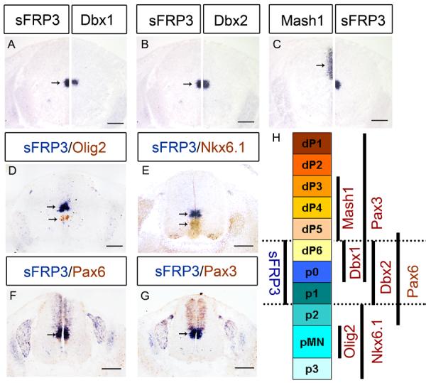
Fig. 3.

sFRP3 was expressed dp6 to p1 domains of ventricular zone at E12.5. A–C: Immediately adjacent sections from E12.5 spinal cord were subjected to in situ hybridization with sFRP3, Dbx1, Dbx2 and Mash1 riboprobes, and half sections were aligned at the midline. D–G: Spinal cords from E12.5 embryos were double-labeled with sFRP3 (blue) and anti-Olig2 (brown in D), anti-Nkx6.1 (brown in E), anti-Pax6 (brown in F) or anti-Pax3 (brown in G). H: Scheme of sFRP3 expression in progenitor domains labeled by various progenitor identity genes. Arrows indicate the ISH positive cells. Scale bar = 100 μm.
