Abstract
Background: The purpose of the present study is to investigate the prevalence of dental anomalies according to gender among children.
Materials & Methods: This cross-sectional study was conducted a group of 600 children, of them 293 (48.8%) were males and 275 (45.8%) females which were taken with proper sampling technique. Type III clinical examination was done to know the prevalence of dental anomalies. The Statistical software namely SPSS version 16.0 was used for data analysis. Chi-square test was used at p value of 0.05 or less.
Results: Impactions (39.2%) were the most common anomaly in this study and most of the impacted teeth were related to maxilla. A significant difference was seen in case of hypodontia, microdontia and talons cusp according to gender in which first two anomalies were more among females and last one among males. Children with one dental anomaly were 25.8%, and 13.4% were having more than one.
Conclusion: The percentage of dental anomalies were high specially impaction and rotated teeth. So these anomalies should be treated earlier to avoid further complications.
How to cite this article: Kathariya MD, Nikam AP, Chopra K, Patil NN, Raheja H, Kathariya R. Prevalence of Dental Anomalies among School Going Children in India. J Int Oral Health 2013; 5(5):10-4.
Key Words: : dental anomalies, prevalence, school children
Introduction
Dental anomalies are one of the anomalies of the human structure that result from disturbances during formation of tooth. These anomalies may affect the size, shape, colour and number of teeth. They can be developmental, congenital or acquired and may be localized to single tooth or involving systemic conditions.1
Congenital abnormalities are typically inherited genetically. Abnormalities in tooth shape, size and structure result from disturbances during the morpho-differentiation stage of tooth development, while ectopic eruption, impaction and rotation of teeth result from developmental disturbances in the eruption pattern of permanent teeth.2
Around 7% of children are born with some of the disturbances in the orofacial system and most commonly are supernumerary teeth, missing teeth, fused teeth and peg lateral incisors.3 Dental anomalies in comparison with more common oral disorders such as dental caries and periodontal diseases have low frequency but their management procedure is more complicated, because they can result in esthetic problems, malocclusion, and leads to the other oral problems.4
In industrialized countries, there are about 10% of children with developmental disturbances, whereas in developing countries like India their percentage is higher, ranging between 15% and 20%. The identification of oral/dental and minor anomalies are of great importance for timely and accurate diagnosis of numerous genetic abnormalities of the craniofacial region.5 Hence this study was done to know the prevalence of dental anomalies among children.
Materials & Methods
The study was conducted among 600 school going children in Karad District of Maharastra, India during a four month period in 2012.
First five schools from the city were randomly selected then this study population was taken with cluster sampling technique. Before scheduling the survey, the official permission was obtained from the Ethical Committee. Official permission was obtained from the Heads of the Institutes from the District. Informed oral consent was obtained prior to examination of each subject.
A pilot survey was conducted in one of the school on 50 randomly selected subjects to know the prevalence of dental anomalies and feasibility of the survey. Children with any kind of medical history such as Down's syndrome, ectodermal dysplasia, cleft lip and cleft palate were excluded from the study.
Clinical examination was done to know the prevalence of dental anomalies as: supernumerary teeth, germination, fusion, macrodontia, microdontia, hypodontia, impaction, talons cusp, peg shaped lateral incisor and taurodontism.
All the subjects were made to sit in a chair under natural light for examination (Type III). The recording clerk was made to sit near to the examiner so that the instructions could be effortlessly recorded.
Data analysis: The Statistical software namely SPSS version 16.0 was used for data analysis. Values were compared using chi-square test. The p value of 0.05 or less was considered as statistically significant.
Results
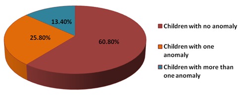
The study population composed of 600 children having 293 (48.8%) males and 275 (45.8%) females. Among all the participants, 60.8% were free from any kind of dental anomaly. Children having only one anomaly were 25.8%, whereas only 13.4% showed presence of more than one anomaly (Graph 1).
Graph 1: Showing frequency of dental anomalies among study population.

There was no significant difference found between dental anomalies according to gender, with the exception of hypodontia, microdontia and talons cusp in which first two anomalies were more among
females and talons cusp was more common among males (P<0.05) as shown in Table 1.
Table 1 - Prevalence of dental anomalies among the study population according to gender.
| Dental Anomalies | Male | Female | p-value | ||
| Supernumerary teeth | 19 | 3.1% | 13 | 2.2% | 0.302** |
| Hypodontia | 11 | 1.8% | 18 | 3.0% | 0.050* |
| Double teeth | 11 | 1.8% | 7 | 1.2% | 0.490** |
| Macrodontia | 6 | 1.0% | 2 | 0.3% | 0.191** |
| Microdontia | 6 | 1.0% | 20 | 3.3% | 0.001* |
| Talons cusp | 26 | 4.3% | 12 | 2.0% | 0.038* |
| Taurodontism | 17 | 2.8% | 16 | 2.7% | 0.346** |
| Impacted | 109 | 18.2% | 126 | 21.0% | 0.489** |
| Rotation | 41 | 6.8% | 38 | 6.3% | 0.438** |
Impactions were the most common anomaly in this study and most of the impacted teeth were related to maxilla and also the number was more among females (21.0%). The data obtained showed that prevalence of double teeth was 3.0%, of which 61.2% were fused and 38.8% were geminated. The cases of microdontia (4.3%) were more than macrodontia (1.3%). 4.8% subjects had hypodontia and the most common teeth were lateral incisor followed by second premolars. Supernumerary teeth were seen among 5.3% of the subjects and mostly in the maxillary arch. The frequency of talons cusp was 6.3% and mainly seen in maxillary canines and incisors. Around 13.2% of the children had rotated teeth which were more found in lower anterior region (Graph 2)
Graph 2: Showing overall prevalence of dental anomalies.

Discussion
Presence of dental anomalies is commonly seen during routine dental check-up. Mostly these anomalies develop earlier than the eruption of dentition, and are often hereditarily. The effect of the dental anomalies leads to functional, aesthetic and occlusal problems.6
Impacted teeth were the most common finding in the present study population. The results were in agreement with earlier studies done by Altug-Atac et al (2007)7, Uslu et al (2009)8 and Kruthika et al (2010)9 among 20,182 Indian patients. However other studies showed lesser results of impacted teeth than the present data.10-11
In the present study 60.8% had no dental anomaly, 25.8% showed presence of one anomaly and 13.4% showed more than one. The findings regarding frequency of dental anomalies were higher than study conducted by Sogra et al in 2012 among Iranian orthodontic patients12 and Gupta et al among Indian population.13
The study showed tooth size discrepancy such as macrodontia, microdontia and peg shaped lateral incisor separately. There was no data related to peg-shaped lateral incisors where as many studies have this finding varied between 0.3 and 8.4%.14-15 Several studies have shown the prevalence of microdontia varies between 0.8 to 8.4% (Neville et al, 2005)16 and our data stated that 4.3% of the subjects had microdontia.
The prevalence of double teeth (fusion and germination) has been reported to be 3% and the results were higher than Kositbowornchai et al in 2010 among Thai patients.17 Where as Sogra et al, had not observed any case of germination and fusion.12
In the present study, supernumerary teeth were seen among 5.3% subjects and mostly in the maxillary arch and these results are more than as observed in study done Gupta et al that showed prevalence 2.40% of participants with supernumerary teeth.13 Other studies also showed that 90% to 98% of supernumerary teeth occur in the maxilla arch.7,18
Apart from third molars the present data showed most common missing tooth as lateral incisors followed by 2nd premolars and similar findings were obtained by Menczer which also showed that lateral incisor is the most common missing teeth followed by 2nd premolars.19But some other studies conducted by Clayton (1956)3 and Castaldi et al (1966)20 showed that 2nd premolar was the most common missing teeth followed by lateral incisor. Mostly dental anomalies are associated with syndromes, even though some occur without any evidence.
Conclusion
The data concluded that 25.8% subjects had atleast one dental anomaly. Impacted teeth was the most common finding followed by rotated teeth. Prevalence of hypodontia was seen more among females and the cases of supernumerary teeth were more among male population and in the maxillary region. The high level of occurrence of these anomalies suggests to find the aetiological factors and earlier treatment of the dental anomaly.
Footnotes
Source of Support: Nil
Conflict of Interest: None Declared
Contributor Information
Mitesh D Kathariya, Department of Pedodontics & Preventive Dentistry, Rural Dental College, Pravara Institute of Medical Sciences (PIMS), Loni, Maharashtra, India.
Atul Pralhad Nikam, Department of Oral Pathology and Microbiology, Rural Dental College, Pravara Institute of Medical Sciences (PIMS), Loni, Maharashtra, India.
Kirti Chopra, Department of Pedodontics & Preventive Dentistry, Rural Dental College, Pravara Institute of Medical Sciences (PIMS), Loni, Maharashtra, India.
Namrata N Patil, Department of Oral Pathology and Microbiology, Saraswati Dhanwantari Dental College and Hospital, Parbhani, Maharashtra, India.
Hitesh Raheja, Vardhmann Mahavir Medical College and Safdarjung hospital, New Delhi, India.
Renuka Kathariya, Department of Conservative Dentistry and Endodontics, Pravara Institute of Medical Sciences (PIMS), Loni, Maharashtra, India.
References
- 1.GB Winter, AH Brook. Enamel hypoplasia and anomalies of the teeth. Dent Clin North Am. 1975;19(1):3–24. [PubMed] [Google Scholar]
- 2.HL Bailit. Dental variation among populations. An anthropologic view. Dent Clin North Am. 1975;19(1):125–139. [PubMed] [Google Scholar]
- 3.JM Clayton. Congenital dental anomalies occurring in 3557 children. J Dent Child. 1956;23:206–208. [Google Scholar]
- 4.J Ghabanchi, AA Haghnegahdar, SH Khodadazadeh, S Haghnegahdar. A Radiographic and Clinical Survey of Dental Anomalies in Patients Referring to Shiraz Dental School. Shiraz Univ Dent J. 2010;10(Suppl):26–31. [Google Scholar]
- 5.V Patel, A Kleinman. Poverty and common mental disorders in developing countries. Bull World Health Organ. 2003;81(8):609–615. [PMC free article] [PubMed] [Google Scholar]
- 6.OO Osuji, J Hardie. Dental anomalies in a population of Saudi Arabian children in Tabuk. Saudi Dent J. 2002;14(1):11–14. [Google Scholar]
- 7.AT Altug-Atac, D Erdem. Prevalence and distribution of dental anomalies in orthodontic patients. Am J Orthod Dentofacial Orthop. 2007;131(4):510–514. doi: 10.1016/j.ajodo.2005.06.027. [DOI] [PubMed] [Google Scholar]
- 8.O Uslu, MO Akcam, S Evirgen, I Cebeci. Prevalence of dental anomalies in various malocclusions. Am J Orthod Dentofacial Orthop. 2009;135(3):328–335. doi: 10.1016/j.ajodo.2007.03.030. [DOI] [PubMed] [Google Scholar]
- 9.KS Guttal, VG Naikmasur, P Bhargav, RJ Bathi. Frequency of Developmental Dental Anomalies in the Indian Population. Eur J Dent. 2010;4(3):263–269. [PMC free article] [PubMed] [Google Scholar]
- 10.K Aitasalo, R Lehtinen, E Oksala. An orthopantomographic study of prevalence of impacted teeth. Int J Oral Surg. 1972;1(3):117–120. doi: 10.1016/s0300-9785(72)80001-2. [DOI] [PubMed] [Google Scholar]
- 11.TH Udom, J Terrence. Prevalence of dental anomalies in orthodontic patients. Aust Dent J. 1998;43(6):395–398. [PubMed] [Google Scholar]
- 12.Y Sogra, GM Mahdjoube, K Elham, TM Shohre. Prevalence of dental anomalies in Iranian orthodontic patients. Journal of Dentistry and Oral Hygiene. 2012;4(2):16–20. [Google Scholar]
- 13.SK Gupta, P Saxena, S Jain, D Jain. Prevalence and distribution of selected developmental dental anomalies in an Indian population. J Oral Sci. 2011;53(2):231–238. doi: 10.2334/josnusd.53.231. [DOI] [PubMed] [Google Scholar]
- 14.I Brin, A Becker, M Shalhav. Position of the maxillary permanent canine in relation to anomalous or missing lateral incisors: a population study. Eur J Orthod. 1986;8(1):12–16. doi: 10.1093/ejo/8.1.12. [DOI] [PubMed] [Google Scholar]
- 15.T Ooshima, R Ishida, K Mishima, S Sobue. The prevalence of developmental anomalies of teeth and their association with tooth size in the primary and permanent dentitions of 1650 Japanese children. Int J Paediatr Dent. 1996;6(2):87–94. doi: 10.1111/j.1365-263x.1996.tb00218.x. [DOI] [PubMed] [Google Scholar]
- 16.DW Neville, DD Damm, CM Allen, JE Bouquot. Abnormalities of teeth: Oral and Maxillofacial Pathology, 2nd ed. Philadelphia, PA: Elsevier. 2005:49–89. [Google Scholar]
- 17.S Kositbowornchai, C Keinprasit, N Poomat. Prevalence and distribution of dental anomalies in pretreatment orthodontic Thai patients. Khon Kaen Uni Dent J. 2010;13:92–100. [Google Scholar]
- 18.RR Parry, VS Iyer. Supernumerary teeth amongst orthodontic patients in India. Br Dent J. 1961;111:257–258. [Google Scholar]
- 19.LF Menczer. Anomalies of the primary dentition. J Dent Child. 1955;22:57–62. [Google Scholar]
- 20.CR Castaldi, BA Bodnarchuk, PD MacRae, WA Zacherl. Incidence of congenital anomalies in permanent teeth of a group of Canadian children aged 6-9. J Can Dent Assoc. 1966;32(3):154–159. [PubMed] [Google Scholar]


