Abstract
Background: The purpose of this in-vitro study was to compare canal transportation and centering ability of Twisted and Hyflex Rotary Files with stainless steel hand k-flexofiles by using Spiral Computed Tomography.
Materials & Methods: A total of 90 freshly extracted human mandibular single rooted Premolar teeth were selected. The crowns were flattened with steel disks and a final dimension of 18-mm WL was achieved for each tooth. Canals were divided randomly into 3 groups of 30 teeth each. Group I:Hyflex files, Group II:Twisted files, Group III:stainless steel hand k-flexofiles. Three sections from apical, mid-root, and coronal levels of the canal were recorded. All the teeth were scanned before and after instrumentation by using Spiral Computed Tomography.
Results: K-files showed highest transportation and less centered when compared to the Twisted and Hyflex rotary files. No significant difference was found between TF and Hyflex CM instruments.
Conclusion: TF and Hyflex files shaped curved root canals without significant shaping errors when compared to the Hand stainless steel k-flexofiles.
How to cite this article: Kumar BS, Pattanshetty S, Prasad M, Soni S, Pattanshetty KS, Prasad S. An in-vitro Evaluation of canal transportation and centering ability of two rotary Nickel Titanium systems (Twisted Files and Hyflex files) with conventional stainless Steel hand K-flexofiles by using Spiral Computed Tomography. J Int Oral Health 2013;5(5):108-15.
Key Words: : canal transportation, centering ability, spiral computed tomography
Introduction
Endodontic triad comprises of three basic principles thorough debridement, sterilization and complete obturation of root canal.1 These all together constitute equally important phases of endodontic treatment.
One of the primary goals of root canal preparation during application of debriding instruments is maintaining the root canal curvature with centering ability and without or less canal transportation.
According to Walton et al (1928)2 the canals prepared with stainless steel instruments only cleaned superfici-ally and the pulp tissue was not removed completely.
Stainless steel files create aberrations, probably as a result of the inherent stiffness of stainless steel, which
is compounded by instrument design and canal shape.3 Tran V.Lam4 reported that most instrumentation techniques with stainless steel instruments in curved canals result in root canal transportation. Various studies has showed that the incidence of transportation and straightening of the Root canal is common with the use of stainless steel files.5
Rotary endodontic instruments has been developed from nickel-titanium alloy that potentially allows
shaping of narrow, curved root canals, without causing aberrations.6
Recently, thermal treatment of NiTi alloy has been used to optimize the mechanical properties of NiTi alloy7-12. Hyflex CM rotary instruments (Coltene-Whaledent, Allstetten, Switzerland) are made from a
new type of NiTi wire, namely CM wire(controlled memory), that has been subjected to proprietary thermomechanical processing.13 It has been
manufactured by a unique process that controls the material's memory, making the files extremely flexible but without the loss of shape memory typical of other NiTi files. Twisted File (TF) (SybronEndo, Orange, CA) was developed by the manufacturer through a special thermomechanical treatment. TF differs from other
rotary NiTi files in that the metal wire is first ground into the triangular cross-sectional shape and then twisted after the grinding. It has been reported to have a higher fracture resistance than other files. The manufacturer claims that the Twisted files are subjected to R-phase heat treatment, followed by
twisting of the metal, and special surface conditioning, which significantly increases the instruments' flexibility and resistance to cyclic fatigue.14 Because of the increased flexibility, the new generation NiTi files may maintain the original canal shape better and minimize canal transportation in the curved root canals.
Materials and Methods
Selection of teeth: 90 freshly extracted human mandibular single rooted premolar teeth were
selectedand stored in 0.1% thymol until use (to lower the permeability of the teeth). The criteria for selection were: Free of caries, free of restoration, Complete root formation, with curved root canal with angle of curvature 10 to 20 degree (moderate curvature) by using schneiders method.15
Preparation of Specimens:
The teeth were cleaned free of debris and calculus. A conventional access cavity was prepared in each tooth with a No.4 round diamond point with high speed to allow direct access to all root canals. A size 10 stainless steel hand k-flexofile was filed into the canal until it was visible at the apical foramen and the working length was recorded 0.5 mm short of this length. For uniform samples,the final length of 18-mm WL was achieved by flattening the crown using steel disks for each tooth.16
All the roots were embedded into transparent acrylic. The ninety teeth were randomly divided into 3 experimental groups containing 30 teeth each.
GROUP-I(n=30):Using Hyflex files
GROUP-II(n=30):Using Twisted files
GROUP-III(n=30):Using Hand k-flexofiles
The root canal shape before instrumentation was determined by scanning all the teeth using Spiral Computed Tomography. The 2 sections were marked 3 mm from the apical end of the root (apical level) and 3
mm below the orifice from the coronal level (15 mm from the apex). A further section at the mid-root level (9 mm from the apex) was marked by dividing the distance between the sections into 2 equal lengths. After initial scans root canals were instrumented by using a step-back technique. All root canals were instrumented to the, WL with sizes 10, 15 K-files by using a step-back technique. Root canals with Apical sizethat were larger than ISO size 15 were discarded.
GROUP I and GROUP-II: (n = 30)-Hyflex CM and TF and a torque-control level of 2.5 Ncm by using an 8:1
reduction handpiece powered by an endodontic motor (XSmart; DentsplyMaillefer) with the speed of 500rpm. Each set of NiTi files for the group I and group II included the following: orifice shaping with .08/25, followed by .06/25 and .04/25 to WL, and then enlargement with .06/25 and .06/30.
GROUP-III: (n = 30)
In these group root canals were prepared using stainless steel hand K flexo files using following steps:-
All the 30 teeth were shaped by using "watch-winding" manipulation with precurved hand stainless steel K-flexofiles instruments from size 20 to size 60 with a step-back technique. Final apical preparation size was 30.
For all groups after the use of each file, canals were irrigated with 3 mL of a 5.25% NaOCl solution. Lubrication was obtained using Glyde (Dentsply, Maillefer) during instrumentation, and after instrume-ntation, 1 mL of 17% ethylenediaminetetraacetic acid was used for 1 minute followed by a final flush of 3 mL of NaOCl. Each instrument was changed after 5 canals. All irrigation procedures were delivered with a 27 gauge needle.
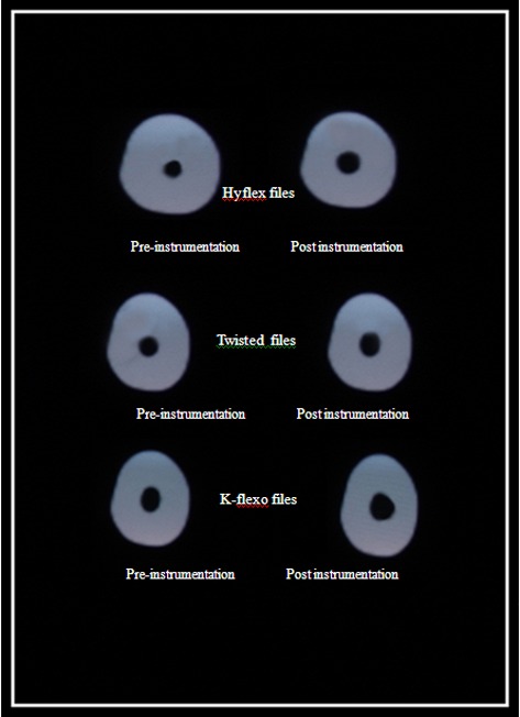
All the instrumented teeth were then scanned with Spiral CT and were recorded (Figure 2).
Fig. II: Showing spiral CT scan pre and post instrumentation of the files.
Evaluation of Canal Transportation
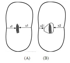
The canal transportation was determined by measuring the shortest distance from the root canal surface of uninstumented canal to the outer root surface (mesial and distal), and then comparing the same measurments obtained from instrumented image. All values were measured and a mean value was taken.16 The following formula was used for the calculation of root canal transportation:
| (a1 – a2) - (b1 – b2) |
, where a1 is the distance from the mesial surface of the root to the mesial surface of the uninstrumented canal, b1 is the distance from distal surface of the root to the distal surface of the uninstrumented canal, a2 is the distance from the mesial surface of the root to the mesial surface of the instrumented canal, and b2 is the distance from distal surface of the root to the distal surface of the instrumented canal (Figure 1). Results other than 0 indicates that transportation has occurred in the canal.16
Fig. I: DIAGRAMMATIC REPRESENTATION SHOWING MEASURESENTS FOR IMAGE CROSS SECTIONS:(A) Uninstrumented CT Scan (B) Instrumented CT Scan.
Evaluation of Centering Ability:
The centering ability was calculated by using the following ratio:
| (a1 – a2)/(b1 – b2) or (b1 – b2)/(a1 – a2), |
If these numbers are not equal, the lower figure is considered as the numerator of the ratio. Result of 1 indicates perfect centering.16
Statistical Analysis
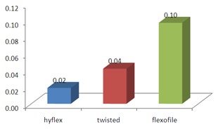
One-way analysis of variance followed by post hoc Tukey honestly significant difference (HSD) test were conducted to explore a significant difference in mean degree of canal transportation between the 3 groups. The level of significance was p = 0.05 (Table 3A & Graph 3A).
TABLE 3A: Showing Mean Value of Canal Transportation at Apical Section.
| Ca | Middle | |
| Groups | Mean ± SD | |
| I. | hyflex | 0.02 ± 0.06 |
| II. | twisted | 0.04 ± 0.06 |
| III. | flexofile | 0.10 ± 0.07 |
Graph 3A: Showing mean value of canal tranportation at apical section.
Results
Comparison between groups for Canal Transportation:
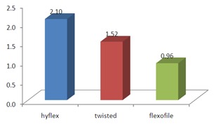
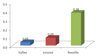
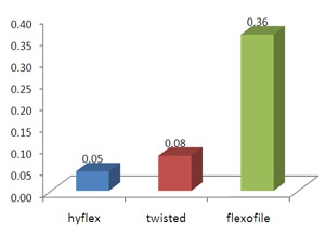
The difference in apical canal transportation between TF and Hyflex CM instruments was not significant (P> .05). In the coronal and the middle thirds of the canals, Hyflex CM files caused slightly less transportation than TF instruments. In the apical third of canals, TF remained better centered (Table 1A & 2A). However, in all of the 3 areas studied, the difference was not significant (P> .05)(Graph 1A & 2A).
TABLE 1A: Showing Mean Value of Canal Transportation at Coronal Section.
| Ct | Coronal | |
| Groups | Mean ± SD | |
| I | hyflex | 0.05 ± 0.05 |
| II. | twisted | 0.08 ± 0.04 |
| III. | flexofile | 0.36 ± 0.10 |
TABLE 2A: Showing Mean Value of Canal Transportation at Middle Section.
| Ct | Middle | |
| Groups | Mean ± SD | |
| I. | hyflex | 0.04 ± 0.05 |
| II. | twisted | 0.09 ± 0.08 |
| III. | flexofile | 0.38 ± 0.44 |
Graph 1A: Showing mean value of canal tranportation at coronal section.
Graph 2A: Showing mean value of canal tranportation at middle section.
Overall, preparation of root canal with Hyflex CM, TF resulted in adequate canal shapes with no major shaping errors.
The mean degree of canal transportation was statistically highest with the manual technique(P< .001).
Comparison between groups for Centering Ratio (Table 2B & 3B)(Graph 2B & 3B) - In the cervical section of the root canal, mean centering ratio was statistically highest with Twisted and Hyflex files (P<.001) and minimal with the manual technique (P< .05).
TABLE 2B: Showing Mean Value of Centering Ability at Middle Section.
| Ct | Middle | |
| Groups | Mean ± SD | |
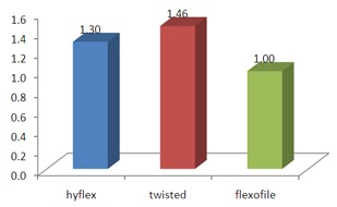
| I. | hyflex | 1.30 ± 0.64 |
| II. | twisted | 1.46 ± 0.40 |
| III. | flexofile | 1.00 ± 0.00 |
TABLE 3B: Showing Mean Value of Centering Ability at Apical Section.
| Ca | Apical | |
| Groups | Mean ± SD | |
| I. | hyflex | 2.10 ± 0.34 |
| II. | twisted | 1.52 ± 0.10 |
| III. | flexofile | 0.95 ± 0.26 |
Graph 2B: Showing mean value of centering ability at middle section.
Graph 3B: Showing mean value of centering ability at apical section.
Discussion
Various methods have been used to compare the canal shape before and after preparation to investigate the efficiency of instruments and techniques developed for root canal preparation.
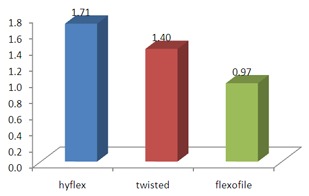
Radiography is one of the methods which only provide a 2-dimensional image.17 The serial sectioning technique18 is a complicated and a physical sectioning of the teeth before preparation can result in loss of material and can cause tissue changes.19 A noninvasive method for the analysis of canal geometry and efficiency of shaping techniques, can be achieved by use of computed tomography imaging technique,20 it can also be used to compare the anatomic structure of root canal before and after instrumentation. (Table 1B & Graph 1B)
TABLE 1B: Showing Mean Value of Centering Ability at Coronal Section.
| Ca | Coronal | |
| Groups | Mean ± SD | |
| I. | hyflex | 1.71 ± 0.33 |
| II. | twisted | 1.40 ± 0.26 |
| III. | flexofile | 0.97 ± 0.26 |
Graph 1B: Showing mean value of centering abilityat coronal section.
Recently, novel thermomechanical processing and new manufacturing technologies have been developed to optimize the microstructure of NiTi alloys. TF and Hyflex CM files have been said to have higher resistance to cyclic fatigue and flexibility than conventional superelasticNiTi files.7 The cutting ability of root canal instruments involves a complex interrelationship of different parameters such as the cross-sectional design, which seems to be a decisive parameter,21 chip-removal capacity of the instrument, the helical and rake angle, metallurgical properties, and also surface treatment of the instrument.4 In the present study a comparison between rotary TF ,Hyflex files and hand stainless steel K-flexofiles files was performed to evaluate canal transportation and centering ratio. For stainless steel Kfiles more canal transportation and less centering ability was seen when compared to the other two rotary files because of its stiffness and by the cutting efficiency.4 (Figure 2) The difference in apical canal transportation between TF and Hyflex CM instruments was not significant (P> .05). In the coronal and the middle thirds of the canals, Hyflex CM files caused slightly less transportation than TF instruments. In the apical third of canals, TF remained better centered (Table 3B)(Graph 3B). However, in all of the 3 areas studied, the difference was not significant. Obvious procedural errors were not detected in this study. However, some canal transportation was detected in the present study. This result was in accordance with study conducted by Gergiet al16 which showed that TF instruments caused less canal transportation than ProTaper instruments by using spiral CT scanning. El Batouty and Elmallah22 found that TF instruments showed a greater tendency to preserve the curvature of curved canals of the mandibular molar than K3 instruments by using 2 dimensional photographic technique. Zhao et al23 showed no statistical difference in canal transportation between TF and Hyflex files, the fact that TF instruments provided a centered preparation while maintaining the original shape of the curved canal is because TF is provided with R-phase treatment and are manufactured by twisting NiTi wire , the surface deoxidation of these file which might explain its high flexibility,24 even hyflex files provided a centered preparation while maintaing the original shape of the curved canals, the HyFlex® CMTM NiTi files has Controlled Memory and are 300% more resistant to cyclical fatigue compared to other NiTi files, which helps reducing the incidence of file separation in the canals. HyFlex® CMTM NiTi files have been manufactured with a unique process that controls the material's memory, making the files extremely flexible.24 This increases the ability of the file to follow the anatomy of the canal very closely, and reduces the risk of ledging, transportation and perforation.
Conclusion
Under the conditions of the present study, Hyflex CM and TF instruments shaped curved root canals in vitro without significant shaping errors.
Footnotes
Source of Support: Nil
Conflict of Interest: None Declared
Contributor Information
B Shiva Kumar, Department of Conservative Dentistry & Endodontics, D A P M R V Dental College, Bangalore, Karnataka, India.
Spoorti Pattanshetty, Impression Dental Clinic, Belgaum, Karnataka, India.
Manju Prasad, Department of Orthodontics & Dento-facial Orthopedics, Krishnadevaraya College of Dental Sciences, Bangalore, Karnataka, India.
Sunny Soni, Department of Conservative Dentistry & Endodontics, D A P M R V Dental College, Bangalore, Karnataka, India.
Kirti S Pattanshetty, Department of Pedodontics & Preventive Dentistry, Dr. D Y Patil Dental College and Hospital, Nerul, Navi Mumbai, Maharashtra, India.
Shiva Prasad, Department of Orthodontics & Dento-facial Orthopaedics, D A P M R V Dental College, Bangalore, Karnataka, India.
References
- 1.M Hülsmann, OA Peters, PM Dummer. Mechanical preparation of root canals: shaping goals, techniques and means. Endod Topics. 2005;10(1):30–76. [Google Scholar]
- 2.RE Walton. Histologic evaluation of different methods of enlarging the pulpcanal space. J Endod. 1976;2(10):304–311. doi: 10.1016/S0099-2399(76)80045-3. [DOI] [PubMed] [Google Scholar]
- 3.I Musani, V Goyal, A Singh, C Bhat. Evaluation and Comparison of Biological Cleaning Efficacy of Two Endofiles and Irrigants as Judged by Microbial Quantification in Primary Teeth – An In Vivo Study. Int J Pediatr Dent. 2009;2(3):15–22. doi: 10.5005/jp-journals-10005-1013. [DOI] [PMC free article] [PubMed] [Google Scholar]
- 4.TV Lam, DJ Lewis, DR Atkins, RH Macfarlane, RM Clarkson, MG Whitehead, PJ Brockhurst, AJ Moule. Changes in root canal morphology in simulated curved canals over-instrumented with a variety of stainless steel and nickel titanium files. Aust Dent J. 1999;44(1):12–19. doi: 10.1111/j.1834-7819.1999.tb00530.x. [DOI] [PubMed] [Google Scholar]
- 5.E Schäfer. Effects of four instrumentation techniques on curved canals: acomparison study. J Endod. 1996;22(12):685–689. doi: 10.1016/S0099-2399(96)80065-3. [DOI] [PubMed] [Google Scholar]
- 6.S Ozer. Outcome of root canal treatments prepared with tri-auto zx and hand filing. An in vivo study. Biotechnol Biotechnol Eq. 2006;20(1):157–162. [Google Scholar]
- 7.G Gambarini, NM Grande, G Plotino, F Somma, M Garala, M De Luca, L Testarelli. Fatigue resistance of engine-driven rotary nickel-titanium instruments producedby new manufacturing methods. J Endod. 2008;34(8):1003–1005. doi: 10.1016/j.joen.2008.05.007. [DOI] [PubMed] [Google Scholar]
- 8.E Johnson, A Lloyd, S Kuttler, K Namerow. Comparison between a novelnickel-titanium alloy and 508 nitinol on the cyclic fatigue life of ProFile 25/.04 rotary instruments. J Endod. 2008;34(11):1406–1409. doi: 10.1016/j.joen.2008.07.029. [DOI] [PubMed] [Google Scholar]
- 9.TR Kramkowski, J Bahcall. An in vitro comparison of torsional stress andcyclic fatigue resistance of ProFile GT and ProFile GT Series X rotarynickel-titanium files. J Endod. 2009;35(3):404–407. doi: 10.1016/j.joen.2008.12.003. [DOI] [PubMed] [Google Scholar]
- 10.Y Gao, V Shotton, K Wilkinson, G Phillips, WB Johnson. Effects of raw materialand rotational speed on the cyclic fatigue of ProFile Vortex rotary instruments. J Endod. 2010;36(7):1205–1209. doi: 10.1016/j.joen.2010.02.015. [DOI] [PubMed] [Google Scholar]
- 11.Y Shen, W Qian, H Abtin, Y Gao, M Haapasalo. Fatigue testing of controlledmemory wire nickel-titanium rotary instruments. J Endod. 2011;37(7):997–1001. doi: 10.1016/j.joen.2011.03.023. [DOI] [PubMed] [Google Scholar]
- 12.G Gambarini, G Plotino, NM Grande, D Al-Sudani, M De Luca, L Testarelli. Mechanical properties of nickel-titanium rotary instruments produced with a new manufacturing technique. Int Endod J. 2011;44(4):337–341. doi: 10.1111/j.1365-2591.2010.01835.x. [DOI] [PubMed] [Google Scholar]
- 13.L Testarelli, G Plotino, D Al-Sudani, V Vincenzi, A Giansiracusa, NM Grande, G Gambarini. Bending properties of a new nickel-titanium alloy with a lower percent by weight of nickel. J Endod. 2011;37(9):1293–1295. doi: 10.1016/j.joen.2011.05.023. [DOI] [PubMed] [Google Scholar]
- 14.G Gambarini, R Gerosa, M De Luca, M Garala, L Testarelli. Mechanical properties of a new and improved nickel-titanium alloy for endodontic use: an evaluation of file flexibility. Oral Surg Oral Med Oral Pathol Oral Radiol Endod. 2008;105(6):798–800. doi: 10.1016/j.tripleo.2008.02.017. [DOI] [PubMed] [Google Scholar]
- 15.Zhu Ya-qin, Gu Ying-xin, MSD, Du Rong, Li Chen. Reliability of two methods on measuring root canal curvature. Int Chin J Dent. 2003;3:118–121. [Google Scholar]
- 16.R Gergi, JA Rjeily, J Sader, A Naaman. Comparison of canal transportation and centering ability of twisted files, Pathfile-ProTaper system, and stainless steel hand K-files by using computed tomography. J Endod. 2010;36(5):904–907. doi: 10.1016/j.joen.2009.12.038. [DOI] [PubMed] [Google Scholar]
- 17.SE Dowker, GR Davis, JC Elliott. X-ray microtomography: non destructive three-dimensional imaging for in vitro endodontic studies. Oral Surg Oral Med Oral Pathol Oral Radiol Endod. 1997;83(4):510–516. doi: 10.1016/s1079-2104(97)90155-4. [DOI] [PubMed] [Google Scholar]
- 18.CM Bramante, A Berbert, RP Borges. A methodology for evaluation of root canal instrumentation. J Endod. 1987;13(5):243–245. doi: 10.1016/S0099-2399(87)80099-7. [DOI] [PubMed] [Google Scholar]
- 19.JM Gambill, M Alder, CE del Rio. Comparison of nickel-titanium and stainless steel hand-file instrumentation using computed tomography. J Endod. 1996;22(7):369–375. doi: 10.1016/S0099-2399(96)80221-4. [DOI] [PubMed] [Google Scholar]
- 20.JS Rhodes, TR Ford, JA Lynch, PJ Liepins, RV Curtis. Micro-computedtomography: a new tool for experimental endodontology. Int Endod J. 1999;32(3):165–170. doi: 10.1046/j.1365-2591.1999.00204.x. [DOI] [PubMed] [Google Scholar]
- 21.E Schäfer, M Oitzinger. Cutting efficiency of five different types of rotary nickel-titanium instruments. J Endod. 2008;34(2):198–200. doi: 10.1016/j.joen.2007.10.009. [DOI] [PubMed] [Google Scholar]
- 22.KM El Batouty, WE Elmallah. Comparison of canal transportation and changes in canal curvature of two nickel-titanium rotary instruments. J Endod. 2011;37(9):1290–1292. doi: 10.1016/j.joen.2011.05.024. [DOI] [PubMed] [Google Scholar]
- 23.D Zhao, Y Shen, B Peng, M Haapasalo. Micro-computed tomography evaluation ofthe preparation of mesiobuccal root canals in maxillary first molars with Hyflex CM, Twisted Files, and K3 instruments. J Endod. 2013;39(3):385–388. doi: 10.1016/j.joen.2012.11.030. [DOI] [PubMed] [Google Scholar]
- 24.Y Shen, HM Zhou, YF Zheng, B Peng, M Haapasalo. Current challenges andconcepts of the thermomechanical treatment of nickel-titanium instruments. J Endod. 2013;39(2):163–172. doi: 10.1016/j.joen.2012.11.005. [DOI] [PubMed] [Google Scholar]


