Abstract
Purpose: A multiparticular floating-pulsatile drug delivery system was developed for time and site specific drug release of piroxicam. A blend of floating and pulsatile principles of drug delivery system would have the advantage that a drug can be released in the upper GI tract after a definite time period. Methods: Hollow microspheres were prepared by the emulsion solvent diffusion method using Eudragit S as an enteric acrylic polymer with piroxicam at various polymer/drug ratios in a mixture of dichloromethane and ethanol. Developed formulations were evaluated for yield, encapsulation efficiency, particle size, shape, apparent density, buoyancy studies and dissolution studies. Results: The obtained microballoons were spherical with no major surface irregularity and mean particle size ranging from 250 to 380 for different batches. Formulations show a slight amount of relaese ranging from 0.7 to 11% in acidic medium (SGF) with complete release of drug in simulated intestinal fluid (SIF) in less than 3 h. Encapsulation efficiency of different formulations varied from 90 to 98%. The optimum loading amount of drug in the particles was found to impart suitable floatable properties to the microballoons. With increasing polymer/drug ratio, buancy of the microballoons increases accompanied by simultaneous reduction of apparent particle density. Conclusion: A pulsatile release of piroxicam was demonstrated by a simple drug delivery system which could be useful in chronopharmacotherapy of rheumatoid arthritis.
Keywords: Piroxicam, floating microparticles, pulsatile systems
Introduction
Pulsatile drug delivery system (PDDS) is based on principle of rapid release of a certain amount of drug within short time period after a predetermined off-release period, lag time.1 Such novel drug delivery has been attempted for: (i) chronopharmacotherapy of diseases which show circadian rhythms in their pathophysiology2; (ii) avoiding degradation of active ingredients in upper GI tract, e.g. proteins and peptides3; (iii) for time programmed administration of hormones and many drugs such as isosorbidedinitrate, respectively to avoid suppression of normal secretion of hormones in body that can be hampered by constant release of hormone from administered dosage form and development of resistance4,5; (iv) to avoid pharmacokinetic drug–drug interactions between concomitantly administered drugs6, etc. Chronopharmacotherapy for rheumatoid arthritis has been recommended to ensure that the highest blood levels of the drug coincide with peak pain and stiffness.7 Morning stiffness associated with pain at the time of awakening is a diagnostic criterion of the rheumatoid arthritis and these clinical circadian symptoms are supposed to be outcome of altered functioning of hypothalamic–pitutary–adrenocortical axis.8,9 A pulsatile drug delivery system that can be administered at night (before sleep) but that release drug in early morning would be a promising chronopharmaceutic system.
Oral pulsatile drug delivery systems that release drug after a lag period of 6–7 h usually release drug in large intestine 10 however, the viscous contents of lower part of GI tract cause hindrance to the drug diffusion and also enzymatic degradation of some drugs makes it an unfavorable site for drug release.11 Moreover, the majority of drugs are preferentially absorbed from the upper part of the small intestine 12 hence, drug release at site of better absorption can improve therapeutic efficacy of drug. This is of more concern for drug delivery that is meant for pulse drug release after a lag period of 5–6 h following oral administration of dosage form. A blend of floating and pulsatile principles of drug delivery system would have the advantage that a drug can be released in the upper GI tract after a definite time period of no drug release (lag period). The major objectives of the present study were to develop a simple, multiparticulate, floating-pulsatile drug delivery system for obtaining no drug release during floating time followed by pulse drug release in small intestine. Additionally, multiple unit dosage forms provide many relative advantages over single unit dosage forms such as predictable GI transit time, greater product safety, etc.. 13
The essential features of this conceptual delivery system exhibit as a (a) multiparticulate system, (b) combination of gastroretentive and pulsatile principles in same dosage form, (c) single-step formulation process (d) idealistic drug release profile that is effective at morning time intended for a particular pathological condition based on chronotherapy, and (e) having low drug release in stomach suited for non-steroidal anti-inflammatory drug (NSAID) class of drug. Piroxicam, for symptomatic relief in rheumatoid arthritis, exhibits better toleration than indomethacin, naproxen, and enteric- coated acetylsalicylic acid hence; piroxicam was used as a model drug in this study. 14
Materials and Methods
Materials
Piroxicam, PX (Shasun Chemicals & Drugs, India), Eudragit S100, Eu S100 (MW ~ 135 KDa, Rohm GmbH, Germany), polyvinyl alcohol, PVA, (MW ~ 70 KDa, 88% hydrolyzed, Sigma, Germany), and ethanol and dichloromethane (Merck, Germany) were used. The Solvents were of analytical grade.
Preparation of the microballoons
PX (0.1- 0.6g) was dissolved with Eu S100 (0.6 g) in a mixed solution of ethanol (good solvent, 5 ml), and dichloromethane (bridging liquid, 5 ml). The microballoons were prepared by slowly adding the resultant solution (during 10 min) to 200 ml 0.1 N HCl containing 0.75 % of PVA (at 25 °C) placed in a cylindrical vessel (500 ml) equipped with three baffles under agitation (400 rpm) using a propeller type stirrer with four blades. After agitating the system for 20 min the solidified microballoons were recovered by filtration. The resultant products were dried in an oven at 50 °C for 6 h.
Determination of production yield and encapsulation efficiency of microballoons
All microballoon formulations were prepared in triplicate. Microballoons dried at 50 °C were then weighed and the yield of microballoon preparation was calculated using the formula (Eq. (1)):

Eq. (1)
Microballoons were crushed and powdered by using a mortar. Accurately weighed of this powder was extracted in ethanol. The solution was then filtered, a sample of 2ml was withdrawn from this solution and assayed spectrophotometrically to determine the PX content of the microballoons. A calibration curve based on standard solutions of PX in ethanol was used to calculate the PX concentration.
The encapsulation efficiency (%) was calculated by using following Equation (Eq. (2)).
Eq. (2).

where Mactual is the actual PX content in weighed quantity of powder of microballoons and Mtheoretical is the theoretical amount of PX in microballoons calculated from the quantity added in the fabrication process. The means of three assays were reported.
Measurement of micromeritic properties
Size distribution and mean diameter were determined by the sieving method. A total of 25 g of material was sieved using an Erweka vibration sieve (Erweka, Germany) through a nest of sieves. The vibration rate was set at 200 strokes/min and the sieving time was 10 min. The powder fractions retained by the individual sieves were determined and expressed as mass percentages. The morphology of the microballoonss was observed by optical microscopy (Nikon Labophot, Tokyo, Japan). Apparent particle density and roundness was determined by the projective image method as follows. Microballoons were placed on a glass plate and Heywood diameter, microballoon number, circumference of a projective image and area of a projective image were measured by an Image analyzing software (scion image analyzer). Subsequently, the apparent particle density and roundness were calculated
according to Eq. (3) and Eq. (4), respectively.
Apparent particle density w/v=w/∑(πd3n/6) Eq. (3)
where W = weight of microballoons, V = volume of microballoons, d = Heywood diameter, and n = number of microballoons.
Roundness of microballoons = L2/4πs Eq. (4)
where L = circumference of a projective image and S = area of a projective image. When the roundness of microballoons was close to 1, the microballoons closely resembled spherical particles.
Flowability of the crystals was assessed by determining the angle of repose and the compressibility index (Carr index). The angle of repose was measured by a fixed funnel method. 15 The Carr index 16,17 is a measure of the propensity of a powder to consolidate. Changes occurring in packing arrangement during the taping procedure are expressed as the Carr index (CI).
CI= [(tapped density – bulk density) / tapped density] × 100 Eq. (5)
The Carr index reflects the packability of the powders, and there is also a correlation between the Carr index and the flowability of the crystals. The results presented are mean value of six determinations.
Differential scanning calorimetery (DSC)
The thermal characteristics of the microballoons and pure drug were determined using a differential scanning calorimeter (DSC60, Shimadzu, Japan). After calibration with indium and lead standards, samples of the crystals (3–5 mg) were heated (range 25-300°C) at 10 °C/min in hermetically sealed aluminum pans.
X-ray powder diffraction (XRPD)
In order to investigate polymorphism X-ray was used. The cavity of the metal sample holder of the X-ray diffractometer was filled with the ground sample powder and then smoothed with a spatula. X-ray diffraction pattern of PX samples was obtained using the X-ray diffractometer (Siemens, Model D5000, Germany) at 40 kV, 30 mA and a scanning rate of 0.06°/min over the range 2θ=5–40, using CuKα1 radiation of wavelength 1.5405A°.
Buoyancy percentage
Microballoons (100 mg) were spread over the surface of a USP XXIV dissolution apparatus (type II) filled with 900 mL 0.1 mol L-1HCl containing 0.02% Tween 20.18 The medium was agitated with a paddle rotating at 100 rpm for 8 h. The floating and the settled portions of microballoons were recovered separately. The microballoons were dried and weighed. Buoyancy percentage was calculated as the ratio of the mass of the microballoons that remained floating and the total mass of the microballoons.
In vitro release studies
The in vitro dissolution of microballoons was determined with a USP rotating paddle method (900 ml 0.1 N HCl or pH 7.4 phosphate buffer; 100 rpm; 37oC; n =3). At preset time intervals aliquots were withdrawn and replaced by an equal volume of dissolution medium to maintain constant volume. After suitable dilution, the samples were analyzed spectrophotometrically at 333.2 and 349.2 nm for the 0.1 N HCl and pH 7.4 phosphate buffer, respectively.
Results and discussion
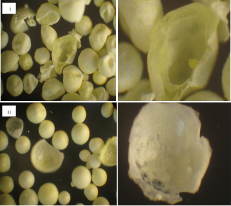
The microballoons prepared in this study, as observed in Figure 1, were near spherical in shape with a central hollow core surrounded by a shell as shown in the photograph of a broken half of a microballoon. The cavity was supposedly formed as follows according to the previous reports.19-21 As soon as the polymer solution was added to the aqueous medium, the ethanol diffused rapidly from the droplets of the polymer solution because of higher solubility of ethanol in water than dichloromethane. As ethanol diffuses through the aqueous solution, accompanied by simultaneous diffusion of water inside the sphere, the solubility of the polymer in the droplet is drastically reduced and reduces the coprecipitation of drug and polymer at the interface of the emulsion droplet, forming a solid shell covering the droplet. Dichloromethane evaporation appeared to be especially related to cavity formation in microballoons and properties of formed solid shell. Dichloromethane remaining as the central core diffused slowly due to its low water solubility. Therefore, the diffusion of dichloromethane began late, after the initial solidification, and formed a central hollow structure. As shown in photomicrographs the shape and shell thickness of the microballoons were different among the formulations. Preparation at higher polymer/drug ratio provided microballoons with a thin shell as to crumble upon touching and roundness close to 1 while the microballoons prepared at lower polymer/drug ratio showed thicker shell, probably arising as a trace of solvent evaporation.
Figure 1.
Particle morphology of microballoons (I): polymer: drug (1:1) and (II) polymer: drug (:1), right photographs (×100) left photographs (×500).
These results could be explained by considering the mechanism of formation of microballons as previously noted. Microballoons shells were conveniently formed by coprecipitation of drug and polymers, as well as successive evaporation of the inner solvent.
It is assumable that any factor which could modify the time needed to reach the physical state-transition boundaries of the polymeric solution (solution-gel-glass) during the hardening process will be effective in the polymer precipitation and consequently in solvent evaporation rate. 22The polymer -to- drug ratio can be a critical factor during microballoon formation. The increase in polymer/drug ratio leads to a shorter time for the composition of the polymer solution to reach the viscous (gelation) boundary, resulting in a rapid film-like membrane formation on the droplets periphery. If the film-like polymeric membrane is quickly solidified, the microballoon structure is more fixed and thus solvent evaporation is delayed.22As previously explained a slow rate of evaporation of dichloromethane led to formation of porous microballoons as dichloromethane remained in the pockets preventing formation of a hard polymer shell.
Thus it can be concluded that thin and porous structure of the microballoons shell prepared at higher polymer /drug ratio was attributed to slower rate of evaporation of dichloromethan as a consequence of rapid polymer shell formation.
On the other hand, the slower solidification of the polymeric shell at the droplet periphery due to decreasing polymer/drug ratio, delays creation of shell barrier to mass transfer, increases solvent evaporation rate and consequently increases the rigidity of the polymer shell.
Microballoons displaying diameters of between 250 and 380 µm (Table 1). Theparticle size increased with increasing the polymer / drug ratio. This is due to the increase in viscosity of the solution and the decrease in stirring efficiency. Also with increasing polymer/drug ratio, the hardening time of the microballoons was shortened. Consequently, a shorter time was provided for the breakup of droplets and larger microballoons were formed. Flowability results are shown in Table 1 as expressed in term of angle of repose and the compressibility index. All formulations showed high flowability as angle of repose ranged between 17.5 to 23 o and the compressibility index ranged between7% to 11%. The excellent flow property indicates that the floating microballoons produced are non-aggregated (Table 1).
Table 1. Mean diameter, angle of repose, Carr’s Index, yield and encapsulation efficiency of samples.
|
samples |
Mean diameter ± σ (µm) |
sphericity |
Angle of repose (o) |
Carr’s Index (%) |
Yield (%) |
Encapsulation efficiency (%) |
Buancy 8h (%) |
Apparent particle density (g/cm3) |
|
|
Untreated PX |
10.75±0.36 | - | 35.8±1.3 | 32.3±0.6 | - | - | - | - | |
| Eu:PX | 1:1 | 250±110 | 0.98±0.02 | 17.5±0.9 | 7.0± 0.2 | 73.8±0.8 | 90.8±0.5 | 62.5±1.0 | 0.5±0.03 |
| 2:1 | 290±80 | 0.96±0.03 | 20.7±1.0 | 10.8±0.3 | 70.6±1.0 | 92.5±0.6 | 77.7±0.9 | 0.29±0.03 | |
| 4:1 | 310±260 | 0.93±0.03 | 19.1±1.5 | 9.1±0.1 | 68.8±0.9 | 95.4±0.4 | 80.2±0.8 | 0.22±0.04 | |
| 6:1 | 380±220 | 0.92±0.02 | 23.0±0.6 | 11.0±0.3 | 65.5±1.1 | 98.3±0.4 | 85.4±1.1 | 0.19±0.04 | |
Yield and encapsulation efficiency
The percentage yield of floating microballoons determined by weighing after drying was73, 70, 68 and 65% for microballoons prepare at 1:1, 2:1, 4:1 and 6:1 polymer/drug ratios, respectively. Decreasing yield of microballoons by increasing polymer/drug ratio (p< 0.05) may be due to higher viscosity of polymer which resulted in agglomeration of droplets and adhering to the vessel wall and propeller. As can be seen in Table 1, the encapsulation efficiencies were high in all formulations (> 90%).The high encapsulation efficiencies probably result from the poor water solubility of the drug in the external aqueous phase. Slight loss of drug may be due to some dissolution prior to drop formation or during the hardening process in polyvinyl alcohol solution mixed with diffused ethanol.23The ratio ‘polymer / drug’ affected the resulting encapsulation efficiency. With more polymer/drug ratios, higher encapsulation efficiencies were produced. For instance, PX encapsulation efficiency was increased from 90.8% to 98.3 % as polymer:drug ratio was increased from 1:1 to 6:1.
This can either be attributed to the increased probability for the drug to be entrapped within the microballoons with increasing ratio of polymer to drug, and/or to the decreasing drug loss into the external water phase under these conditions. These results can be also related to hardening process of microballoons. Increasing polymer/drug ratio leads to a more viscous droplets and consequently faster solidification rate of microballoons. Therefore, the microballoon structure is more fixed and thus solvent and nonsolvent counter- diffusion is delayed. As a result, less water may be allowed to diffuse into the droplets and consequently less amount of drug will be carried by the solvent into the aqueous phase.22
DSC and X-ray diffraction results
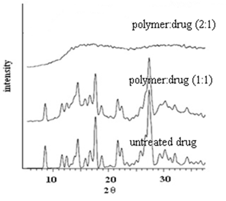
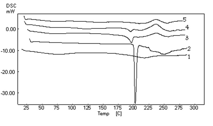
The DSC curves and X-ray diffraction pattern of the microballoons are shown in Figure 2 and Figure 3, respectively. As shown in Figure 2, the melting peak of the drug in the microballoons disappeared with increasing the ratio of polymer/drug from 1:1 to 2:1, although the melting peak was observed in the physical mixtures of the drug and polymer with the same ratio. These indicated that PX had been disordered in the microballoons at 2:1 polymer/ drug ratio, suggesting that PX had been highly dispersed at this ratio, so as amorphous state.
Figure 2.
DSC scans of 1) polymer 2) untreated drug 3) polymer:drug (1:1), 4) physical .
Figure 3.

The X-ray diffraction patterns of untreated drug and microballoons prepared at polymer: drug ratios of 1:1 and 2:1.
This was further supported by the X-ray analysis data given in Figure 3, which shows the representative diffraction patterns for the pure drug, the microballoons prepared at 1:1 and 2:1 polymer/drug ratios. Characteristic peaks of piroxicam appeared at a diffraction angle of 2θ, at 8.99, 15.76, 23.02 and 25.85°. These values were comparable to those reported for form I of PX. 24Crystalline peaks were observed in the 1:1polymer/drug ratio but no crystalline peaks of PX were found in the microballoons at 2:1 polymer /drug ratio. The scans and XRPD patterns of microballoons produced at 4:1 and 6:1 polymer/drug ratios are similar to 2:1 ratio and hence not all of them are shown. In summary, the DSC and XRPD data indicated no sign of interaction between the drug and the polymer and also showed that the PX inside the microballoons was changed only in its crystallinity.
Floating properties
To assess the floating properties, microballoons were placed in 0.1 HCl containing 0.02% polysorbate, in order to simulate surface tension of human gastric juice (35-50mN/m2).18 Buoyancy of beads is directly related to performance of floating-pulsatile drug delivery system since lag time for beads is equivalent to their floating time.
Apparent particle density and buoyancy of microballoons prepared at various polymer/drug ratios were measured. It is noticeable that during the floating test, no swelling or gelation of the microballoons was found. In accordance with polymer/drug ratio (1:1, 2:1, 4:1 and 6:1), buoyancy and apparent particle density of the resulting microballoons varied distinctly as presented in Table 1. According to results, by increasing the polymer/drug ratio, the buoyancy of the microballoons largely increased, accompanied by decreased particle density. For instance, the microballoons prepared at 6:1 polymer/drug showed highest buoyancy as a consequence of lowest particle density. Therefore, it was concluded that the buoyancy of the microballoons appeared to be more greatly affected by the apparent particle density. Decreasing apparent particle density with increasing polymer /drug ratio could be explained by considering the mechanism of formation of microballons as previously noted. As can be seen in Figure 1, preparation at higher polymer/drug ratio provided larger microballoons with thinner shell. As previously described, this was attributed to slow rate of evaporation of dichloromethane as a consequence of rapid polymer shell formation.
Moreover, in other researches, it was observed that the microballoons of larger size, showed the longer floating time. Therefore, increasing buoyancy of microballons by increasing the polymer/drug ratio may also be attributed to larger particle size by increasing polymer/drug ratio (Table 1).25,26
In vitro drug release
Release of the drug from Eu S100 based microballoons was evaluated in pH 1.2 and 7.4. Release studies in SGF for 6 h showed only 0.7–11% cumulative drug release from beads of different batches (Figure 4).
Figure 4.
Release of drug from microballoons into 0.1 N HCl (± SD., n=3).
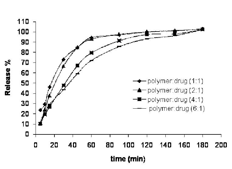
Since the acrylic polymer used is not soluble in acidic pH and starts to dissolve above pH 727, no marked amount of drug was released from the microballoons in SGF. Drug release from Eu S100 in SGF took place only through diffusion and drug release in SIF (pH 7.4) might have involved both mechanisms i.e. diffusion and erosion.
All the batches of beads showed fast disintegration and drug release in SIF due to pH dependent dissolving of Eudragit S. Complete drug dissolution from beads of each batch occurred during 3h in SIF, which would be useful in in vivo drug absorption from large surface area of small intestine. Small intestinal transit time for a drug formulation or for a meal is known to be almost constant at about 3 h.28 As shown in Figure 5, although the drug release from beads of all batches was sufficiently rapid in SIF, the composition of bead showed significant effect on drug release rate and the drug release was inversely related to polymer/drug ratio. For example, batches prepared by using 6:1or 4:1 polymer/drug ratios showed marked slower drug release than beads prepared by using 2:1or 1:1 polymer/drug ratios. This significant effect of polymer/drug ratio on drug release in SIF may be attributed to formation of stronger and thicker gel formation with increase in polymer/drug that restrict the drug diffusion through the beads.
Figure 5.

Release of drug from microballoons into pH 7.4 phosphate buffer (± SD., n=3).
Conclusion
As results showed, properties (buoyancy, apparent particle density, size and sphericity) of microballoons varied according to the polymer/drug ratio. Microballoons prepared at polymer/drug ratio higher than 2:1 were so brittle as to crumble upon touching and the buoyancy was higher probably due to lower apparent particle densities of these microballoons. The floating test revealed that approximately 85% of the microballoons with 6:1 polymer/drug ratio were still floating after 8h. During the floating test, no swelling or gelation of the microballoons was found, which suggested that they were dispersed individually in the stomach without adhesion to the mucosa. This property should avoid local irritation due to a localized overdose, which frequently occurs with the bioadhesion system. The beads showed fast drug release in SIF after sufficient floating time in SGF where no noticeable drug released. In conclusion, the piroxicam-loaded Eu S100 microballoons, prepared using a solvent diffusion method, have the qualities suitable for their successful use as pulsatile drug delivery system.
Acknowledgment
The financial support from the Drug applied Research Center of Tabriz University of Medical Sciences is greatly acknowledged.
Conflict of interest
The authors report no conflicts of interest. The authors alone are responsible for the content and writing of the paper.
References
- 1.Kikuchi A, Okano T. Pulsatile drug release control using hydrogels. Adv Drug Deliv Rev. 2002;54:53–77. doi: 10.1016/s0169-409x(01)00243-5. [DOI] [PubMed] [Google Scholar]
- 2.Sawada T, Kondo H, Nakashima H, Sako K, Hayashi M. Time-release compression-coated core tablet containing nifedipine for chronopharmacotherapy. Int J Pharm. 2004;280:103–111. doi: 10.1016/j.ijpharm.2004.05.004. [DOI] [PubMed] [Google Scholar]
- 3.Rubinstein A, Tirosh B, Baluom M, Nassar T, David A, Radai R, Gliko-kabir I, Friedman M. The rationale for peptide drug delivery to the colon and the potential of polymeric carriers as effective tools. J Control Release. 1997;46:59–73. [Google Scholar]
- 4.Terasawa E, Keen KL, Mogi K, Claude P. Pulsatile release of luteinizing hormone-release (LHRH) in cultured LHRH neurons derived from the embryolic olfactory placode of the rhesus monkey. Endocrinology. 1999;140:1432–1441. doi: 10.1210/endo.140.3.6559. [DOI] [PubMed] [Google Scholar]
- 5.Charloux A, Gronfier C, Lonsdorfer-wolf E, Piquard F, Brandenberger G. Aldosterone release during the sleep-wake cycle in humans. Am J Physiol. 1999;276:E43–E49. doi: 10.1152/ajpendo.1999.276.1.E43. [DOI] [PubMed] [Google Scholar]
- 6.Sawada T, Sako K, Yoshihara K, Nakamura K, Yokohama S, Hayashi M. Timed-release formulation to avoid drug–drug interaction between diltiazem and midazolam. J Pharm Sci. 2003;92:790–797. doi: 10.1002/jps.10336. [DOI] [PubMed] [Google Scholar]
- 7.Stehlin I. A time to heal: chronotherapy tunes in to body’s rhythms. FDA Consumer. 1997;31:16. [Google Scholar]
- 8.Crofford LJ, Kalogeras KT, Mastorakos G, Magiakou MA, Kanik KS, Gold PW, Chrousos GP, Wilder RL. Circadian relationships between interleukin (IL)-6 and hypothalamic-pitutary-adrenal axis hormones: failure of IL-6 to cause sustained hypercortisolism in patients with early untreated rheumatoid arthritis. J Clin Endocr Metab. 1997;82:1279–1283. doi: 10.1210/jcem.82.4.3852. [DOI] [PubMed] [Google Scholar]
- 9.Cutolo M, Seriolo B, Craviotto C, Pizzorni C, Sulli A. Circadian rhythms in RA. Ann Rheum Dis. 2003;62:593–596. doi: 10.1136/ard.62.7.593. [DOI] [PMC free article] [PubMed] [Google Scholar]
- 10.Niwa K, Takaya T, Morimoto T, Takada K. Preparation and evaluation of a time-controlled release capsule made of ethylcellulose for colon delivery of drugs. J Drug Target. 1995;3:83–89. doi: 10.3109/10611869509059209. [DOI] [PubMed] [Google Scholar]
- 11.Hoffman A, Stepensky D, Lavy E, Eyal S, Klausner E, Friedman M. Pharmacokinetic and pharmacodynamic aspects of gastroretentive dosage forms. Int J Pharm. 2004;277:141–153. doi: 10.1016/j.ijpharm.2003.09.047. [DOI] [PubMed] [Google Scholar]
- 12.Rouge N, Buri P, Doelker E. Drug absorption sites in the gastrointestinal tract and dosage forms for site-specific delivery. Int J Pharm. 1996;136:117–139. [Google Scholar]
- 13.Ritschel WA. Targeting in the gastrointestinal tract: new approaches. Methods Find Exp Clin Pharmacol. 1991;13:313–336. [PubMed] [Google Scholar]
- 14.Heynen G. Toleration and safety of piroxicam. Eur J Rheumatol Inflamm. 1987;8:86–93. [PubMed] [Google Scholar]
- 15.Pilpel N. The flow properties of magnesia. J Pharm Pharmacol. 1964;16:705–16. doi: 10.1111/j.2042-7158.1964.tb07394.x. [DOI] [PubMed] [Google Scholar]
- 16.Carr RL. Evaluation flow properties of solids. Chem Eng. 1965;72:163–8. [Google Scholar]
- 17.Carr RL. Classify flow properties of solids. Chem Eng. 1965;72:69–72. [Google Scholar]
- 18.El-gibaly I. Development and in vitro evaluation of novel floating chitosan microcapsules for oral use: comparison with non floating chitosan microballoons. Int J Pharm. 2002;249:7–21. doi: 10.1016/s0378-5173(02)00396-4. [DOI] [PubMed] [Google Scholar]
- 19.Sato Y, Kawashima Y, Takeuchi H, Yamamoto H. Physicochemical properties to determine the buoyancy of hollow microballoons (microballoons) prepared by the emulsion solvent diffusion method. Eur J Pharm Biopharm. 2003;55:297–304. doi: 10.1016/s0939-6411(03)00003-1. [DOI] [PubMed] [Google Scholar]
- 20.Kawashima Y, Niwa T, Takeuchi H, Hino T, Itoh Y. Preparation of multiple unit hollow microballoons (microballoons) with acrylic resin containing tranilast and their drug release characteristics (in vitro) and floating behavior (in vivo) J Control Release. 1991;16:279–290. [Google Scholar]
- 21.Kawashima Y, Niwa T, Takeuchi H, Hino T, Itoh Y. Hollow microballoons for use as a floating controlled drug delivery system in the stomach. J Pharm Sci. 1992;81:135–140. doi: 10.1002/jps.2600810207. [DOI] [PubMed] [Google Scholar]
- 22.Re MI, Biscans B. Preparation of microballoons of ketoprofen with acrylic polymers by a quasi-emulsion solvent diffusion method. Powder technology. 1999;101:120–133. [Google Scholar]
- 23.Zaniboni HC, Fell JT, Collett JH. Production and characterization of enteric beads. Int J Pharm. 1995;125:151–155. [Google Scholar]
- 24.Vrecer F, Vrbinc M, Meden A. Characterization of piroxicam crystal modifications. Int J Pharm. 2003;256:3–15. doi: 10.1016/s0378-5173(03)00057-7. [DOI] [PubMed] [Google Scholar]
- 25.Jain SK, Awasthi AM, Jain NK, Agrawal GP. Calcium silicate based microballoons of repaglinide for gastroretentive floating drug delivery: Preparation and in vitro characterization. J Control Release. 2005;107:300–309. doi: 10.1016/j.jconrel.2005.06.007. [DOI] [PubMed] [Google Scholar]
- 26.Singh AN, Pathak K. Development and evaluation of dual controlled release microballoons containing riboflavin and citric acid: in vitro and in vivo evaluation. J Microencapsul. 2011;28:442–54. doi: 10.3109/02652048.2011.576788. [DOI] [PubMed] [Google Scholar]
- 27.Yoo JW, Giri N, Lee CH. PH-sensitive Eudragit nanoparticles for mucosal drug delivery. Int J Pharm. 2011;403:262–267. doi: 10.1016/j.ijpharm.2010.10.032. [DOI] [PubMed] [Google Scholar]
- 28.Davis SS, Hardy JG, Fara JW. Transit of pharmaceutical dosage forms through the small intestine. Gut. 1986;27:886–892. doi: 10.1136/gut.27.8.886. [DOI] [PMC free article] [PubMed] [Google Scholar]


