Abstract
Purpose: This study aimed to evaluate the possible estrogenic activity of some ingredients of Nigella sativa including Linoleic acid and Gama-Linolenic acid by vaginal cornification assay. Methods: Forty ovariectomized (OVX) rats, aged 16 weeks were allotted randomly to five groups: negative control (taking 1 ml olive oil/ day); positive control (taking 0.2 mg/kg/day Conjucated Equine Estrogen-CEE); experimental groups (taking 50 mg/kg/day Linoleic acid or 10 mg/kg/day Gamma Linolenic acid or 15mg/kg/day Thymoquinone ). All of supplements administered via intragastric gavage for 21 consecutive days. To assess estrogen like activity, vaginal smear was examined daily and serum estradiol was measured at baseline, after 10 days and at the end of experiment. Results: The significant occurrence of vaginal cornification cell (p<0.05) after Linoleic acid supplementation indicated estrogenic activity of Linoleic acid which was in consistency with serum estradiol level, but this effect was not as much as CEE. Gama-Linolenic acid also exist a few cornified cell in smear which was not significantly differ from those control group. Conclusion: Linoleic acid showed the beneficial effects on OVX rats’ reproductive performance, thereby indicating its beneficial role in the treatment of the postmenopausal symptoms.
Keywords: Estrogenic Effects, Gama-Linolenic acid, Linoleic acid, Ovariectomized Rats, Vaginal Cornification Assay
Introduction
Menopause is the period in a woman’s life when hormonal changes cause menstruation to cease permanently1 and it is a natural part of the aging process. The experience of menopause varies greatly from one woman to another. For some, it is completely symptom free. Others may require assistance to cope with physical and psychological effects of menopause including hot flashes, vaginal atrophy, reductions in cardiovascular health and enhanced risk for developing osteoporosis and Alzheimer’s disease.2 For women requiring assistance, a range of options and supports are available such as lifestyle changes, medical treatments such as Hormone replacement therapy (HRT) and complementary approaches.3 Since some studies showed linkage between HRT use and some women cancers (e.g. Breast and endometrial cancer) and cardiovascular risk, therefore women tended to look for viable and safe alternatives.4 In addition due to the fear of developing cancer and discomfort, many users of HRT exhibit poor compliance.5 As a result, women frequently considered natural estrogenic alternatives for the treatment of menopausal pathologies and symptoms, because natural products offer the hope of improved safety and greater compliance.
Nigellasativa seeds have traditionally been used in Middle Eastern folk medicine as a natural remedy for various diseases as well as a spice for over 2000 years. The seeds of Nigellasativa have been subjected to a range of pharmacological, phytochemical and nutritional investigations in recent years.6-8It has been shown to contain more than 30% (w/w) of a fixed oil with 85% of total unsaturated fatty acid.9 Nigella sativa oil is a rich source of linoleic acid (LA) an omega‐6 fatty acid. Because estrogens have typically been used for the treatment of menopausal symptoms and because Nigella sativa have been shown to have a remarkable number of 23 sterols have been identified in the seed which can improve some symptoms associated with menopause, we investigated the potential estrogenic effects of some of its active ingredients. Although there have been no studies to determine the specific impact of LA and other principles of Nigella sativa on reproductive performance, previous studies have shown essential Fatty Acids (EFA) have been implemented as key nutrients in sustaining reproductive performance.10 In the present study, we evaluated the potential of some active ingredients on Nigella sativa including Linoleic Acid (LA), Gamma-Linolenic Acid (GLA) and thymoquinone (TQ) to exhibit estrogenic effects using vaginal cornification assay.
Materials and Methods
Experimental Design
In order to induce menopause and to investigate reproductive changes following supplementation with different ingredients of Nigella sativa, the rats were ovariectomized under a combination of xylazine and ketamine (10 mg/kg + 75 mg/kg, i.p. respectively) anesthesia. Bilateral ovariectomy was performed via a dorso-lateral approach with a small lateral vertical skin incision.11 Theovariectomized animals were acclimatized at the Animal House of Faculty of Medicine and Health Sciences for one month prior to supplementation. Five experimental rat groups were established with 8 rats per group. The groups were as follows: group 1, negative control (1 ml Olive Oil), group 2, positive control (0.2mg/kg/day CEE diluted in distilled water), group 3 Linoleic acid (daily 50 mg/kg LA which calculated based on yielding Nigella sativa fixed oil (29%) and concentration of LA (57%) in fixed oil), group 4, Thymoquinone (daily 15mg/kg TQ which calculated based on yielding Nigella sativa fixed oil (29%) and concentration of TQ (16.1%) in fixed oil which analyzed and reported by Latiff et al.,12 on the same plant source) and group 5, Gamma Linolenic acid (daily 10 mg/kg GLA which calculated based on yielding Nigella sativa fixed oil (29%) and concentration of GLA (2%) in fixed oil and probability of its production through conversion from LA). All ingredients were diluted in olive oil as vehicle. Dosage of the ingredients were selected based on the optimum desired effect of Nigella sativa and its extracts in the previous experiments,13,14 which was at low dose (300mg/kg BW/day) and were administered by intra-gastric gavage for 3 weeks. Serum estradiol were measured at baseline (day 0), 11th days, and at the end of experiment (21st day) and vaginal epithelium was checked daily.
Animals
Forty female Sprague–Dawley rats weighing between 250 and 350g aged 4 months were used in this study. They were supplied by animal house of Faculty of Medicine and Health Sciences, University Putra Malaysia (Serdang, Selangor, Malaysia). Rats were individually housed in stainless steel cages in a well ventilated room with a 12/12h light/dark cycle at an ambient temperature of 29–32 °C and 50- 60 % relative humidity. Experiments were carried out according to the guidelines for the use of animals and approved by the Animal Care and Use Committee of the Faculty of Medicine and Health Sciences, University Putra Malaysia with UPM/FPSK/PADS/BR/UUH/F01- 00220 reference number for notice of approval. They were fed standard rat chow pellets purchased from As-Sapphire (Selangor, Malaysia) and allowed to drink water ad libitum.
Chemicals
Linoleic Acid (95%), Gamma-Linolenic Acid and thymoquinone (99%) were obtained from Sigma-Aldrich Chemical Co. (St. Louis, MO, USA). Conjugated Equine Estrogen (0.625mg) was purchased from Wyeth, Montreal, Canada and prepared in a dosage of 0.2mg/kg15-17 by dissolving it in distilled water,13-15 and was used as a positive control for the purpose of comparison with the treated groups. All other reagents and chemicals were of analytical grade.
Blood collection
Fasting blood samples were collected under the deep ether anaesthesia by cardiac puncture using sterile disposable syringes at baseline (pre-treatment), day 11 (during treatment) and day 21 (after treatment). The blood samples were then centrifuged at 3000 rpm for 10 minutes to separate the serum. The serum was stored at -80°C until assays were carried out. Estradiol Radioimmunoassay (RIA) kit was purchased from Diagnostic Systems Laboratories (DSL), USA. The principle of the test is the competition of radioactive antigen and non-radioactive antigen for the fixed number of antibody binding sites. All tests were performed according to the manufacturer’s instructions.
Vaginal Smear
Vaginal smears were carried out to monitor cellular differentiation and to evaluate the presence of leukocytes, nucleated epithelial cells, or cornified cells. Vaginal smear samples were collected between 08.00 and 10.00 am daily. The vaginal smears were prepared by washing with 10 µl of normal saline (NaCl 0.9%) and were then thinly spread on a glass slide. They were allowed to dry at room temperature and then stained using Methylene blue dripping. The slides were rinsed in distilled water after 30 minutes and allowed to dry. The smears were studied using the light microscope (40x) and the cell type and their relative numbers were recorded. Vaginal smear cell counts were performed on 100 cells randomly. The percentage of cornified cells was determined according to Terenius18 using the following formula:

Statistical Analysis
Data were expressed as means ± standard deviation. The data were analyzed using SPSS Windows program version 15 (SPSS Institute, Inc., Chicago, IL, USA) statistical packages. The One-Way Analysis of Variance (ANOVA) and General linear Model (GLM) followed by Duncan Multiple Range Test (DMRT) were used to determine which ingredients of Nigella sativa showed optimum effects. A p-value less than 0.05 (p<0.05) was considered to be significant.
Results
Serum estradiol
Over the period of treatment, all groups showed reduction in the level of estradiol except positive control (CEE) which significantly increased (p<0.05). OVX rats supplemented with CEE showed 359% elevation in the estradiol level at the end of experiment. The means of serum estradiol level were not significantly different at baseline (day 0) among groups. In the first 10 days of treatment, serum estradiol level tend to reduce in TQ, GLA and control groups compared to baseline while in LA and CEE groups, the levels increase. There was a significant difference between estradiol level in CEE and other groups. Instead of a tendency to decrease in estradiol levels, the value of serum estradiol in LA (15.74± 4.39) remained much higher than other groups except CEE (Table 1). There was also a significant effect for treatments and duration of treatment (p<0.05) and the interaction effect (p<0.05).
Table 1. Means of serum estradiol (pg/ml) of OVX rats supplemented with various ingredients of Nigella sativa or Conjugated Equine Estrogen.
| Treatment | Day | Total | ||
| 0 | 11 | 21 | ||
| TQ | 13.23± 4.09 a | 9.84± 3.50 a | 7.55± 3.73 a | 9.93± 4.24 A |
| CEE | 14.88± 12.11a | 15.89± 13.37a | 53.51± 34.77 b | 28.09± 28.36 B |
| LA | 16.45± 5.53 a | 19.91± 2.68 a | 15.74± 4.39 a | 17.36± 4.54 A |
| GLA | 12.77± 3.10 a | 9.61± 2.07 a | 5.87± 3.31 a | 9.60± 4.00 A |
| C | 11.72± 7.43 a | 5.94± 4.15 a | 6.26± 5.51 a | 7.98± 6.22 A |
Data are expressed as Mean ± SD.
Treatment TQ=Thymoquinone (15mg/kg/day); LA=linoleic Acid (50mg/kg/day); GLA= Gamma Linolenic Acid (10mg/kg/day); CEE= conjugated equine estrogen (0.2mg/kg/day); and C= control (1 ml Olive Oil/day)groups.
AB: Comparison of the means between rows within column with different superscripts are significantly different at p<0.05.
XY: Comparison of the means between columns within row with different superscripts are significantly different at p<0.05.
ab: Comparison of the means between column and between row with different superscripts are significantly different at p<0.05.
Vaginal epithelial cell cornification
There was no significant difference in the percentage of cornified cells between groups at baseline and results confirmed a menopausal pattern in OVX rats. However after treatment, cornification was observed in all treatment groups which was significantly different from those negative control group (p<0.05) which remained in an atrophic pattern as observed in the absence of estrogen (Figures 1-5). In the first 10 days of treatment, percentage of cornified cells increased significantly (p<0.05) in all groups except control group. Extending the supplementation period to 21 days, consistently increased percentage of cornified cells among LA and CEE groups until end of the treatment period, while control group remained unchanged until the end of the experiment.
Figure 1.
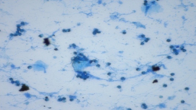
Vaginal smear of ovariectomized rat treated with thymoquinone (15mg/kg) for 3 weeks. A few number of cornified cells and also leukocytes are observed (methylene blue staining, 40x).
Figure 2.
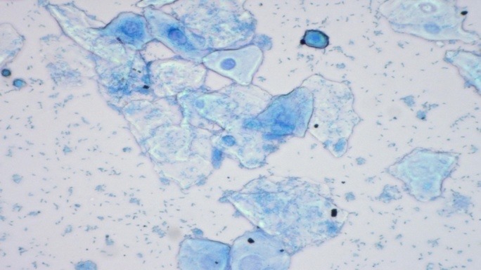
Vaginal smear of ovariectomized rat treated with conjugated equine estrogen (0.2 mg/kg) for 3 weeks. A great number of cornified cells and also nucleated epithelial cells are observed (methylene blue staining, 40x).
Figure 3.
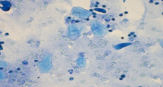
Vaginal smear of ovariectomized rat treated with linoleic acid (50mg/kg) for 3 weeks. A few number of cornifiedcells, nucleated epithelial cells and also leukocytes are observed (methylene blue staining, 40x).
Figure 4.
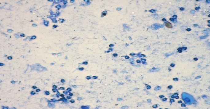
Vaginal smear of ovariectomized rat treated with gamma linoleic acid (10mg/kg) for 3 weeks. A few number of nucleated epithelial cells and also leukocytes are observed (methylene blue staining, 40x).
Figure 5.
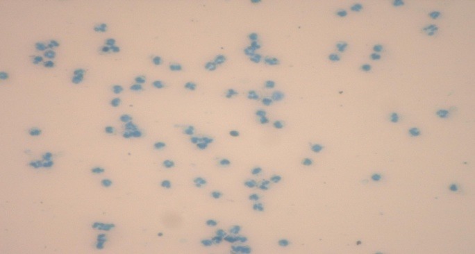
Vaginal smear of ovariectomized rat from control group treated with olive oil for 3 weeks. A great number of leukocytes are observed (methylene blue staining, 40x).
Discussion
In the current study, we compared the possible beneficial effects of active ingredients of Nigella sativa, thymoquinone, linoleic acid and gamma linolenic acid on reproduction function in OVX induced rats. Results indicated that active ingredients of Nigella sativa, linoleic acid, had a weak estrogenic effect as shown in its effect in serum estrogen level and percent of cornified cells. The results, however, fail to show a linear consistent time dependent effect of linoleic acid on the parameter studied. In general the level of estradiol was much higher in linoleic acid group compare to control and other active ingredients. Several studies have suggested that diet, particularly one enriched with either saturated or unsaturated fatty acids can alter serum steroid concentrations in a variety of species, including rodents and humans.19-23 The mechanisms underlying diet-induced alteration of steroid concentration are likely complex. Dietary fat can influence the expression of enzymes that metabolize sex steroid hormones.24,25 Adipose tissue is an important site of steroid hormone biosynthesis.26,27 Moreover, ovarian derived ∆4 androstenedione and testosterone can be aromatized in adipose tissue to estrone and estradiol respectively.28 An additional potential mechanism of dietary influence on sex steroid concentration relates to the status of cholesterol as a precursor to steroid hormones. Diet can influence serum cholesterol29 and high cholesterol is correlated with high serum androgen and estrogen concentrations.30,31 Past studies in rodents, cattle, and humans have indicated that diet might underpin changes in serum hormonal concentrations, including testosterone and estrogen.20,22,32 Female rats fed a diet enriched with n-3 polyunsaturated fatty acids had a 48% increase in serum concentrations of 17β-estradiol compared with rats fed a diet enriched with n-6 fatty acids.18 Similarly, female rats fed a low protein diet had a significant increase in 17β-estradiol compared with those fed a control diet.32 A high saturated fat diet induces an increase in estrogen, estrone, and dehydroepiandrosterone sulfate concentrations in women.33 A controlled clinical trial revealed that girls fed a low fat (LF) diet exhibited higher serum testosterone concentrations during the luteal phase of the cycle but lower estradiol concentrations.22 In contrast, in other study there were no significant correlations between any measure of fat (total fat, saturated fat, and linoleic acid) and serum estrone and estradiol in 325 climacteric US women.34
The insignificant change in the levels of estrogen in the present study suggests that linoleic acid may act directly on the estrogen receptors without enhancing the endogenous estrogen levels.
Linoleic acid is a fatty acid, which is ubiquitous in nature. Some fatty acids have been reported to bind noncompetitively or with mixed-competition to a variety of receptors most likely based on hypodrophobic interactions.35-38 Arachidonic acid, palmitic acid, stearic acid, oleic acid, and docosahexaenoic acid have been reported to bind to the estrogen, progesterone, androgen, and glucocorticoid receptors at weak binding sites different from the endogenous steroid binding site.35 Linoleic acid demonstrated the ability to interact with the opioid receptor and the nucleoside transport protein.38
The relationship between dietary fat and changes in reproductive function is not limited to affects on cholesterol and progesterone. Staples and Thatcher39 proposed an elegant control feedback system that involves not only progesterone, but also affects prostaglandin synthesis and the role estrogen plays in biological (cellular) function as illustrated in Figure 6. Polyunsaturated fatty acids are proposed to decrease the release of prostaglandins that would augment the establishment of a pregnancy. In addition, the PUFA also decrease the effects of estradiol that enhance the action of prostaglandins.
Figure 6.
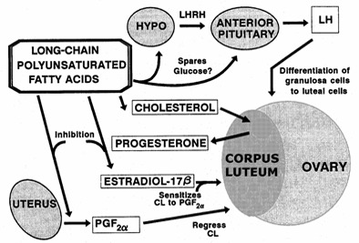
Possible mechanisms for the role of polyunsaturated fatty acids on reproductive function in dairy cows.(Staples and Thatcher, 1999).
Liu et al.,40 reported the identification of linoleic acid as an estrogen receptor ligand capable of displacing estradiol from the ER and binding to the ligand binding domain of the protein using competitive binding assays and pulsed ultra filtration. They evaluated several other fatty acids for binding to the estrogen receptor. Among the 20 fatty acids tested, 13 bound to ER α and six bound to ER β. In general, fatty acids shorter than 16 carbons did not bind to the receptor; however, saturated acids had no obvious selectivity for the receptor compared with unsaturated acids.
Previous studies demonstrated the ability of conjugated linoleic acid to bind to PPAR gamma and alter the expression of some genes regulated by an estrogen response element (ERE).41 Liu and his colleagues40 reported that linoleic acid present in the fruits of V. agnus-castus can bind to estrogen receptors and induce certain estrogen inducible ER mRNA up-regulation. The interaction of linoleic acid with the estrogen receptor did increase the mRNA of estrogen inducible genes in Ishikawa and T47:A18 cells. These data suggest that the likely pathway for upregulation of genes regulated by ERE natural promoters, such as the ones reported in previous studies, is by linoleic acid binding to and activating estrogen receptors. Additional characterization must be completed to determine if Nigella sativa contains more compounds that interact with estrogen receptors and stimulate estrogen inducible genes. Functional assays should be used to determine if linoleic acid bound to nuclear receptors have any effect on the regulation of gene expression.
Conclusion
The observed estrogenic effect following linoleic acid treatment suggests that this fatty acid could possibly act on the estrogen receptors with enhancing the endogenouse estrogen levels. Linoleic acid showed the beneficial effects on OVX rats’ reproductive performance, thereby indicating its beneficial role in the treatment of the postmenopausal symptoms.
Acknowledgement
The authors would like to thank University Putra Malaysia for its financial support of this research project.
Conflict of Interest
The authors report no conflicts of interest.
References
- 1.Andrews WC. The transitional years and beyond. Obstet Gynecol . 1995;85(1):1–5. doi: 10.1016/0029-7844(94)00253-a. [DOI] [PubMed] [Google Scholar]
- 2.Colditz GA, Hankinson SE, Hunter DJ, Willett WC, Manson JE, Stampfer MJ. et al. The use of estrogens and progestins and the risk of breast cancer in postmenopausal women. New Engl J Med . 1995; 332:1589–93. doi: 10.1056/NEJM199506153322401. [DOI] [PubMed] [Google Scholar]
- 3.Bones O. Menopause: What causes the menopause? Symptoms, Health risks.Medical NewsToday 17th July 2006. [Google Scholar]
- 4.Messina MJ, Persky V, Setchell KD, Barnes S. Soy intake and cancer risk: a review of the in vitro and in vivo data. Nutr Cancer . 1994;21:113–31. doi: 10.1080/01635589409514310. [DOI] [PubMed] [Google Scholar]
- 5.De Lignieres B. Hormone replacement therapy: clinical benefits and side effects. Maturitas . 1996; 23 Suppl:S31–6. doi: 10.1016/s0378-5122(96)90012-2. [DOI] [PubMed] [Google Scholar]
- 6.Coban S, Yildiz F. The effects of Nigella sativa on bile duct ligation induced-liver injury in rats. Cell Biochem Funct . 2010; 28(1):83–8. doi: 10.1002/cbf.1624. [DOI] [PubMed] [Google Scholar]
- 7.Al-Nazawi MH, El-Bahr SM. Hypolipidemic and Hypocholestrolemic Effect of Medicinal Plant Combination in the Diet of Rats: Black Cumin Seed (Nigella sativa) and Turmeric (Curcumin) J Anim Vet Adv . 2012;11:2013–9. [Google Scholar]
- 8.Akhtar M, Maikiyo AM. Ameliorating effects of two extracts of Nigella sativa in middle cerebral artery occluded rat. J Pharm Bioallied Sci . 2012;4(1):70–5. doi: 10.4103/0975-7406.92740. [DOI] [PMC free article] [PubMed] [Google Scholar]
- 9.Houghton PJ, Zarka R, Heras BDL, Hoult JRS. Fixed oil of Nigella sativa and derived thymoquinone inhibit eicosanoid generation in leukocytes and membrane lipid peroxidation. Planta Medica . 1994;61:33–6. doi: 10.1055/s-2006-957994. [DOI] [PubMed] [Google Scholar]
- 10.Zeitlin L, Segev E, Fried A, Wientroub S. Effects of long-term administration of N-3 polyunsaturated fatty acids (PUFA) and selective estrogen receptor modulator (SERM) derivatives in ovariectomized (OVX) mice. J Cell Biochem . 2003; 90(2):347–60. doi: 10.1002/jcb.10620. [DOI] [PubMed] [Google Scholar]
- 11.Parhizkar S, Rashid I, Latiffah AL. Incision Choice in Laparatomy: a Comparison of Two Incision Techniques in Ovariectomy of Rats. World ApplSci J . 2008;4(4):537–40. [Google Scholar]
- 12.Latiffah AL, Hassanzadeh Ghahramanloo K, Hanachi P. Comparative analysis of essential oil composision of Iranian and Indian Nigella sativa extracted by using Supercritical Fluid Extraction (SFE) and solvent extraction. Clin Biochem . 2011; 44(13):S20. doi: 10.2147/DDDT.S87251. [DOI] [PMC free article] [PubMed] [Google Scholar]
- 13.Parhizkar S, Latiff LA, Rahman SA, Dollah MA, Hanachi P. Assessing estrogenic activity of Nigella sativa in ovariectomized rats using vaginal cornification assay. Afr J Pharm Pharacol . 2011; 5(2):137–42. [Google Scholar]
- 14.Parhizkar S, Latiffah AL, Sabariah AR, Dollah MA. Preventive effect of Nigella sativa on metabolic syndrome in Menopause Induced Rats. J Med Plants Res . 2011; 5(8):1478–84. [Google Scholar]
- 15.Genazzani AR, Stomati M, Bernardi F, Luisi S, Casarosa E, Puccetti S. et al. Conjugated equine estrogens reverse the effects of aging on central and peripheral allopregnanolone and beta-endorphin levels in female rats. FertilSteril. 2004;81(1):757–66. doi: 10.1016/j.fertnstert.2003.08.022. [DOI] [PubMed] [Google Scholar]
- 16.Oropeza MV, Orozco S, Ponce H, Campos MG. Tofupill lacks peripheral estrogen-like actions in the rat reproductive tract. Reprod Toxicol . 2005;20:261–6. doi: 10.1016/j.reprotox.2005.02.007. [DOI] [PubMed] [Google Scholar]
- 17.Araujo LBF, Soares JM, Simoes RS, Santos RS, Calió PL, Oliveira-Filho, et al. Effect of conjugated equine estrogens and tamoxifen administration on thyroid gland histomorphology of the rat. Clinics 2006; 61: 321-6. [DOI] [PubMed] [Google Scholar]
- 18.Terenius L. The Allen-Doisy test for estrogens reinvestigated. Steroids . 1971;17:653–61. doi: 10.1016/0039-128x(71)90081-x. [DOI] [PubMed] [Google Scholar]
- 19.Talavera F, Park CS, Williams GL. Relationships among dietary lipid intake, serum cholesterol and ovarian function in Holstein heifers. J AnimSci. 1985;60:1045–51. doi: 10.2527/jas1985.6041045x. [DOI] [PubMed] [Google Scholar]
- 20.Hilakivi-Clarke L, Cho E, Cabanes A, DeAssis S, Olivo S, Helferich W. et al. Dietary modulation of pregnancy estrogen levels and breast cancer risk among female rat offspring. Clin Cancer Res . 2002;8:3601–10. [PubMed] [Google Scholar]
- 21.Woods MN, Barnett JB, Spiegelman D, Trail N, Hertzmark E, Longcope C. et al. Hormone levels during dietary changes in premenopausal African–American women. J Natl Cancer Inst . 1996; 88:1369–74. doi: 10.1093/jnci/88.19.1369. [DOI] [PubMed] [Google Scholar]
- 22.Dorgan JF, Hunsberger SA, McMahon RP, Kwiterovich PJ, Lauer RM, Van Horn L. et al. Diet and sex hormones in girls: findings from a randomized controlled clinical trial. J Natl Cancer Inst. 2003;95:132–45. doi: 10.1093/jnci/95.2.132. [DOI] [PubMed] [Google Scholar]
- 23.Whyte JJ, Alexenko AP, Davis AM, Ellersieck MR, Fountain ED, Rosenfeld CS. Maternal diet composition alters serum steroid and free fatty acid concentrations and vaginal pH in mice. J Endocrinol. 2007;192:75–81. doi: 10.1677/JOE-06-0095. [DOI] [PubMed] [Google Scholar]
- 24.Zhou Y, Lin S, Chang HH, Du J, Dong Z, Dorrance AM. et al. Gender differences of renal CYP-derived eicosanoid synthesis in rats fed a high-fat diet. Am J Hypertens. 2005;18:530–7. doi: 10.1016/j.amjhyper.2004.10.033. [DOI] [PubMed] [Google Scholar]
- 25.Dieudonne MN, Sammari A, Dos Santos E, Leneveu MC, Giudicelli Y, Pecquery R. Sex steroids and leptin regulate 11beta-hydroxysteroid dehydrogenase I and P450 aromatase expressions in human preadipocytes: sex specificities. J Steroid Biochem Mol Biol. 2006;99:189–96. doi: 10.1016/j.jsbmb.2006.01.007. [DOI] [PubMed] [Google Scholar]
- 26.Belanger C, Luu-The V, Dupont P, Tchernof A. Adipose tissue intracrinology: potential importance of local androgen/estrogen metabolism in the regulation of adiposity. Horm Metab Res . 2002;34:737–74. doi: 10.1055/s-2002-38265. [DOI] [PubMed] [Google Scholar]
- 27.Simpson ER. Sources of estrogen and their importance. J Steroid Biochem Mol Biol . 2003; 86:225–30. doi: 10.1016/s0960-0760(03)00360-1. [DOI] [PubMed] [Google Scholar]
- 28.Lambrinoudaki I, Christodoulakos G, Rizos D, Economou E, Argeitis J, Vlachou S. et al. Endogenous sex hormones and risk factors for atherosclerosis in healthy Greek postmenopausal women. Eur J Endocrinol . 2006;154:907–16. doi: 10.1530/eje.1.02167. [DOI] [PubMed] [Google Scholar]
- 29.Menotti A. Diet, cholesterol and coronary heart disease. A perspective. Acta Cardiol . 1999; 54:169–72. [PubMed] [Google Scholar]
- 30.Shelley JM, Green A, Smith AM, Dudley E, Dennerstein L, Hopper J. et al. Relationship of endogenous sex hormones to lipids and blood pressure in mid-aged women. Ann Epidemiol. 1998;8:39–45. doi: 10.1016/s1047-2797(97)00123-3. [DOI] [PubMed] [Google Scholar]
- 31.Kumagai S, Kai Y, Sasaki H. Relationship between insulin resistance, sex hormones and sex hormone-binding globulin in the serum lipid and lipoprotein profiles of Japanese postmenopausal women. J Atheroscler Thromb . 2001;8:14–20. doi: 10.5551/jat1994.8.14. [DOI] [PubMed] [Google Scholar]
- 32.Fernandez-Twinn DS, Ozanne SE, Ekizoglou S, Doherty C, James L, Gusterson B, Hales CN. The maternal endocrine environment in the low-protein model of intra-uterine growth restriction. Br J Nutr . 2003;90:815–22. doi: 10.1079/bjn2003967. [DOI] [PubMed] [Google Scholar]
- 33.Nagata C, Nagao Y, Shibuya C, Kashiki Y, Shimizu H. Fat intake is associated with serum estrogen and androgen concentrations in postmenopausal Japanese women. J Nutr . 2005;135:2862–5. doi: 10.1093/jn/135.12.2862. [DOI] [PubMed] [Google Scholar]
- 34.Newcomb PA, Klein R, Klein BEK, Haffner S, Mares-Perlman J, Cruickshanks KJ. et al. Association of dietary and life-style factors with sex hormones in postmenopausal women. Epidemiol . 1995;6:318–21. doi: 10.1097/00001648-199505000-00022. [DOI] [PubMed] [Google Scholar]
- 35.Kato J. Arachidonic acid as a possible modulator of estrogen, progestin, androgen, and glucocorticoid receptors in the central and peripheral tissues. J Steroid Biochem . 1989;34:219–27. doi: 10.1016/0022-4731(89)90085-x. [DOI] [PubMed] [Google Scholar]
- 36.Vallette G, Vanet A, Sumida C, Nunez EA. Modulatory effects of unsaturated fatty acids on the binding of glucocorticoids to rat liver glucocorticoid receptors. Endocrinol. 1991;129:1363–9. doi: 10.1210/endo-129-3-1363. [DOI] [PubMed] [Google Scholar]
- 37.Kang JX, Leaf A. Effects of long-chain polyunsaturated fatty acids on the contraction of neonatal rat cardiac myocytes. Proc Natl Acad Sci USA . 1994;91:9886–90. doi: 10.1073/pnas.91.21.9886. [DOI] [PMC free article] [PubMed] [Google Scholar]
- 38.Ingkaninan K, von Frijtag D, Kunzel JK, IJzerman AP, Verpoorte R. Interference of linoleic acid fraction in some receptor binding assays. J Nat Prod . 1999;62:912–4. doi: 10.1021/np9805490. [DOI] [PubMed] [Google Scholar]
- 39.Staples CR, Thatcher WW. Fat supplementation may improve fertility of lactating dairy cows. Proceedings of the Southeast Dairy Herd Management Conference. 1999. Macon, GA. 56.
- 40.Liu M, Xu X, Rang W, Li Y, Song Y. Influence of ovariectomy and 17β-estradiol treatment on insulin sensitivity, lipid metabolism and post-ischemic cardiac function. Int J cardiol. 2004;97(3):485–93. doi: 10.1016/j.ijcard.2003.11.046. [DOI] [PubMed] [Google Scholar]
- 41.Stoll BA. Linkage between retinoid and fatty acid receptors: Implications for breast cancer prevention. Eur J Cancer Prev . 2002;11:319–25. doi: 10.1097/00008469-200208000-00002. [DOI] [PubMed] [Google Scholar]


